

Cardiac computed tomography in cardio-oncology: an update on recent clinical applications
- PMID: 33555007
- PMCID: PMC8453276
- DOI: 10.1093/ehjci/jeaa351
Cardiac computed tomography in cardio-oncology: an update on recent clinical applications
Abstract
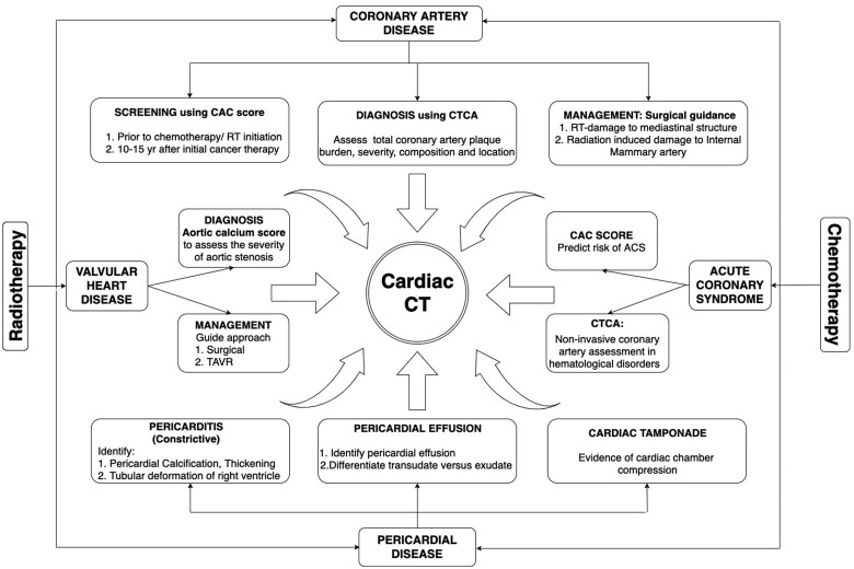
Chemotherapy and radiotherapy have drastically improved cancer survival, but they can result in significant short- and long-term cardiovascular complications, most commonly heart failure from chemotherapy, whilst radiotherapy increases the risk of premature coronary artery disease (CAD), valve, and pericardial diseases. Cardiac computed tomography (CT) with calcium scoring has a role in screening asymptomatic patients for premature CAD, cardiac CT angiography (CTCA) allows the identification of significant CAD, also in the acute settings where concerns exist towards invasive angiography. CTCA integrates the diagnostic work-up and guides surgical/percutaneous management of valvular heart diseases and allows the assessment of pericardial conditions, including detection of effusion and pericardial calcification. It is a widely available and fast imaging modality that allows a one-step evaluation of CAD, myocardial, valvular, and pericardial disease. This review aims to provide an update on its current use and accompanying evidence-base for cardiac CT in the management of cardio-oncology patients.
Keywords: cardiac CT; cardio-oncology; coronary artery disease; pericardial disease; valvular disease.
Published on behalf of the European Society of Cardiology. All rights reserved. © The Author(s) 2021. For permissions, please email: journals.permissions@oup.com.
Figures
References
-
- Celutkiene J, Pudil R, Lopez-Fernandez T, Grapsa J, Nihoyannopoulos P, Bergler-Klein J et al. Role of cardiovascular imaging in cancer patients receiving cardiotoxic therapies: a position statement on behalf of the Heart Failure Association (HFA), the European Association of Cardiovascular Imaging (EACVI) and the Cardio-Oncology Council of the European Society of Cardiology (ESC). Eur J Heart Fail 2020;22:1504–24. - PubMed
-
- Achenbach S, Marwan M, Ropers D, Schepis T, Pflederer T, Anders K et al. Coronary computed tomography angiography with a consistent dose below 1 mSv using prospectively electrocardiogram-triggered high-pitch spiral acquisition. Eur Heart J 2010;31:340–6. - PubMed
Publication types
MeSH terms
LinkOut - more resources
Full Text Sources
Other Literature Sources
Medical
Miscellaneous
