Review
. 2021 Feb 23;15(2):2068-2098.
doi: 10.1021/acsnano.0c09115. Epub 2021 Feb 8.

Harnessing Endogenous Stimuli for Responsive Materials in Theranostics

Affiliations
Review

Harnessing Endogenous Stimuli for Responsive Materials in Theranostics

Alexander B Cook et al. ACS Nano. .

Abstract

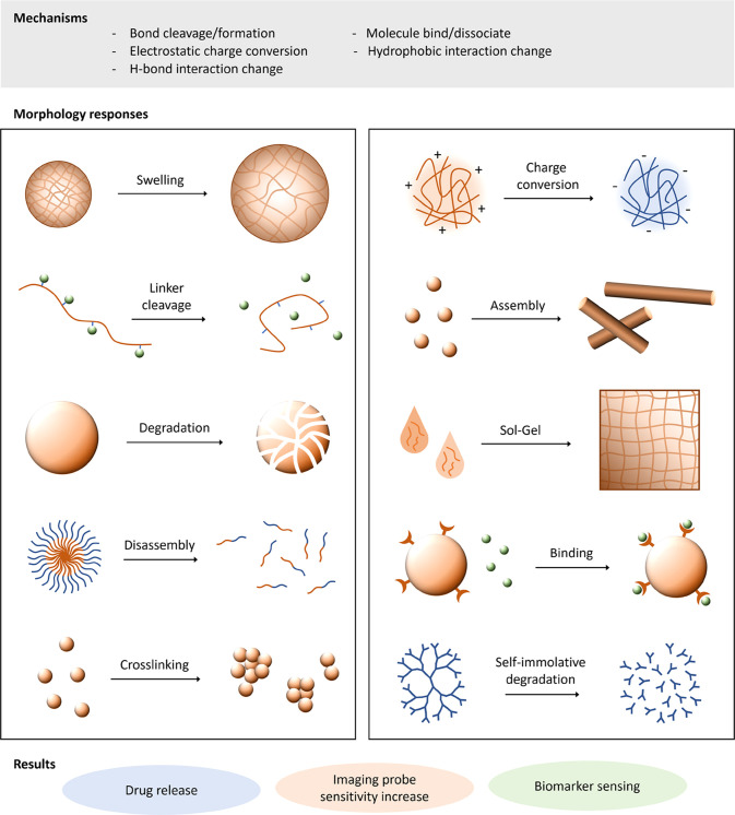
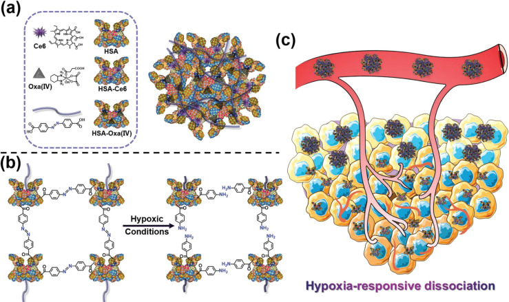
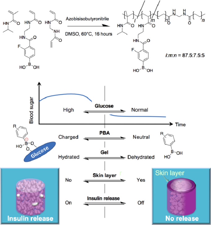
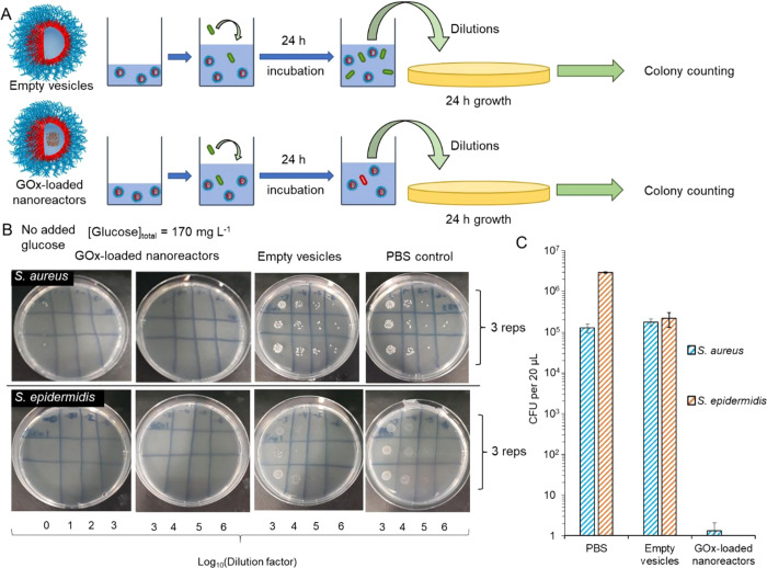
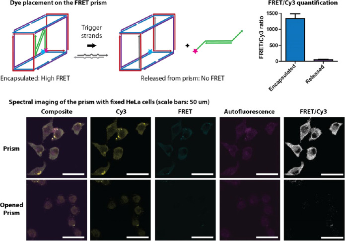
Materials that respond to endogenous stimuli are being leveraged to enhance spatiotemporal control in a range of biomedical applications from drug delivery to diagnostic tools. The design of materials that undergo morphological or chemical changes in response to specific biological cues or pathologies will be an important area of research for improving efficacies of existing therapies and imaging agents, while also being promising for developing personalized theranostic systems. Internal stimuli-responsive systems can be engineered across length scales from nanometers to macroscopic and can respond to endogenous signals such as enzymes, pH, glucose, ATP, hypoxia, redox signals, and nucleic acids by incorporating synthetic bio-inspired moieties or natural building blocks. This Review will summarize response mechanisms and fabrication strategies used in internal stimuli-responsive materials with a focus on drug delivery and imaging for a broad range of pathologies, including cancer, diabetes, vascular disorders, inflammation, and microbial infections. We will also discuss observed challenges, future research directions, and clinical translation aspects of these responsive materials.

Keywords: biological stimuli; enzymes; formulations; materials; nanomedicine; nanoparticles; pH; responsive polymers.


Conflict of interest statement

The authors declare no competing financial interest.

Figures

Figure 1
Figure 1
Schematic showing design synergies between materials’ physiochemical properties and biological environments. Biomaterials varying in shape and size from centimeters to nanometers can be applied in tissue/cell environments having structural features across similar length scales, while also expressing various cues able to be recognized by endogenous stimuli-responsive materials (including enzymes, pH, redox, glucose, hypoxia, ATP, and nucleic acids).
Figure 2
Figure 2
Material response to endogenous stimuli trigger can be diverse. Response can be due to changes in covalent bonding, electrostatic charge, H-bonding interactions, or other intermolecular interactions, leading to morphology changes in materials from polymer conjugates to nanoparticles, self-assembled systems, or hydrogels. These materials’ response mechanisms have seen applications in drug release, biomarker sensing, and activatable imaging agents.
Figure 3
Figure 3
Study of supramolecular aggregation-induced emission nanodots, with MMP responsiveness for image-guided cancer treatment. Nanoparticles formed via host–guest assembly and multiple stages of tumor-microenvironment enzymatic activation and GSH-triggered drug release are shown schematically. Reproduced with permission from ref (87). Copyright 2020 American Chemical Society.
Figure 4
Figure 4
Thrombin-responsive PEG-based heparin hydrogels used in the treatment of coagulation-related diseases. (a) Thrombin generation from prothrombin. (b) Selective peptide linker cleavage, releasing heparin. (c) Heparin-catalyzed thrombin deactivation. (d) Gel degradation halted on inactivation of thrombin in a self-regulated release mechanism. Reproduced with permission from ref (92). Copyright 2013 Springer Nature.
Figure 5
Figure 5
Application of cathepsin B in theranostic responsive systems. (A) Designed FRET imaging response of polymer conjugate on exposure to enzyme cathepsin B (papain). (B) Cy5 FRET ratio of conjugate when incubated with different concentrations of papain. (C) Cy5 FRET ratio change over time (4 h with 5 × 10⁻⁶ M papain). (D) In vivo FRET imaging of mice bearing A2780 human ovarian tumor after intravenous administration of polymer conjugate. Reproduced with permission from ref (102). Copyright 2017 Wiley VCH.
Figure 6
Figure 6
Application of lipase in theranostic responsive systems. (a) Antibacterial properties of a lipase-responsive theranostic wound dressing system against P. aeruginosa and E. coli over time. (b) Color change after 4 h incubation with bacteria. (c) Schematic of the enzyme activation system. Reproduced with permission from ref (112). Copyright 2019 American Chemical Society.
Figure 7
Figure 7
Design strategy for an enzyme-responsive polymer–drug conjugate which forms 5–10 nm assemblies. (a) Schematic illustrating the proposed polycation-induced transcytosis tumor penetration. (b) Conjugate chemical structure and charge-switching responsive behavior. (c) Zeta potential changes on exposure to membrane γ-glutamyl transpeptidase. Reproduced with permission from ref (119). Copyright 2019 Springer Nature.
Figure 8
Figure 8
Summary of the functional groups and linkers used in the development of pH-sensitive materials. (A) pH-responsive cationic polymers: (i) poly(2-dimethylaminoethyl methacrylate) (PDMAEMA), (ii) poly(2-diethylaminoethyl methacrylate) (PDEAEMA), (iii) poly(2-diisopropylaminoethyl methacrylate) (PDPAEMA), (iv) poly(4-vinylpyridine) (PVP), (v) poly(4-(1H-imidazol-1-yl)butyl methacrylate (PImBuMA), (vi) poly(lysine) (PLys), (vii) poly(histidine) (PHis), (viii) poly(ethylenimine) (PEI), (ix) chitosan, and (x) poly(β-amino ester) (PbAE). (B) pH-responsive anionic polymers: (i) poly(acrylic acid) (PAA), (ii) poly(2-carboxyethyl acrylate) (PCEA), (iii) poly(2-propylacrylic acid) (PPAA), (iv) poly(4-vinylbenzoic acid) (PVBA), (v) poly(aspartic acid) (PAsA), (vi) poly(glutamic acid) (PGA), (vii) poly(vinylsulfonic acid) (PVSA), (viii) poly(vinylphenylboronic acid) (PVPBA), and (ix) hyaluronic acid. (C) pH-cleavable linkers: (i) ortho-esters, (ii) ketals/acetals, (iii) hydrazones, (iv) imines, (v) maleic acid amide derivatives (including cis-aconityl shown), (vi) silyl ethers, and (vii) trityl derivatives.
Figure 9
Figure 9
Tumor microenvironment-targeted delivery of antimiRs using a peptide with a low-pH-induced transmembrane structure (pHLIP). Distribution of pHLIP labeled with Alexa Fluor 750, 36 h after systemic administration to (a) nude mouse with miR-155 flank tumors and (b) mouse with lymphadenopathy. (c) Schematic of pHLIP-mediated antimiR delivery. Reproduced with permission from ref (128). Copyright 2015 Springer Nature.
Figure 10
Figure 10
Application of pH-responsive polymer nanoparticles in pain management. (a) Schematic of pH-responsive polymer nanoparticles (from P(PEGMA-co-DMAEMA) shell blocks and P(DIPMA-co-DEGMA) or P(BMA) core-forming blocks) which target the neurokinin 1 receptor in endosomes to treat chronic pain. (b) Particle characterization data. (c) Particle TEM images. (d) Particle pH-responsive behavior characterization. Reproduced with permission from ref (175). Copyright 2019 Springer Nature.
Figure 11
Figure 11
(a) Schematic showing the generation of hypoxia-responsive human serum albumin (HSA)-based nanosystems with photosensitizer chlorin e6, oxaliplatin prodrug, and hypoxia-responsive azobenzene cross-linking groups. (b) Illustration of the hypoxia response mechanism. (c) Tumor activation schematic. Reproduced with permission from ref (202). Copyright 2019 Wiley VCH.
Figure 12
Figure 12
Boronate gel-based insulin delivery system. Schematic showing the chemical structure of the boronate gel-based insulin delivery system and optimal glucose-responsive insulin delivery under physiological conditions (threshold concentration of glucose at normoglycemic (100 mg/dL), above which the gel delivers insulin). Reproduced with permission from ref (253). Copyright 2017 The Authors, some rights reserved; distributed under a Creative Commons Attribution-NonCommercial License 4.0 (CC BY-NC).
Figure 13
Figure 13
Glucose oxidase-loaded polymer vesicles. (a) Testing protocol for GOx-loaded polymer vesicles and empty vesicles against Gram-positive bacteria. (b) Nutrient agar plates showing associated viability of S. aureus and S. epidermidis after 24 h incubation. (c) Colony forming units quantification of each bacterial species. Reproduced with permission from ref (263). Copyright 2020 American Chemical Society.
Figure 14
Figure 14
Nucleic acid-based container device. Schematic of the design, assembly, and properties of the nucleic acid origami container device, including FRET quantification of the Cy3 ratio before and after cargo release and images obtained of the nucleic acid system incubated with cells before and after cargo release. Reproduced with permission from ref (297). Copyright 2016 American Chemical Society.

References

    1. Blum A. P.; Kammeyer J. K.; Rush A. M.; Callmann C. E.; Hahn M. E.; Gianneschi N. C. Stimuli-Responsive Nanomaterials for Biomedical Applications. J. Am. Chem. Soc. 2015, 137 (6), 2140–2154. 10.1021/ja510147n. - DOI - PMC - PubMed
    1. Alexander C.; Shakesheff K. M. Responsive Polymers at the Biology/Materials Science Interface. Adv. Mater. 2006, 18 (24), 3321–3328. 10.1002/adma.200502640. - DOI
    1. Mura S.; Nicolas J.; Couvreur P. Stimuli-Responsive Nanocarriers for Drug Delivery. Nat. Mater. 2013, 12 (11), 991–1003. 10.1038/nmat3776. - DOI - PubMed
    1. Rosen H.; Abribat T. The Rise and Rise of Drug Delivery. Nat. Rev. Drug Discovery 2005, 4 (5), 381–385. 10.1038/nrd1721. - DOI - PubMed
    1. Mura S.; Couvreur P. Nanotheranostics for Personalized Medicine. Adv. Drug Delivery Rev. 2012, 64 (13), 1394–1416. 10.1016/j.addr.2012.06.006. - DOI - PubMed

Publication types

Substances