. 2021 Mar 8;22(3):1065-1079.
doi: 10.1021/acs.biomac.0c01085. Epub 2021 Feb 8.

The Effects of Stably Tethered BMP-2 on MC3T3-E1 Preosteoblasts Encapsulated in a PEG Hydrogel

Affiliations

The Effects of Stably Tethered BMP-2 on MC3T3-E1 Preosteoblasts Encapsulated in a PEG Hydrogel

Sarah A Schoonraad et al. Biomacromolecules. .

Abstract

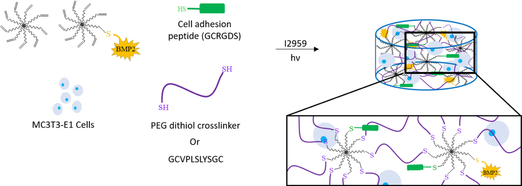
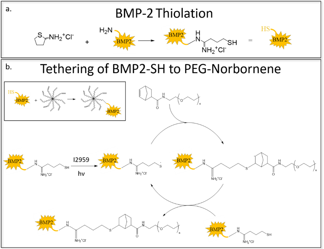
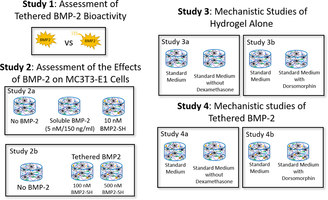
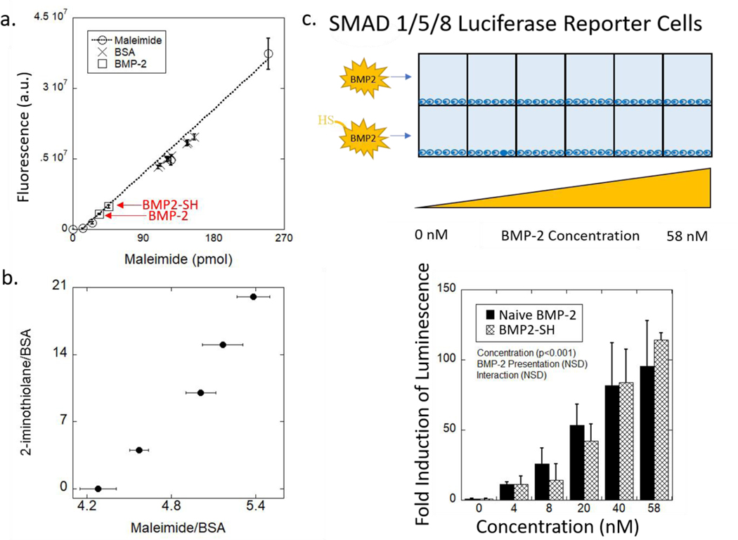
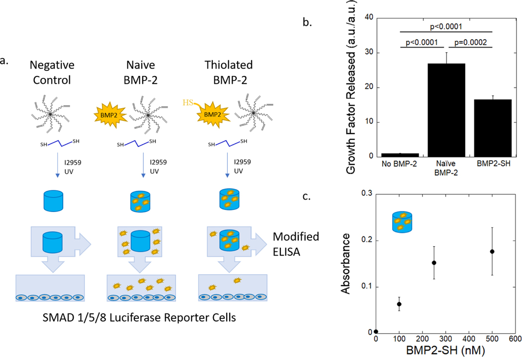
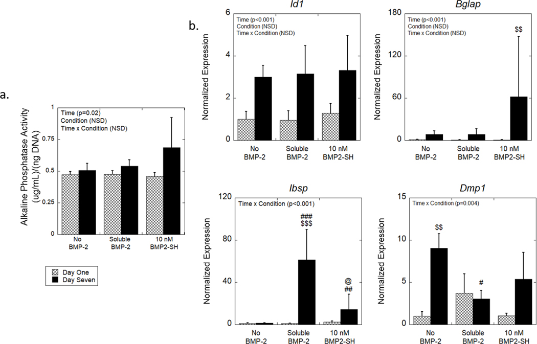
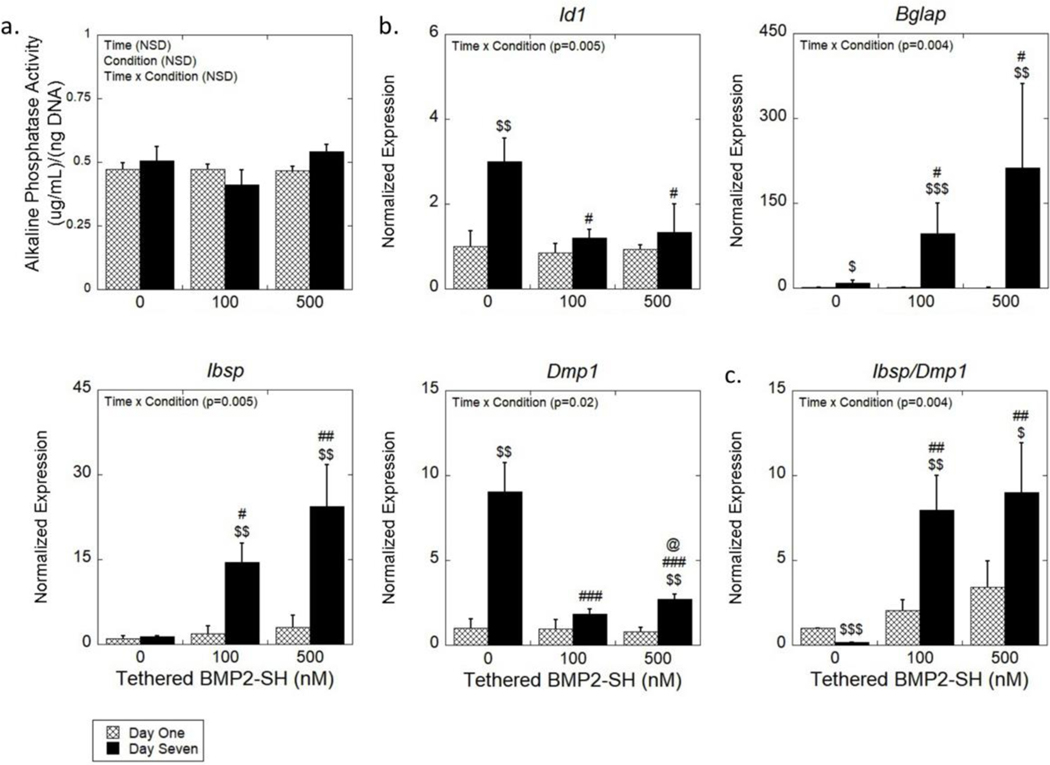
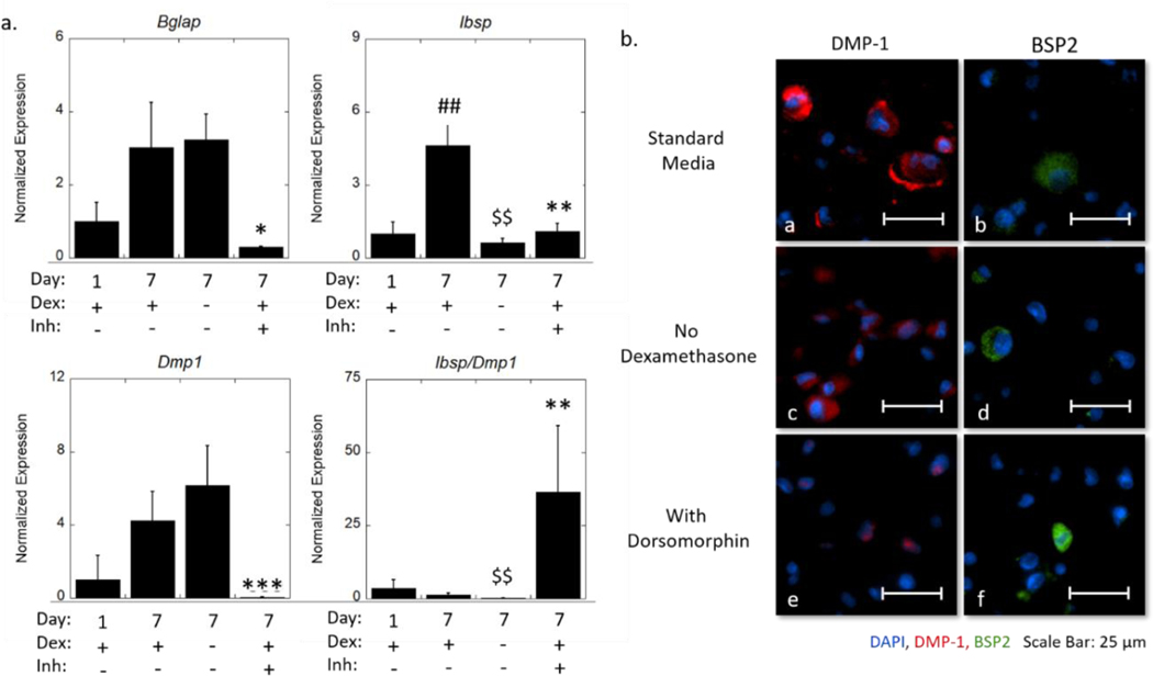
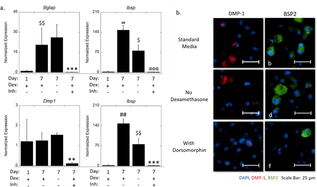
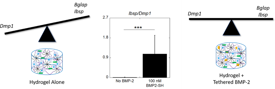
Bone morphogenetic protein-2 (BMP-2) is a clinically used osteoinductive growth factor. With a short half-life and side effects, alternative delivery approaches are needed. This work examines thiolation of BMP-2 for chemical attachment to a poly(ethylene glycol) hydrogel using thiol-norbornene click chemistry. BMP-2 retained bioactivity post-thiolation and was successfully tethered into the hydrogel. To assess tethered BMP-2 on osteogenesis, MC3T3-E1 preosteoblasts were encapsulated in matrix metalloproteinase (MMP)-sensitive hydrogels containing RGD and either no BMP-2, soluble BMP-2 (5 nM), or tethered BMP-2 (40-200 nM) and cultured in a chemically defined medium containing dexamethasone for 7 days. The hydrogel culture supported MC3T3-E1 osteogenesis regardless of BMP-2 presentation, but tethered BMP-2 augmented the osteogenic response, leading to significant increases in osteomarkers, Bglap and Ibsp. The ratio, Ibsp-to-Dmp1, highlighted differences in the extent of differentiation, revealing that without BMP-2, MC3T3-E1 cells showed a higher expression of Dmp1 (low ratio), but an equivalent expression with tethered BMP-2 and more abundant bone sialoprotein. In addition, this work identified that dexamethasone contributed to Ibsp expression but not Bglap or Dmp1 and confirmed that tethered BMP-2 induced the BMP canonical signaling pathway. This work presents an effective method for the modification and incorporation of BMP-2 into hydrogels to enhance osteogenesis.


Figures

Scheme 1.
Scheme 1.
The formation of the BMP-2 tethered hydrogel with MC3T3-E1 cells. Hydrogels were formed from a precursor solution containing 8-arm PEG-norbornene, 8-arm PEG-norbornene-BMP-2, crosslinker (PEG-dithiol or matrix- metalloproteinase (MMP)-sensitive crosslinker), cell adhesion peptide, and MC3T3-E1 cells. The BMP-2 characterization studies utilized hydrogels with the PEG-dithiol crosslinker without cells. Close-up box highlights the crosslinked network. Not drawn to scale.
Scheme 2.
Scheme 2.
(a) The addition of a free thiol to BMP-2 by reaction with 2-iminothiolane. (b) The radical-mediated, photoclick, thiol-norbornene reaction between norbornene-functionalized PEG and thiol-functionalized BMP-2 (i.e., BMP2-SH). BMP-2 is tethered via a stable bond.
Figure 1.
Figure 1.
An overview of the experimental design to assess the effectiveness of immobilizing BMP-2, via a thiol-norbornene click reaction, into PEG hydrogels. Study 1 assessed the bioactivity of thiolated BMP-2 in solution (study 1a) and the efficacy of tethering the functionalized growth factor to the PEG network (study 1b). A cell-reporter assay for SMAD 1/5/8 signaling and a modified-ELISA were used. Study 2 assessed the effects of BMP-2 (soluble and tethered) (study 2a) and concentration effects of tethered BMP-2 (study 2b) on MC3T3-E1 cells encapsulated in a PEG hydrogel and cultured in defined osteogenic medium (study 2a). Study 3 assessed the contribution of dexamethasone, which is present in the chemically defined medium (i.e., standard medium), on MC3T3-E1 cells encapsulated in the PEG hydrogel. The hydrogels were cultured in standard medium or standard medium without dexamethasone. Study 4 investigated if tethered BMP-2 signals via its receptors, that activates the SMAD 1/5/8 pathway, to induce the osteogenic response of the encapsulated cells. MC3T3-E1 cells were encapsulated in the PEG network and cultured in standard medium or standard medium with the inhibitor, dorsomorphin. In Studies 2–4, cell-laden hydrogels were cultured for up to seven days.
Figure 2.
Figure 2.
(a) Fluorescence measured as a function of picomoles of fluorescently tagged-maleimide is used to quantify the average number of free thiols added to BMP-2 and BSA. Red notation on graph identifies data point related to the BMP2-SH used in this study. Data represent average with standard deviation error bars (n = 2). (b) Molar ratio of 2-iminothiolane to BSA as a function of the average number of molecules of maleimide added per macromolecule of BSA. Data represent average with standard deviation error bars (n = 2–4). (c) Schematic of the experimental system to test for biological activity of BMP2-SH as compared to naive BMP-2 growth factor using SMAD 1/5/8 reporter cells. Fold-induction of luminescence of the reporter cells as a function of concentration for naive BMP-2 and BMP2-SH relative to the 0 nM condition. p > 0.05 between each condition at all concentrations. Data represent average with standard deviation error bars; n = 3. Data were analyzed using a two-way ANOVA and Tukey’s post-hoc test.
Figure 3.
Figure 3.
(a) Schematic of experimental design to test for efficacy of immobilizing BMP2-SH in hydrogel using SMAD 1/5/8 reporter cells and a modified-ELISA. Hydrogels were formed from a precursor solution containing no BMP-2, naive BMP-2, or BMP2-SH and then photopolymerized. The precursor solution contained 8-arm PEG-norbornene, PEG-dithiol, and photoiniator (I2959). Once formed, the hydrogels were swelled in cell-culture media for 24 hours to release any untethered BMP-2 or BMP2-SH. Luciferase reporter cells were treated with released BMP. (b) Luminescence of the reporter cells as a function of BMP-2 and BMP2-SH released from hydrogels. Data are normalized to the no BMP-2 condition. (n = 4). Data were analyzed using a one-way ANOVA and a Bonferroni’s post-hoc comparison test.(c) Absorbance measured from a modified ELISA quantifies the presence of tethered BMP2-SH on the surface of the PEG hydrogel (n = 3). Data represent average with standard deviation error bars.
Figure 4.
Figure 4.
The effects of BMP-2 presentation on osteogenesis of MC3T3-E1 cells encapsulated in MMP-sensitive hydrogels. (a) ALP activity normalized to DNA content as a function of time. (b) Relative expression of osteogenic genes normalized to no BMP-2 condition at day one. Data are represented as the mean with standard deviation error bars (n = 3). Data were analyzed by two-way ANOVA. Statistical significance determined by student’s t-test within a condition between days indicated by $. Statistical significance on the same day as determined by one-way ANOVA indicated by # for those relative to the no BMP-2 condition and by @ for those relative to the soluble condition. One symbol indicates p < 0.05, two symbols indicate p < 0.01, and three symbols indicate p < 0.0001. NSD = Not statistically different.
Figure 5.
Figure 5.
(a) The ALP activity normalized to DNA content as a function of time. Data are represented as the mean with standard deviation error bars (n = 3). (b) Expression of osteogenic genes (Id1, Bglap, Ibsp, Dmp1) of encapsulated MC3T3-E1 cells normalized to gene expression of no BMP-2 condition at day one. (c) A ratio of the gene expression of Ibsp and Dmp1 of encapsulated MC3T3-E1. Data represented as the mean with standard deviation error bars (n = 3). Data were analyzed by two-way ANOVA. Statistical significance determined by student’s t-test within a condition between days indicated by $. Statistical significance on the same day as determined by one-way ANOVA indicated by # for those relative to the no BMP-2 condition and by @ for those relative to the 100 nM condition. One symbol indicates p < 0.05, two symbols indicate p < 0.01, and three symbols indicate p < 0.0001. NSD = Not statistically different.
Figure 6.
Figure 6.
(a) Expression of osteogenic genes (Id1, Bglap, Dmp1) normalized to gene expression of no BMP-2 in standard medium at day one and a ratio of expression of Ibsp and Dmp1 of encapsulated MC3T3-E1 cells. Data are represented as the mean with standard deviation error bars (n=3). (b) Representative widefield fluorescent microscopy images for DMP-1 (red) and BSP2 (green) at day seven cultured in standard medium (a-b), medium without dexamethasone (c-d), and medium with dorsomorphin (e-f). Nuclei are counterstained blue. Scale bar = 25 μm. Data were analyzed by student’s t-test. Statistical significance between day seven standard media and day one indicated by #. Statistical difference between day seven no dexamethasone and day seven standard media indicated by $. Statistical significance between day seven with dorsomorphin and day seven standard media indicated by *. One symbol indicates p < 0.05, two symbols indicate p < 0.01, and three symbols indicate p < 0.001.
Figure 7.
Figure 7.
(a) Expression of osteogenic genes (Id1, Bglap, Dmp1) normalized to gene expression of no BMP-2 in standard medium at day one and a ratio of expression of Ibsp and Dmp1 of encapsulated MC3T3-E1 cells. Data are represented as the mean with standard deviation error bars (n=3). (b) Representative widefield fluorescent microscopy images for DMP-1 (red) and BSP2 (green) at day seven cultured in standard medium (a-b), medium without dexamethasone (c-d), and medium with dorsomorphin (e-f). Nuclei are counterstained blue. Scale bar = 25 μm. Data were analyzed by student’s t-test. Statistical significance between day seven standard media and day one indicated by #. Statistical difference between day seven no dexamethasone and day seven standard media indicated by $. Statistical significance between day seven with dorsomorphin and day seven standard media indicated by *. One symbol indicates p < 0.05, two symbols indicate p < 0.01, and three symbols indicate p < 0.001.
Figure 8.
Figure 8.
The different effects of the hydrogel environment without and with tethered BMP-2 on MC3T3-E1 osteogenic differentiation. The balance in expression of osteogenic markers Ibsp and Bglap compared to the osteocytic marker Dmp1 shows distinctly different osteogenic fates depending on the inclusion of tethered BMP-2. A ratio of expression of Ibsp and Dmp1 of encapsulated MC3T3-E1 cells. Data are represented as the mean with standard deviation error bars (n=6). Data were analyzed by student’s t-test. Statistical significance *** indicates p < 0.001.

Similar articles

Cited by

References

    1. Caballero Aguilar LM; Silva SM; Moulton SE Growth Factor Delivery: Defining the next Generation Platforms for Tissue Engineering. J. Control. Release 2019, 306 (May), 40–58. 10.1016/j.jconrel.2019.05.028. - DOI - PubMed
    1. Taipale J; Keski-Oja J. Growth Factors in the Extracellular Matrix. FASEB J. 1997, 11 (1), 51–59. - PubMed
    1. Chen FM; Zhang M; Wu ZF Toward Delivery of Multiple Growth Factors in Tissue Engineering. Biomaterials 2010, 31 (24), 6279–6308. 10.1016/j.biomaterials.2010.04.053. - DOI - PubMed
    1. Tabata Y. The Importance of Drug Delivery. Pharm. Sci. Technolo. Today 2000, 3 (3), 80–89. - PubMed
    1. Lutolf MP; Weber FE; Schmoekel HG; Schense JC; Kohler T; Müller R; Hubbell JA Repair of Bone Defects Using Synthetic Mimetics of Collagenous Extracellular Matrices. Nat. Biotechnol 2003, 21 (5), 513–518. 10.1038/nbt818. - DOI - PubMed

Publication types

LinkOut - more resources