. 2021 Feb 8;17(2):e1009282.
doi: 10.1371/journal.ppat.1009282. eCollection 2021 Feb.

Multimerization- and glycosylation-dependent receptor binding of SARS-CoV-2 spike proteins

Affiliations

Multimerization- and glycosylation-dependent receptor binding of SARS-CoV-2 spike proteins

Kim M Bouwman et al. PLoS Pathog. .

Abstract

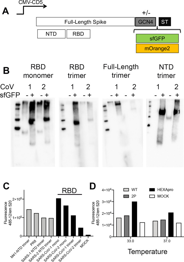
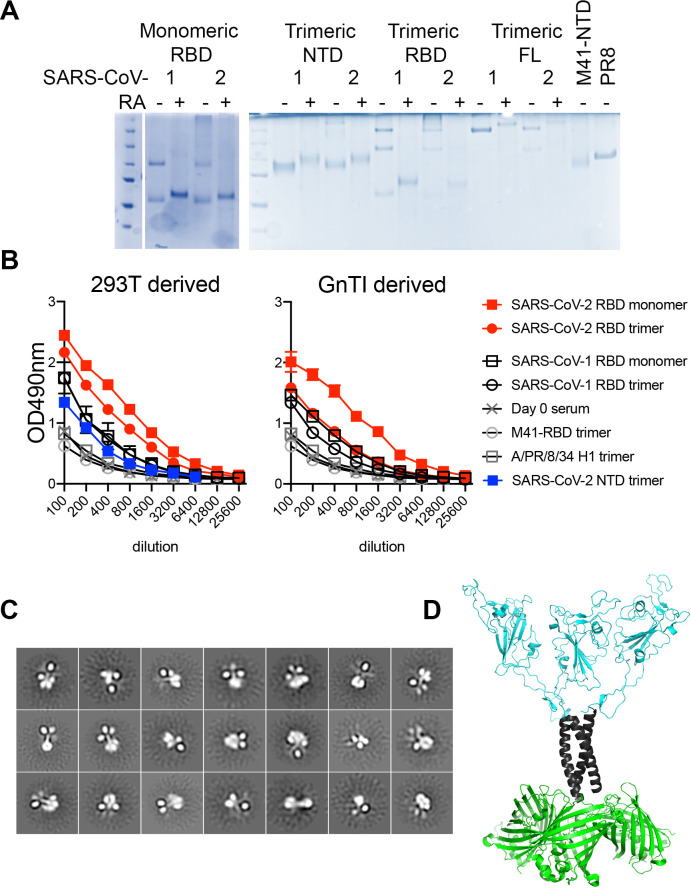
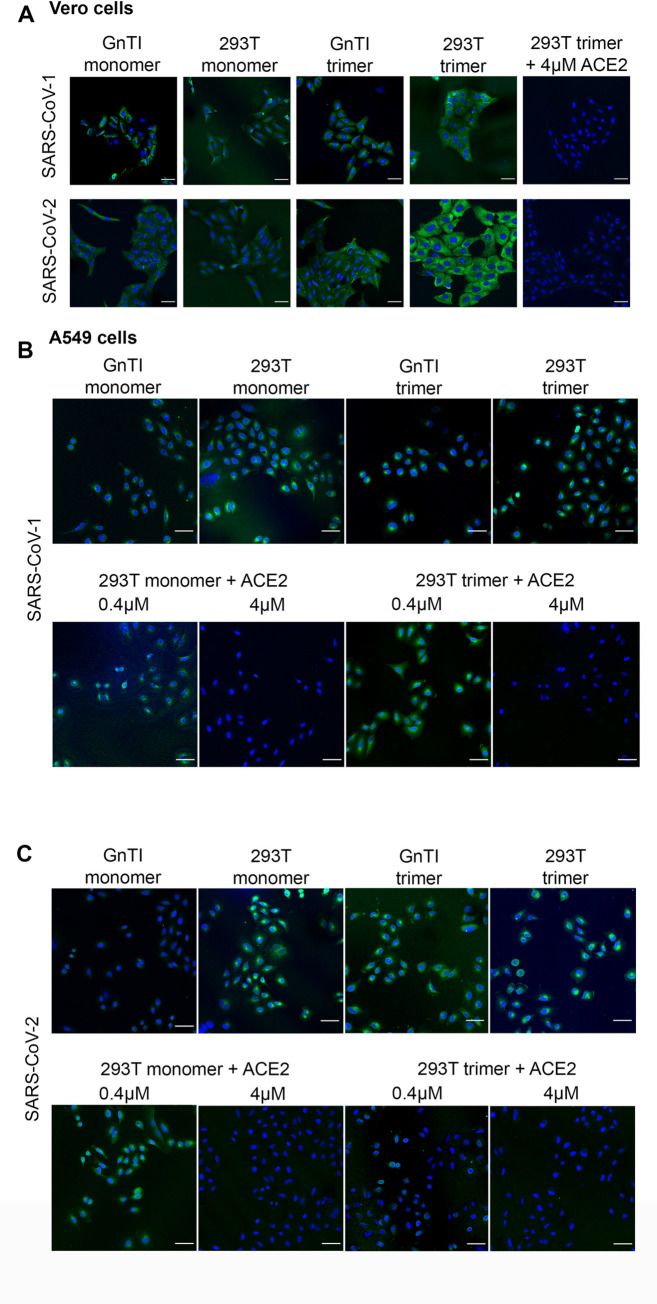
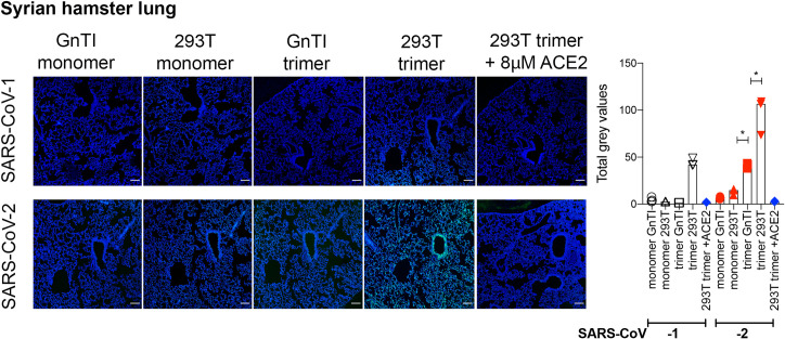
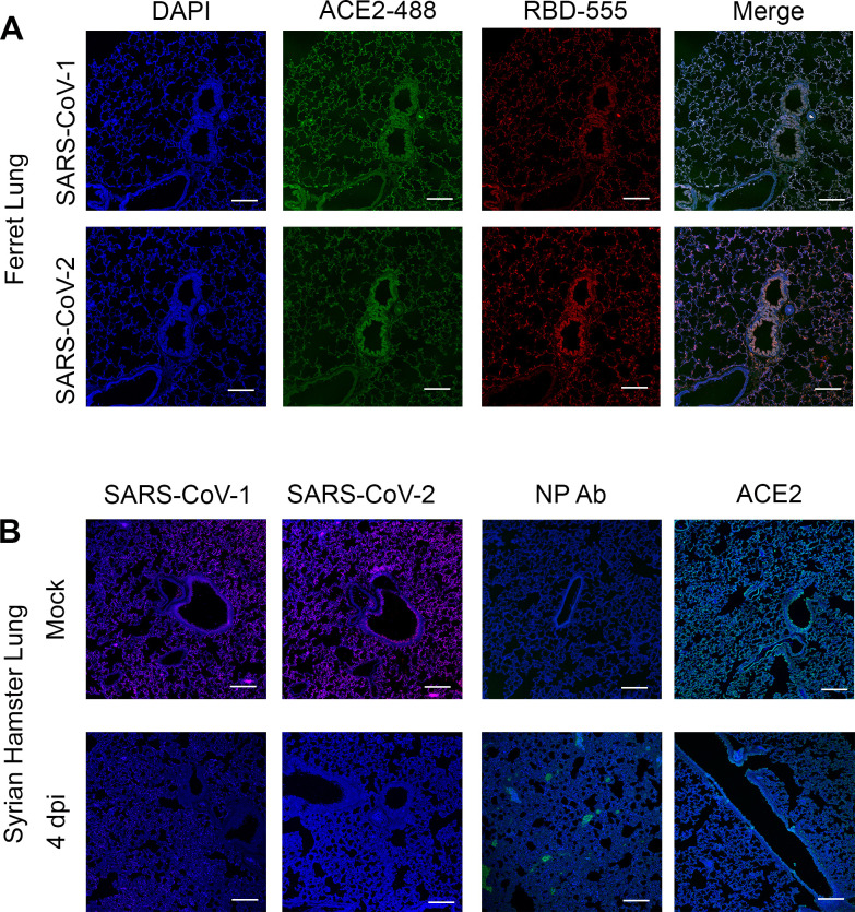
Receptor binding studies on sarbecoviruses would benefit from an available toolkit of recombinant spike proteins, or domains thereof, that recapitulate receptor binding properties of native viruses. We hypothesized that trimeric Receptor Binding Domain (RBD) proteins would be suitable candidates to study receptor binding properties of SARS-CoV-1 and -2. Here we created monomeric and trimeric fluorescent RBD proteins, derived from adherent HEK293T, as well as in GnTI-/- mutant cells, to analyze the effect of complex vs high mannose glycosylation on receptor binding. The results demonstrate that trimeric, complex glycosylated proteins are superior in receptor binding compared to monomeric and immaturely glycosylated variants. Although differences in binding to commonly used cell lines were minimal between the different RBD preparations, substantial differences were observed when respiratory tissues of experimental animals were stained. The RBD trimers demonstrated distinct ACE2 expression profiles in bronchiolar ducts and confirmed the higher binding affinity of SARS-CoV-2 over SARS-CoV-1. Our results show that complex glycosylated trimeric RBD proteins are attractive to analyze sarbecovirus receptor binding and explore ACE2 expression profiles in tissues.


Conflict of interest statement

The authors have declared that no competing interests exist.

Figures

Fig 1
Fig 1. Expression of coronavirus spike proteins using sfGFP and mOrange2 fusions.
(A) Spike expression plasmids with and without GCN4 trimerization motif fused with either sfGFP or mOrange2. Schematic representation of the used spike expression cassette. The spike open reading frame is under the control of a CMV-promotor and was cloned in frame with DNA sequences coding for the CD5 signal peptide. At the C-terminus a GCN4 trimerization domain followed by sfGFP or mOrange2, and a TEV cleavable Strep-tag II. (B) Expression analyses of non and sfGFP fused monomeric and trimeric RBD, the full-length ectodomain spike, and the NTD proteins. Denatured samples of the cell culture supernatant were subjected to SDS-PAGE and western blot analyzes stained with an anti-Streptag-HRP antibody. (C) Quantification. sfGFP emission was directly measured in the supernatants. (D) Full-length spike adaptation to the hexapro variant. Full-length spike expression vectors containing the wildtype, 2P, or Hexapro were expressed at 33 or 37C and fluorescence was measured in cell culture supernatant 4 days post-transfection.
Fig 2
Fig 2. Molecular analyses.
(A) SDS-PAGE analyses of purified RBD proteins. 500 ng of purified proteins were loaded on a gel with or without preheating for 30 minutes at 98C in the presence of a reducing agent. (B) Antigenic analyses using 21-day post-infection macaque serum. 2μg/ml SARS-CoV-RBD proteins were coated on 96-well plates. Proteins were detected with macaque serum for 2hrs at RT. An anti-human IgG-HRP was used to detect RBD specific antibodies. Both 293T and GnTI-/- derived proteins were analyzed. As negative controls, IBV-M41-NTD and HA-PR8D were included. (C) Negative stain EM of trimeric RBD fused to sfGFP. Negative-stain 2D class averages of soluble RBD proteins demonstrate that they are well-folded trimers. The C-terminal helices and fusion proteins are visible in some class averages. (D) Structural model. Based on the crystal structure of a SARS-CoV-2 RBD in the up conformation, with a GNC4 trimerization domain with 3 sfGFP domains added.
Fig 3
Fig 3. Binding of RBD proteins to VERO and A549 cells.
(A) SARS-CoV-RBD proteins were applied at 50μg/ml onto VERO cells and where indicated pre-incubated with recombinant ACE2 protein. (B) Same for SARS-CoV-1 on A549 cells. (C) Same for SARS-CoV-2. SARS-CoV proteins were detected using an anti-Streptag and a goat-anti-mouse antibody sequentially. sfGFP fused SARS-CoV-RBD proteins were applied from GnTI-/- monomers to 293T trimers, left to right. Scalebar is 50μm.
Fig 4
Fig 4. Binding of SARS-COV-RBD proteins and ACE2 antibody to mouse and ferret lung serial tissue slides.
(A) ACE2 antibody, SARS-CoV-1, and -2 on mouse lung tissue slides. Scalebar is 100μm. (B) ACE2 antibody and antibody only control on ferret lung tissue slides. Scalebar is 100μm. (C) SARS-CoV-RBD fluorescent protein localization in ferret lung tissue. SARS-CoV-RBD proteins were applied at 50μg/ml and detected using an anti-Streptag and goat-anti-mouse antibodies sequentially. DAPI was used as a nucleic stain. Scalebar is 100μm. (D) SARS-CoV-RBD trimer produced in HEK 293T cells pre-incubated with recombinant ACE2 before application on ferret lung tissue slides. (E) Quantification. The intensity of gray pixels of stained ferret lung tissue slides was measured with ImageJ version 1.52p. * indicates P<0.05 as determined by an unpaired two-tailed students T-test in the GraphPad software.
Fig 5
Fig 5. Binding of SARS-CoV-RBD proteins to serial Syrian hamster tissue slides.
SARS-COV-RBD proteins (50μg/ml) were applied to Syrian hamster lung tissue slides and detected using additional anti-Streptag and goat-anti-mouse antibodies sequentially. Where indicated SARS-CoV-RBD proteins were pre-incubated with recombinant ACE2 protein. Scalebar is 100μM. For the quantification, the intensity of gray pixels of stained Syrian Hamster lung tissue slides was measured with ImageJ version 1.52p. * indicates P<0.05 as determined by an unpaired two-tailed students T-test in the GraphPad software.
Fig 6
Fig 6
(A) ACE2 antibody, SARS-CoV-1, and -2 co-stainings on ferret lung tissue slides. Scalebar is 100μm. (B) ACE2 antibody, antibody only control, and SARS-CoV-1 and -2 on Syrian hamster tissue slide from mock and infected animals 4 days post-infection. Scalebar is 100μm.

References

    1. Chen X, Li R, Pan Z, Qian C, Yang Y, et al. Human monoclonal antibodies block the binding of SARS-CoV-2 spike protein to angiotensin converting enzyme 2 receptor. Cell Mol Immunol. 2020. 10.1038/s41423-020-0426-7 - DOI - PMC - PubMed
    1. Wang Q, Zhang Y, Wu L, Niu S, Song C, et al. Structural and Functional Basis of SARS-CoV-2 Entry by Using Human ACE2. Cell. 2020. 10.1016/j.cell.2020.03.045 - DOI - PMC - PubMed
    1. Letko M, Marzi A, Munster V. Functional assessment of cell entry and receptor usage for SARS-CoV-2 and other lineage B betacoronaviruses. Nature Micro. 2020;5:562–569. 10.1038/s41564-020-0688-y - DOI - PMC - PubMed
    1. Walls AC, Park YJ, Tortorici MA, Wall A, McGuire AT, et al. Structure, Function, and Antigenicity of the SARS-CoV-2 Spike Glycoprotein. Cell. 2020;181:281–292 e286. 10.1016/j.cell.2020.02.058 - DOI - PMC - PubMed
    1. Amanat F, Stadlbauer D, Strohmeier S, Nguyen THO, Chromikova V, et al. A serological assay to detect SARS-CoV-2 seroconversion in humans. Nature Med. 2020. 10.1101/2020.03.17.20037713 - DOI - PMC - PubMed

Publication types

MeSH terms

Substances