Review
. 2021 Apr 26:39:251-277.
doi: 10.1146/annurev-immunol-093019-110159. Epub 2021 Feb 8.

Microglia and Central Nervous System-Associated Macrophages-From Origin to Disease Modulation

Affiliations
Review

Microglia and Central Nervous System-Associated Macrophages-From Origin to Disease Modulation

Marco Prinz et al. Annu Rev Immunol. .

Abstract

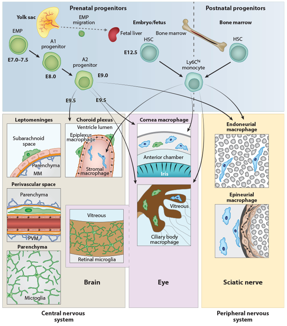
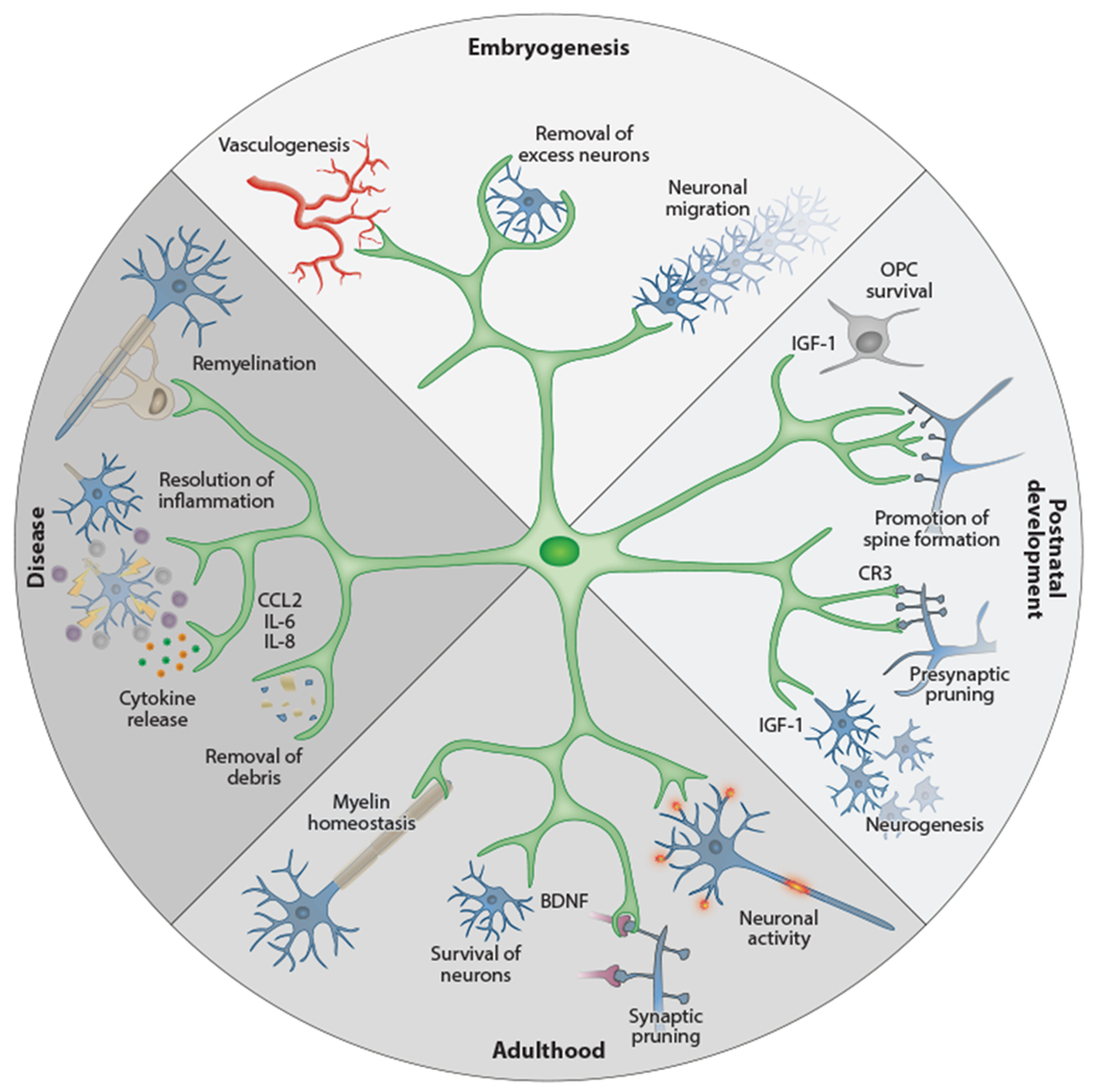
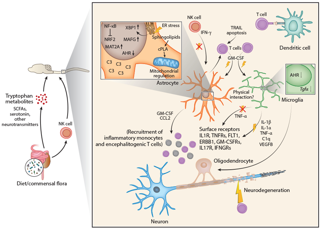
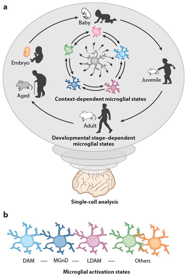
The immune system of the central nervous system (CNS) consists primarily of innate immune cells. These are highly specialized macrophages found either in the parenchyma, called microglia, or at the CNS interfaces, such as leptomeningeal, perivascular, and choroid plexus macrophages. While they were primarily thought of as phagocytes, their function extends well beyond simple removal of cell debris during development and diseases. Brain-resident innate immune cells were found to be plastic, long-lived, and host to an outstanding number of risk genes for multiple pathologies. As a result, they are now considered the most suitable targets for modulating CNS diseases. Additionally, recent single-cell technologies enhanced our molecular understanding of their origins, fates, interactomes, and functional cell statesduring health and perturbation. Here, we review the current state of our understanding and challenges of the myeloid cell biology in the CNS and treatment options for related diseases.

Keywords: development; fate mapping; macrophages; microglia; multiple sclerosis; single-cell profiling.


Figures

Figure 1
Figure 1
Anatomy and origin of microglia and CNS-associated macrophages. Cellular sources and anatomical location of myeloid cells found in the CNS, peripheral nervous system, or eye in the mouse. Microglial cells, PVMs, MMs, and retinal microglia are exclusively derived from prenatal sources and have no exchange with HSC-derived monocytes during postnatal stages. In contrast, choroid plexus stromal macrophages, ciliary body macrophages, corneal macrophages in the eye, endoneurial macrophages, and epineurial macrophages in the sciatic nerve have a mixed origin, from prenatal (yolk sac and fetal liver) and postnatal (bone marrow) sources. A transient early wave of myeloid cell development called primitive hematopoiesis takes place at E7.0–E7.5 (upper left). At this time, c-Kit+ EMP cells develop in the blood islands of the yolk sac. Their progeny (myeloid precursor cells) mature, expand, and migrate. CX3CR1+ A2 myeloid progenitor cells are derived from c-Kit+ CX3CR1+ A1 progenitors and seed the brain at E9.5, where they differentiate into microglial cells, PVMs, ATMs, and retinal microglia, respectively. During further development, some EMPs travel to the fetal liver (which is part of the definitive hematopoiesis), where they differentiate into monocytes. Myelopoiesis is thought to be restricted to bone marrow starting from birth (right). Abbreviations: CNS, central nervous system; EMP, erythromyeloid progenitor; HSC, hematopoietic stem cell; ATM, meningeal macrophage; PVM, perivascular macrophage.
Figure 2
Figure 2
Physiological functions of microglia during development and adulthood. Microglial cells show a remarkable diversity of functions at different stages of ontogeny. During embryogenesis they are involved in promoting vessel development and in the removal of superfluous neurons, and they guide neuronal migration. At postnatal time points, however, they support OPC survival and development (e.g., by IGF-1 production), support neurogenesis in defined brain areas, and promote neuronal spine formation. After finalization of myelinization and establishment of neuronal circuits, their functions shift to ensure the survival of neurons (e.g., by production of BDNF) and oligodendrocytes. Upon perturbation, microglia transform their homeostatic program to an activated state that involves enhanced phagocytosis and production of soluble factors such as cytokines, chemokines, and surface markers. These mechanisms allow the removal of cellular debris and promote regeneration (e.g., remyelination). Abbreviation: OPC, oligodendrocyte precursor cell.
Figure 3
Figure 3
Interactions between CNS-resident cells and the immune system during CNS inflammation. T cell activation is induced by myelin autoantigen presentation via dendritic cells and leads to the production and secretion of proinflammatory cytokines such as GM-CSF and IL-17. These cytokines exert proinflammatory effects on CNS-resident glial cells that respond to them, such as microglia and astrocytes, and promote neurodegeneration. In response to proinflammatory cytokine signals, microglia and astrocytes cross talk through multiple cues. In both astrocytes and microglia, proinflammatory cytokine signaling activates the transcription factor NF-kB and downregulates expression of the transcription factor AHR. NF-kB also inhibits activation of the antioxidant transcription factor NRF2 in astrocytes. Consequently, microglia upregulate and secrete proinflammatory molecules including IL-1β, IL-1α, TNF-α, C1q, and VEGFB while downregulating expression of anti-inflammatory TGF-α. Astrocytes sense these cues through cell surface receptors (IL1R, TNFRs, FLT1, ERBB1, GM-CSFRs, IL-17R, IFNGRs) and induce the expression of pathogenic transcriptional cascades including XBP1 (activated by ER stress), MAFG, and MAT2A alongside other pathogenic molecules like sphingolipid-induced cPLA2/MAVS, and complement component C3. Once activated, pathogenic astrocytes can secrete proinflammatory molecules including GM-CSF and CCL2, which lead to the recruitment of proinflammatory monocytes and encephalitogenic T cells to further promote CNS inflammation. Microglia and astrocytes may also engage in physical interactions during CNS inflammation, yet prominent examples remain to be elucidated. IFN-γ is reported to suppress CNS inflammation in mice and may mediate these functions in astrocytes and microglia through as-yet undetermined mechanisms. Control of microglial and astrocyte activation in the CNS has been linked to molecules derived from the commensal flora such as short-chain fatty acids, tryptophan metabolites, and neurotransmitters. Please note, however, that dietary metabolites such as tryptophan can be produced by the host as well as the commensal flora. Abbreviations: AHR, aryl hydrocarbon receptor; CNS, central nervous system; cPLA2, cytosolic phospholipase A2; C3, complement component 3; ER, endoplasmic reticulum; GM-CSF, granulocyte-macrophage colony-stimulating factor; IFNGR, interferon gamma receptor; NK, natural killer; SCFA, short-chain fatty acid; TNFR, tumor necrosis factor receptor; XBP1, X-box binding protein 1.
Figure 4
Figure 4
Developmental and context-dependent diverse states of microglia in mice and humans, (a) Different single-cell analyses (scRNA-seq, CyTOF, etc.) revealed that microglia possess highly plastic capacity, thereby altering the molecule (RNA, protein, etc.) expression profile during the course of homeostatic development, aging, or disease progression in a context-dependent manner, (b) Activated microglia states are highly divergent. For instance, scRNA-seq analyses deciphered that DAMs (153) or MGnDs (154) found at sites of neurodegeneration represent transcriptionally distinct profiles with high expression levels of Apoe, Clec7a, Itgax, Cst7, and Spp1 and reduced expression of P2ry12 and Tmem 119. In contrast, LDAMs (157) enriched in the central nervous system of aged mice, in which they accumulate lipid droplets, are defective in phagocytosis with massive production of reactive oxygen species and secretion of proinflammatory cytokines and chemokines such as IL-10, IL-6, CCL3, CCL4, and TNF-α. Abbreviations: CyTOF, cytometry by time-of-flight; DAM, disease-associated microglia; LDAM, lipid-droplet-accumulating microglia; MGnD, neurodegenerative microglia; scRNA-seq, single-cell RNA sequencing.

References

    1. Colonna M, Butovsky O 2017. Microglia function in the central nervous system during health and neurodegeneration. Annu. Rev. Immunol. 35:441–68 - PMC - PubMed
    1. Ginhoux F, Guilliams M 2016. Tissue-resident macrophage ontogeny and homeostasis. Immunity 44(3):439–49 - PubMed
    1. Herz J, Filiano AJ, Smith A, Yogev N, Kipnis J 2017. Myeloid cells in the central nervous system. Immunity 46(6):943–56 - PMC - PubMed
    1. Kierdorf K, Masuda T, Jordao MJC, Prinz M 2019. Macrophages at CNS interfaces: ontogeny and function in health and disease. Nat. Rev. Neurosci. 20(9): 547–62 - PubMed
    1. Prinz M, Erny D, Hagemeyer N 2017. Ontogeny and homeostasis of CNS myeloid cells. Nat. Immunol. 18(4): 3 85–92 - PubMed

Publication types