Review
. 2021 Jun 20:90:137-164.
doi: 10.1146/annurev-biochem-080320-110356. Epub 2021 Feb 8.

Repair of DNA Double-Strand Breaks by the Nonhomologous End Joining Pathway

Affiliations
Review

Repair of DNA Double-Strand Breaks by the Nonhomologous End Joining Pathway

Benjamin M Stinson et al. Annu Rev Biochem. .

Abstract

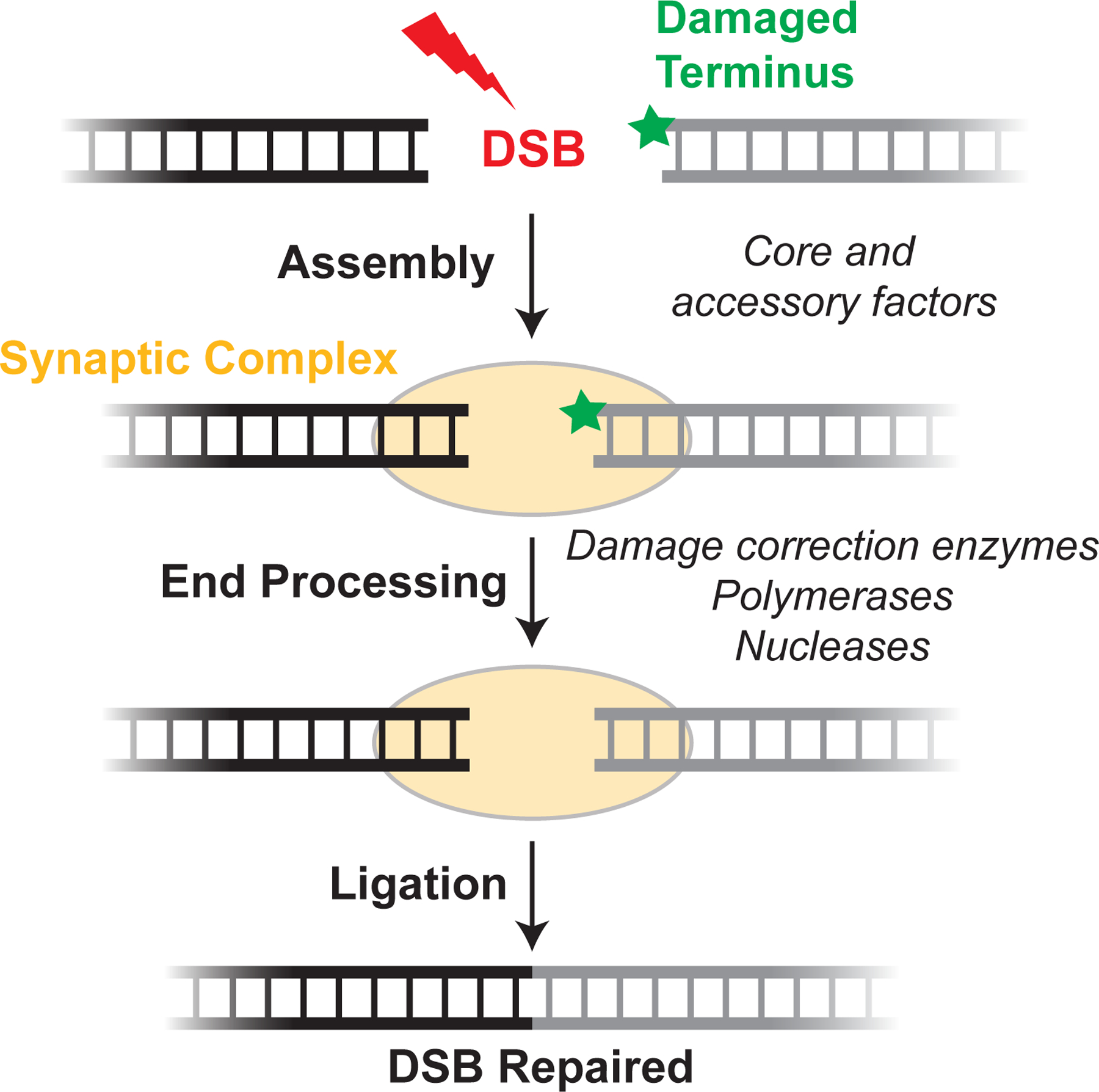
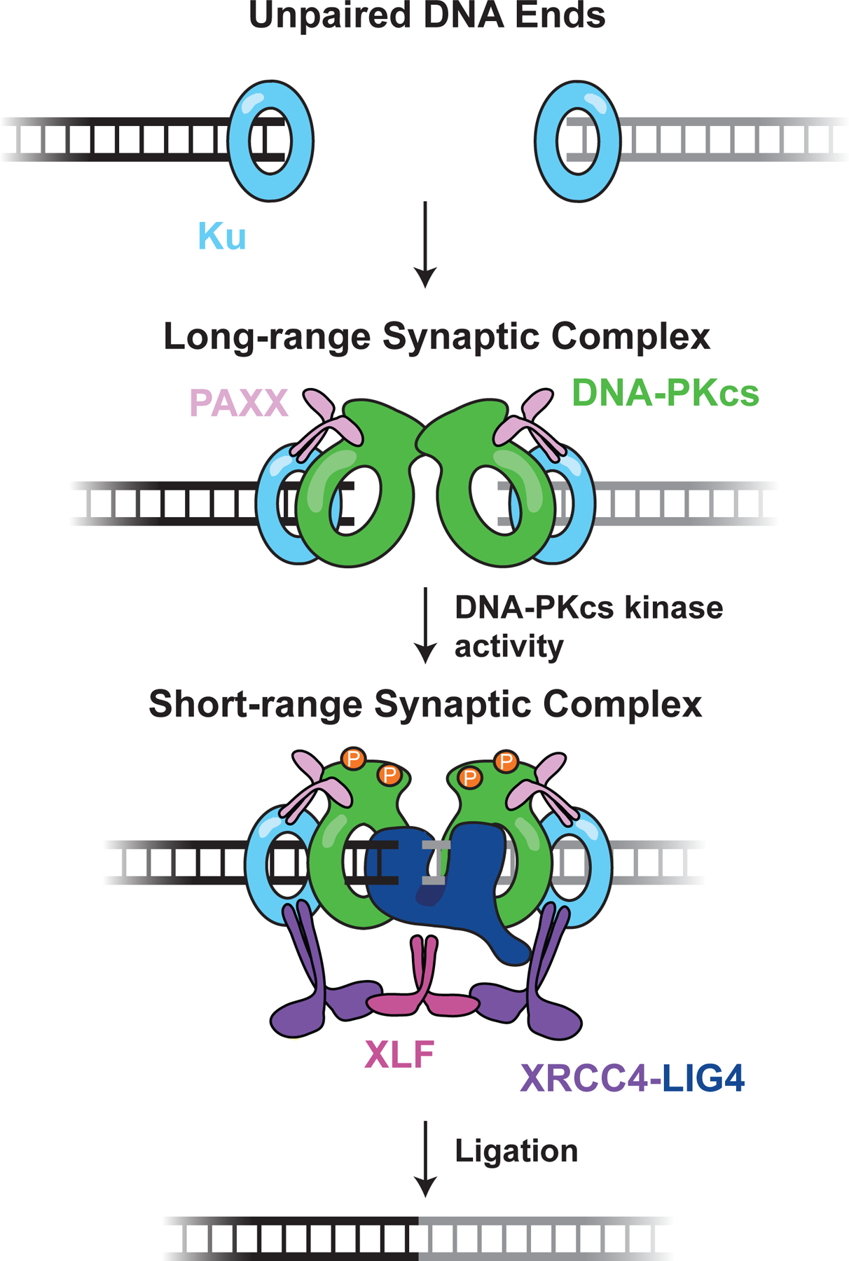
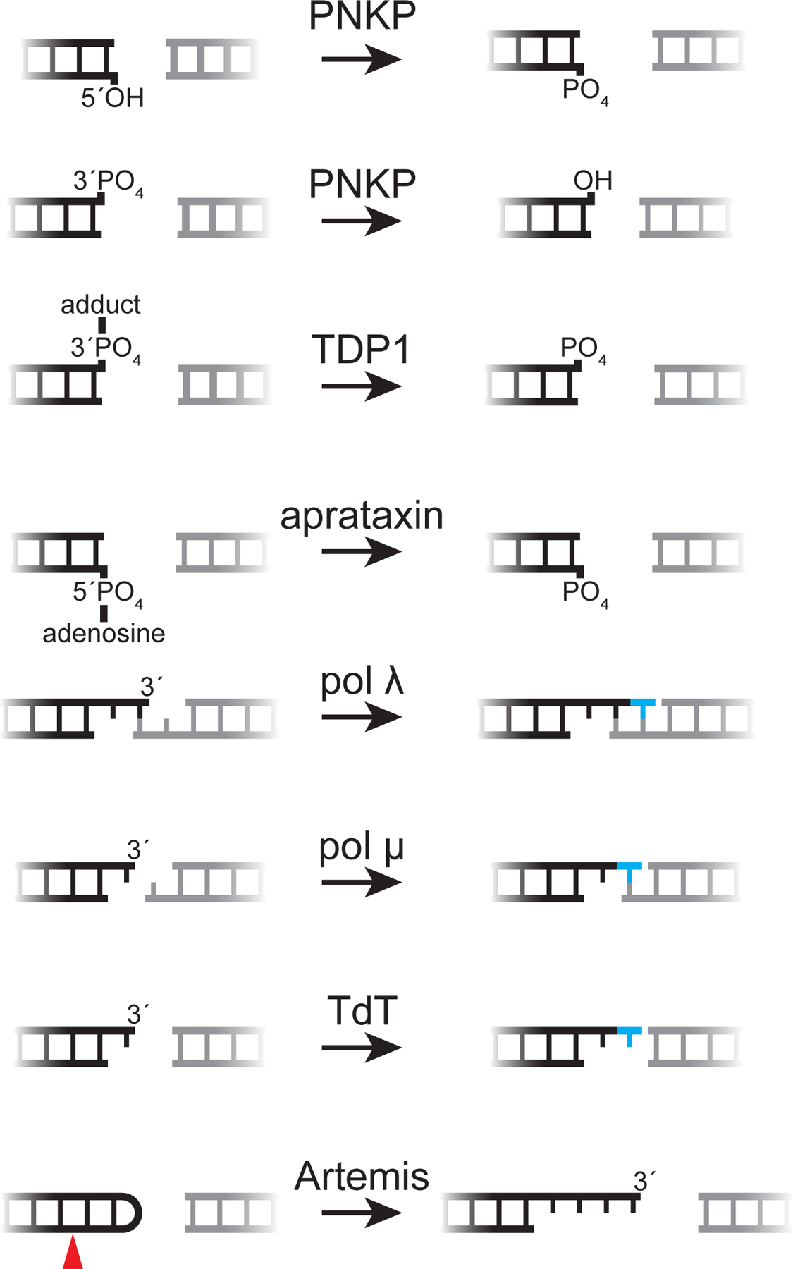
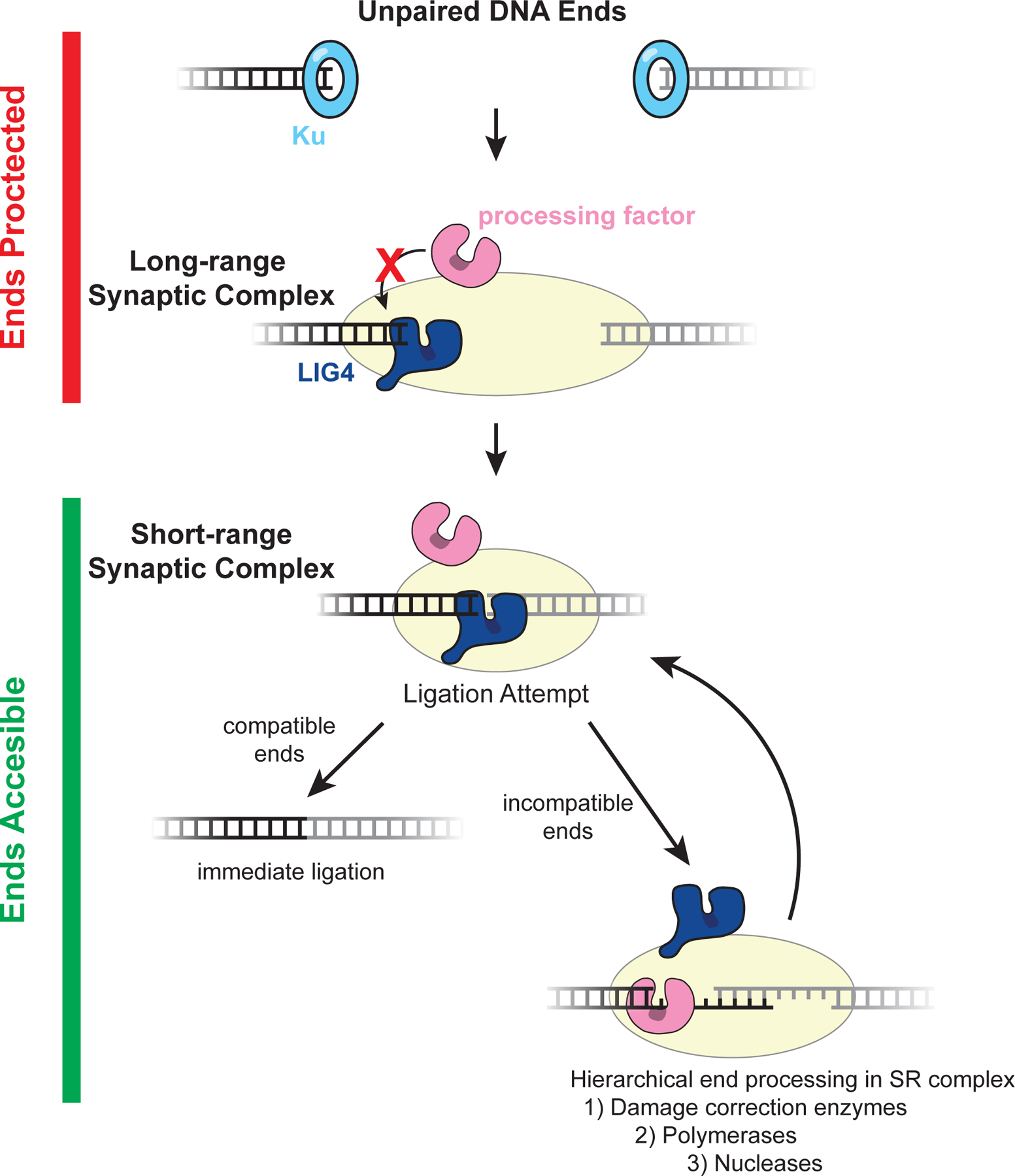
DNA double-strand breaks pose a serious threat to genome stability. In vertebrates, these breaks are predominantly repaired by nonhomologous end joining (NHEJ), which pairs DNA ends in a multiprotein synaptic complex to promote their direct ligation. NHEJ is a highly versatile pathway that uses an array of processing enzymes to modify damaged DNA ends and enable their ligation. The mechanisms of end synapsis and end processing have important implications for genome stability. Rapid and stable synapsis is necessary to limit chromosome translocations that result from the mispairing of DNA ends. Furthermore, end processing must be tightly regulated to minimize mutations at the break site. Here, we review our current mechanistic understanding of vertebrate NHEJ, with a particular focus on end synapsis and processing.

Keywords: DNA double-strand break; DNA end processing; DNA end synapsis; DNA repair; nonhomologous end joining.


Conflict of interest statement

DISCLOSURE STATEMENT

The authors are not aware of any affiliations, memberships, funding, or financial holdings that might be perceived as affecting the objectivity of this review.

Figures

Figure 1:
Figure 1:. An overview of the NHEJ pathway
Upon DSB formation, core and accessory NHEJ factors (Table 1) recognize DNA ends and tether them together in a synaptic complex (see Section 4 for details). If the DNA ends are not compatible for immediate ligation, they are modified by processing enzymes (Table 1). until ligation can occur (see Section 5 for details).
Figure 2:
Figure 2:. NHEJ and programmed DSBs
(A) Simplified example of V(D)J recombination at the IgH locus. RAG introduces DSBs adjacent to a D segment and J segment according to the 12/23 rule (see text), and NHEJ facilitates D to J joining. Subsequent V to DJ joining occurs through a similar process and assembles the V(D)J exon. (B) Generation and repair of DSBs during V(D)J recombination. V-to-D joining is shown as an example. See text for details. 12- and 23-RSSs, open and filled purple triangles, respectively. (C) Simplified example of CSR at the IgH locus. AID activity at the μ and ε switch regions leads to DSBs that are repaired by NHEJ in a productive class switching event to the IgE isotype.
Figure 3:
Figure 3:. Single-molecule assays for synapsis
(A) Inter-molecular synapsis single-molecule FRET (smFRET) assay. Green, donor fluorophore; red, acceptor fluorophore. (B) Intra-molecular synapsis smFRET assay. Proteins labeled with unique fluorophores (blue) can be imaged to quantify their stoichiometry and dynamics with the synaptic complex. (C) Magnetic forceps allow application of force to a DNA scaffold during synapsis.
Figure 4:
Figure 4:. End synapsis during NHEJ
DNA ends are rapidly bound by Ku, which recruits further NHEJ factors. Ku, DNA-PKcs, and PAXX enable formation of the long-range synaptic complex, in which the DNA ends are tethered together but separated by >80 Å. Transition to the short-range synaptic complex, in which DNA ends are directly juxtaposed, requires XLF, XRCC4-LIG4, and DNA-PKcs kinase activity. Putative DNA-PKcs autophosphorylation in the short-range complex is shown as orange circles. Here, LIG4 is shown directly engaging both DNA ends to promote short-range synapsis, but other structural contributions are possible (see text). The figure depicts the protein and enzymatic requirements for formation of each synaptic state; the precise composition and factor stoichiometry of each state is largely unknown, beyond the observation that a single XLF dimer promotes short range synapsis (55).
Figure 5:
Figure 5:. Processing enzymes and their DNA substrates
An array of end processing enzymes enables NHEJ to repair DSBs with diverse end structures, as shown here. Artemis is shown opening a V(D)J recombination hairpin intermediate (red arrow, cleavage site); its cognate substrates during repair of spontaneous breaks are unclear.
Figure 6:
Figure 6:. Model of error-prone processing regulation
Unpaired ends and those in the long-range synaptic complex are protected from aberrant or premature processing by Ku and DNA-PKcs. Although NHEJ processing factors are recruited, they are unable to act on DNA ends in these states (146). It is possible that other core NHEJ factors also protect DNA ends from processing. For example, as depicted here, LIG4 may directly engage a single DNA end prior to short-range synapsis, thereby preventing processing through steric occlusion. A second LIG4 molecule (not depicted) could protect the second end. Subsequently, direct juxtaposition of ends in the LIG4-dependent short-range synaptic enables a ligation attempt. If DNA ends are compatible, they can be ligated immediately without processing. If DNA ends are incompatible for ligation, they undergo processing in the short-range synaptic complex. Processing enzymes act hierarchically with the depicted priority to promote conservative modifications, and LIG4 frequently re-engages DNA ends to ligate them after the minimum number of processing events required for compatibility. Together, these features allow NHEJ to repair diverse DNA ends and minimize unnecessary genomic alterations.

References

    1. Bunting SF, Nussenzweig A. 2013. End-joining, translocations and cancer. Nature reviews Cancer 13(7):443–454 - PMC - PubMed
    1. Lieber MR. 2010. The mechanism of double-strand DNA break repair by the nonhomologous DNA end-joining pathway. Annu. Rev. Biochem 79:181–211 - PMC - PubMed
    1. Karanam K, Kafri R, Loewer A, Lahav G. 2012. Quantitative live cell imaging reveals a gradual shift between DNA repair mechanisms and a maximal use of HR in mid S phase. Molecular cell 47(2):320–329 - PMC - PubMed
    1. Haber JE. 2018. DNA Repair: The Search for Homology. Bioessays 40(5):e1700229. - PMC - PubMed
    1. Jasin M, Rothstein R. 2013. Repair of strand breaks by homologous recombination. Cold Spring Harb Perspect Biol 5(11):a012740. - PMC - PubMed

Publication types

LinkOut - more resources