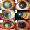
Pathobiology and treatment of viral keratitis
- PMID: 33556334
- PMCID: PMC8043992
- DOI: 10.1016/j.exer.2021.108483
Pathobiology and treatment of viral keratitis
Abstract
Keratitis is one of the most prevalent ocular diseases manifested by partial or total loss of vision. Amongst infectious (viz., microbes including bacteria, fungi, amebae, and viruses) and non-infectious (viz., eye trauma, chemical exposure, and ultraviolet exposure, contact lens) risk factors, viral keratitis has been demonstrated as one of the leading causes of corneal opacity. While many viruses have been shown to cause keratitis (such as rhabdoviruses, coxsackieviruses, etc.), herpesviruses are the predominant etiologic agent of viral keratitis. This chapter will summarize current knowledge on the prevalence, diagnosis, and pathobiology of viral keratitis. Virus-mediated immunomodulation of host innate and adaptive immune components is critical for viral persistence, and dysfunctional immune responses may cause destruction of ocular tissues leading to keratitis. Immunosuppressed or immunocompromised individuals may display recurring disease with pronounced severity. Early diagnosis of viral keratitis is beneficial for disease management and response to treatment. Finally, we have discussed current and emerging therapies to treat viral keratitis.
Keywords: Antiviral drugs; Host-virus interaction; Human herpesvirus; Inflammation; Ocular infection; Viral keratitis.
Copyright © 2021 Elsevier Ltd. All rights reserved.
Conflict of interest statement
Figures


References
-
- Agrawal V, Biswas J, Madhavan HN, Mangat G, Reddy MK, Saini JS, Sharma S, Srinivasan M, 1994. Current perspectives in infectious keratitis. Indian J Ophthalmol 42, 171–192. - PubMed
-
- Ahlfors K, Ivarsson SA, Bjerre I, 1986. Microcephaly and congenital cytomegalovirus infection: a combined prospective and retrospective study of a Swedish infant population. Pediatrics 78, 1058–1063. - PubMed
-
- Albers I, Kirchner H, Domke-Opitz I, 1989. Resistance of human blood monocytes to infection with herpes simplex virus. Virology 169, 466–469. - PubMed
-
- Alford CA, Stagno S, Pass RF, Britt WJ, 1990. Congenital and perinatal cytomegalovirus infections. Rev. Infect. Dis 12 Suppl 7, 745. - PubMed
Publication types
MeSH terms
Substances
Grants and funding
LinkOut - more resources
Full Text Sources
Other Literature Sources

