. 2021 May:152:105291.
doi: 10.1016/j.nbd.2021.105291. Epub 2021 Feb 5.

In vivo aggregation of presynaptic alpha-synuclein is not influenced by its phosphorylation at serine-129

Affiliations

In vivo aggregation of presynaptic alpha-synuclein is not influenced by its phosphorylation at serine-129

Leah J Weston et al. Neurobiol Dis. 2021 May.

Abstract

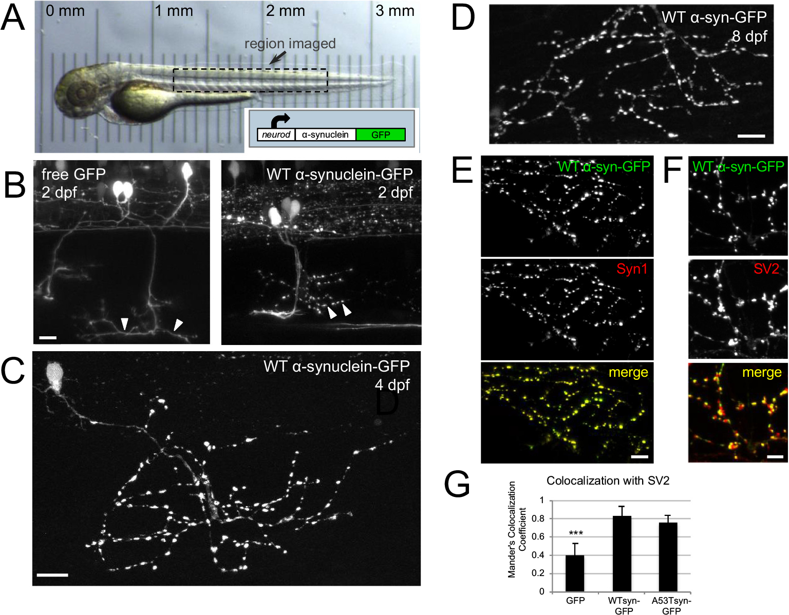
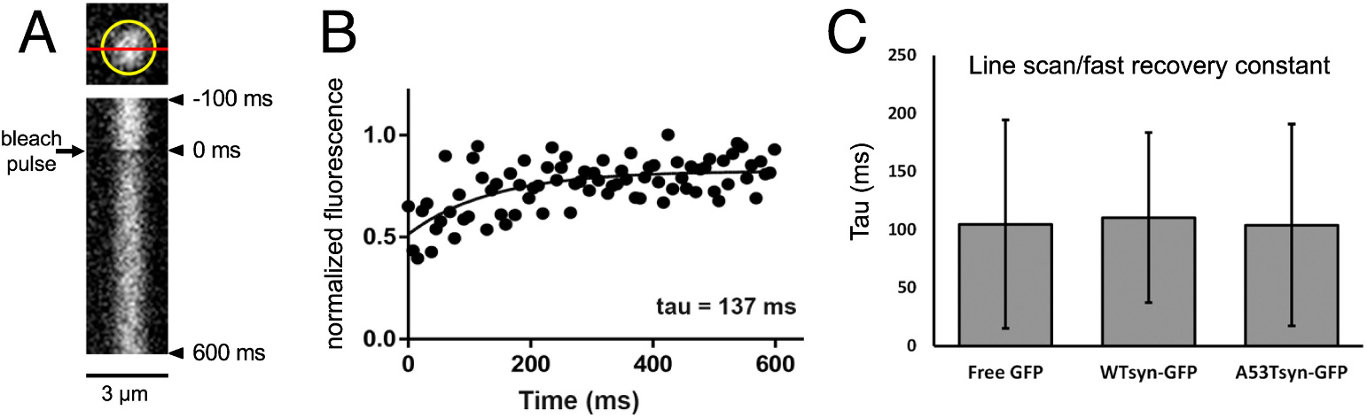
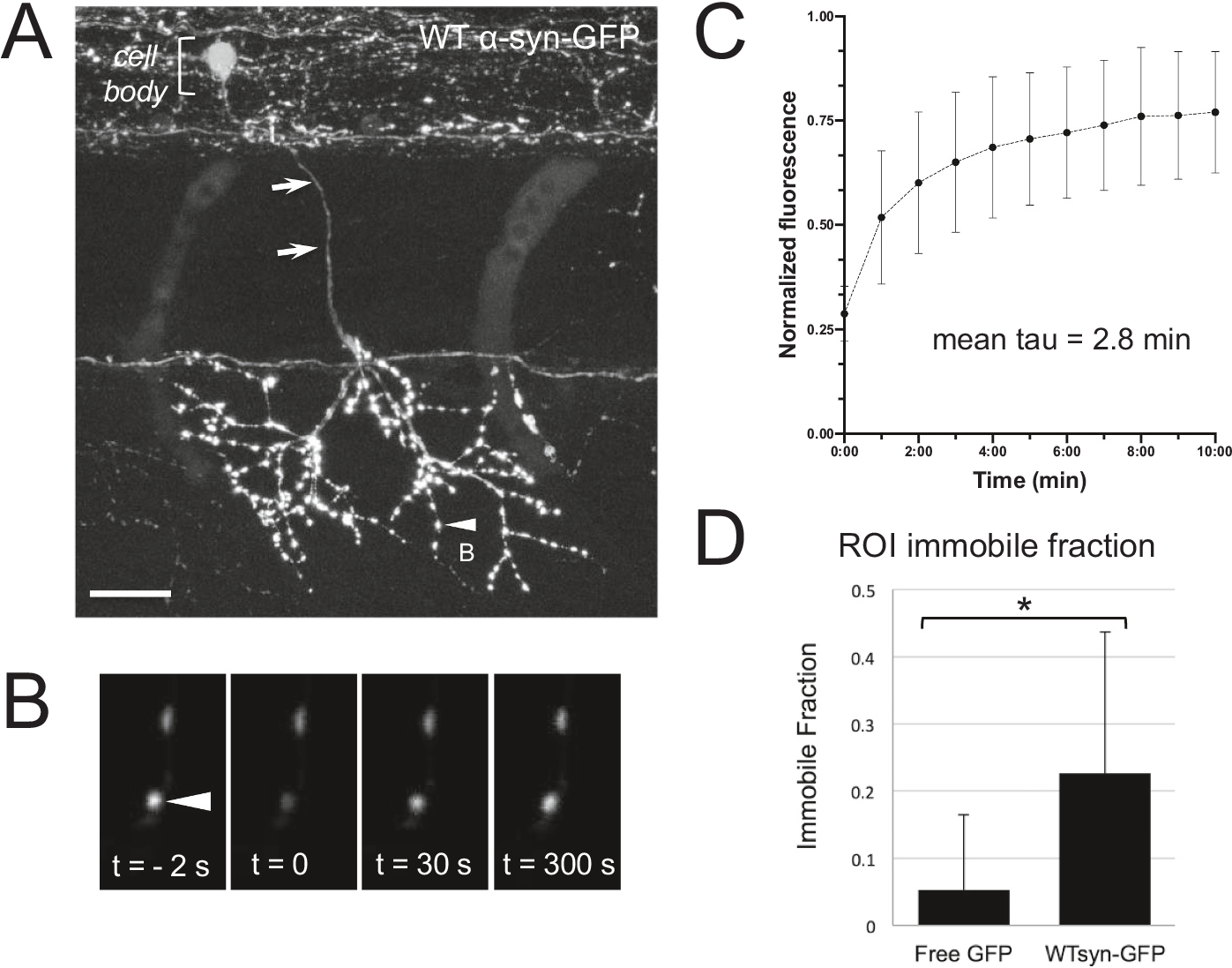
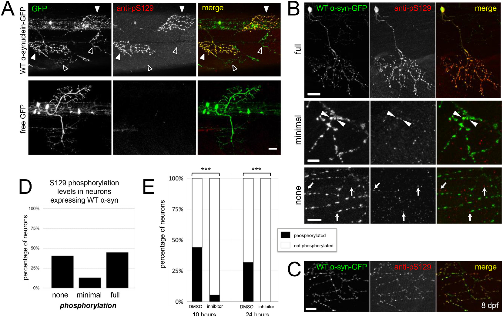
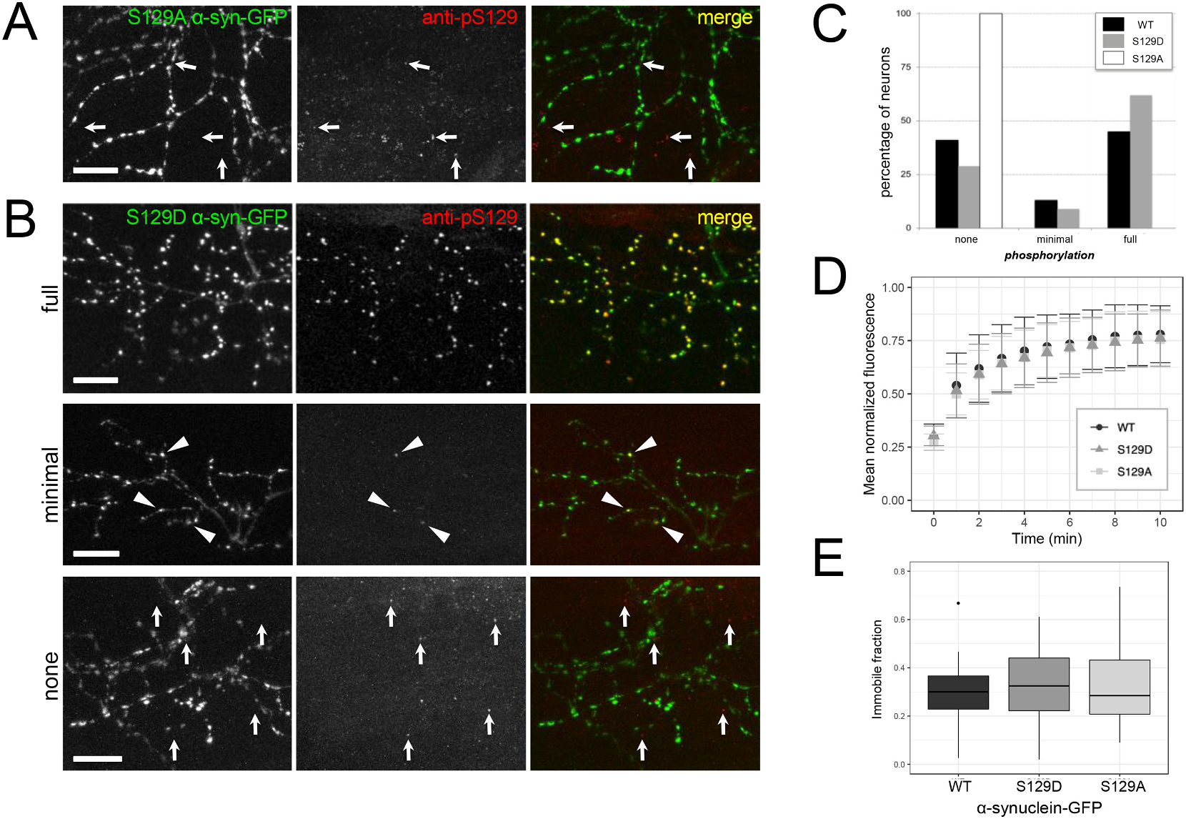
Abnormal aggregation of the α-synuclein protein is a key molecular feature of Parkinson's disease and other neurodegenerative diseases. The precise mechanisms that trigger α-synuclein aggregation are unclear, and it is not known what role aggregation plays in disease pathogenesis. Here we use an in vivo zebrafish model to express several different forms of human α-synuclein and measure its aggregation in presynaptic terminals. We show that human α-synuclein tagged with GFP can be expressed in zebrafish neurons, localizing normally to presynaptic terminals and undergoing phosphorylation at serine-129, as in mammalian neurons. The visual advantages of the zebrafish system allow for dynamic in vivo imaging to study α-synuclein, including the use of fluorescence recovery after photobleaching (FRAP) techniques to probe protein mobility. These experiments reveal three distinct terminal pools of α-synuclein with varying mobility, likely representing different subpopulations of aggregated and non-aggregated protein. Human α-synuclein is phosphorylated by an endogenous zebrafish Polo-like kinase activity, and there is a heterogeneous population of neurons containing either very little or extensive phosphorylation throughout the axonal arbor. Both pharmacological and genetic manipulations of serine-129 show that phosphorylation of α-synuclein at this site does not significantly affect its mobility. This suggests that serine-129 phosphorylation alone does not promote α-synuclein aggregation. Together our results show that human α-synuclein can be expressed and measured quantitatively in zebrafish, and that disease-relevant post-translational modifications occur within neurons. The zebrafish model provides a powerful in vivo system for measuring and manipulating α-synuclein function and aggregation, and for developing new treatments for neurodegenerative disease.

Keywords: Alpha-synuclein; FRAP; Parkinson's disease; Phosphorylation; Polo-like kinase; Protein aggregation; Zebrafish; in vivo imaging.


Figures

Fig. 1.
Fig. 1.
Human α-synuclein-GFP expresses in zebrafish neurons and localizes to presynaptic terminals. A) In vivo transmitted light image of zebrafish at 2 dpf; box indicates general region targeted for imaging. Translucency of larva is demonstrated by micrometer placed below; reprinted from Brockway et al., 2019. Inset shows schematic of neurod:α-synuclein-GFP DNA. B) In vivo imaging at 2 dpf shows that free GFP fills axons smoothly, while α-synuclein-GFP labeling appears more punctate at presumptive synaptic terminals. Arrowheads indicate regions of axons containing synaptic terminals at 2 dpf, which become more mature by 4 dpf (C). C) WT α-synuclein-GFP expression shown at 4 dpf in fixed larvae. D) α-synuclein-GFP expression persists until at least 8 dpf (image from fixed fish). E) Expression of α-synuclein-GFP colocalizes with synuclein protein. Ventrally projecting axons of motor neuron expressing wild-type α-synuclein-GFP (top), anti-Syn1 staining (middle), and merge (bottom). Scale bars 10 μm in B-E. F-G) α-synuclein-GFP expression in motor axons correlates more strongly with presynaptic terminal marker SV2 than does free GFP. F) High magnification view of motor axons expressing wild-type α-synuclein-GFP (top), SV2 antibody staining (middle), and two-channel merge (bottom). Scale bar 5 μm. G) Average Mander’s colocalization coefficient calculated on confocal stacks shows that WT α-synuclein-GFP and A53T α-synuclein-GFP colocalize significantly more strongly with SV2 (0.83 ± 0.11, n = 4 regions from 3 larvae; and 0.77 ± 0.07, n = 5 regions from 4 larvae, respectively) than does free GFP (0.41 ± 0.12, n = 9 regions from 4 larvae; one-way ANOVA, post-hoc Tukey tests p < 0.002 and p < 0.004). In all panels, caudal is right and dorsal is up. Panels B–F are maximum intensity projections of stacks with the following depths: B) left 45 μm; B) right 36 μm; C) 56.9 μm; D) 24 μm; E) 80 μm; F) 80 μm.
Fig. 2.
Fig. 2.
Fast synaptic terminal in vivo FRAP imaging reveals a pool of α-synuclein-GFP with identical recovery to free GFP, likely representing freely diffusing α-synuclein. A) (Top) Schematic of a line-bleached terminal (circled in yellow; single plane). The red line indicates the area bleached and measured over a recovery period of 600 ms. (Bottom) representative x-t plot of a line-bleached terminal expressing WT α-synuclein-GFP. The horizontal axis represents microns, aligned with the red line drawn above. The vertical axis represents time (increasing downward). Line through terminal was scanned every 7.5 ms, thus each y-axis pixel represents 7.5 ms. Bleach pulse occurs at 0 ms. B) Representative graph of individual terminal recovery in (A), plotting presynaptic terminal fluorescence intensity (normalized to baseline fluorescence) over time, fit with a single exponential function. C) Group data of recovery tau (mean) for presynaptic terminals expressing free GFP (105 ± 90 ms; n = 13 terminals from 4 larvae), wild-type α-synuclein-GFP (110 ± 73 ms; n = 15 terminals from 3 larvae), or A53T α-synuclein-GFP (104 ± 87 ms; n = 22 terminals from 3 larvae).
Fig. 3.
Fig. 3.
Slow synaptic terminal in vivo FRAP imaging reveals a more slowly recovering pool and a non-recovering pool of α-synuclein-GFP. A) Motor neuron expressing WT α-synuclein-GFP in 3 dpf zebrafish, with cell body in spinal cord (upper left bracket) and axon (arrows) descending ventrally to innervate muscle. Image in (A) is a maximum projection of a stack (70.5 μm thick) that was acquired after animal was fixed, following live imaging on highlighted synaptic terminal (arrowhead) shown enlarged in (B). Nonlinear adjustments were done in the larger panel only (A), in order to highlight thin axon descending from spinal cord (arrows). Scale bar in A 20 μm. B) Time series of in vivo ROI bleach experiment on presynaptic terminal indicated in (A). Only a single plane of section is shown. Arrowhead indicates the terminal measured before and after the bleach pulse and during the recovery phase. C) Group data of fluorescence recovery over time for WT α-synuclein-GFP. Average tau is 2.8 min (n = 26 terminals from 11 larvae). D) Group data of mean immobile fraction in terminals expressing free GFP (5 ± 11%; n = 22 terminals, 3 larvae) or wild-type (21 ± 25%; n = 27 terminals, 4 larvae). * represents p < 0.05, post-hoc Tukey test.
Fig. 4.
Fig. 4.
Alpha-synuclein-GFP is phosphorylated by endogenous kinase in a subset of neurons. A) Immunostaining for anti-phospho-S129 shows that expressed α-synuclein is phosphorylated in 4 dpf larvae (upper row, white arrowheads), but no stain is present when free GFP is expressed (lower row). Open arrowheads show some synuclein-expressing axons with little to no phospho-S129 staining. GFP channel is shown at left, anti-phospho-S129 in the center, and merge at right. Scale bar 20 μm. B) α-synuclein-GFP expressed in primary motor neurons and stained at 4 dpf is either fully phosphorylated throughout the axonal arbor (upper row example), minimally phosphorylated (middle row; a small fraction of terminals throughout arbor, indicated with arrowheads), or not phosphorylated (bottom row). GFP channel is shown at left, anti-phospho-S129 in the center, and merge at right. Arrows in bottom row indicate immunostaining background puncta that are not overlapping with GFP channel. Scale bar 20 μm in upper row, 5 μm in middle row, and 10 μm in bottom row. In panels A-C, caudal is right and dorsal is up. C) Motor axon at 8 dpf shows that expressed α-synuclein is phosphorylated at older ages. GFP channel is shown at left, anti-phospho-S129 in the center, and merge at right. Scale bar 10 μm. All images are maximum intensity projections of stacks with the following depths: A) 28.7 μm top; 118 μm bottom; B) 46.7 μm, 19 μm, and 30.4 μm (top, middle, and bottom respectively); C) 24 μm. D) Quantification of primary motor neurons from 4 dpf zebrafish expressing wild type α-synuclein-GFP shows that 41.2% of neurons do not exhibit phospho-S129 staining, 13.2% show little phosphorylation, and 45.6% are strongly phosphorylated (n = 69 neurons assessed from 8 larvae) E) Following exposure of zebrafish expressing wild type α-synuclein-GFP to 100 μm of PLK inhibitor BI 2536 (beginning at 3 dpf), phosphorylation of α-synuclein at S129 decreased significantly from 43% to 5.4% at 10 h (DMSO: n = 18/41 neurons from 6 larvae; BI 2536: 2/37 neurons from 6 larvae; chi-squared p < 0.001) and from 31.7% to 0% at 24 h (DMSO: n = 13/41 neurons from 5 larvae; BI 2536: 0/53 neurons from 5 larvae; chi-squared p < 0.001) as compared to DMSO control.
Fig. 5.
Fig. 5.
Genetic modification of serine 129 to alanine abolishes phosphorylation, but modification to alanine or aspartate does not affect immobile fraction. A) S129A α-synuclein-GFP expressed in motor neurons and stained at 4 dpf does not exhibit phospho-S129 staining. GFP channel is shown at left, anti-phospho-S129 in the center, and merge at right. Arrows indicate immunostaining background puncta that are not overlapping with GFP channel. B) Similar to WT α-synuclein-GFP, S129D α-synuclein-GFP expressed in motor neurons and stained at 4 dpf is either fully phosphorylated throughout the axonal arbor (upper row example), minimally phosphorylated (middle row; a small fraction of terminals throughout arbor, indicated with arrowheads), or not phosphorylated (bottom row). In bottom row, arrows indicate immunostaining background puncta that are not overlapping with GFP channel. GFP channel is shown at left, anti-phospho-S129 in the center, and merge at right. Scale bars 10 μm; caudal is right and dorsal is up. All images are maximum intensity projections of stacks with the following depths: A) 39.2 μm B) 65.2 μm, 30.9 μm, and 66.6 μm (top, middle, and bottom respectively). C) Quantification of phosphorylation in motor neurons expressing each form of α-synuclein shows that neurons expressing wild type α-synuclein-GFP are distributed as 41.2% not phosphorylated, 13.2% minimally phosphorylated, and 45.6% fully phosphorylated (n = 69 neurons from 8 larvae). Neurons expressing S129D α-synuclein-GFP show a similar distribution with 28.6% not phosphorylated, 9.5% minimally, and 61.9% fully phosphorylated (n = 103 neurons from 14 larvae). In contrast, S129A α-synuclein-GFP expressed in motor neurons is not phosphorylated (100%; n = 130 neurons from 10 larvae). D) Group data of fluorescence recovery over time. Error bars represent standard deviation. Average tau for WT α-synuclein-GFP was 2.8 min (n = 26 terminals from 11 larvae); S129D was 2.7 min (n = 39 terminals from 13 larvae), and S129A was 2.3 min (n = 29 terminals from 7 larvae; one-way ANOVA, p = 0.638). E) The average immobile fraction for WT α-synuclein-GFP was 29.3% (n = 26 terminals from 11 larvae), S129D was 32.5% (n = 39 terminals from 13 larvae), and S129A was 32.8%; (n = 29 terminals from 7 larvae; one-way ANOVA, p = 0.641). In E, bars indicate range of data within 1.5 IQR; outlier outside this range shown as a point.

References

    1. Anderson JP, Walker DE, Goldstein JM, de Laat R, Banducci K, Caccavello RJ, Barbour R, Huang J, Kling K, Lee M, Diep L, Keim PS, Shen X, Chataway T, Schlossmacher MG, Seubert P, Schenk D, Sinha S, Gai WP, Chilcote TJ, 2006. Phosphorylation of Ser-129 is the dominant pathological modification of α-Synuclein in familial and sporadic Lewy body disease. J. Biol. Chem. 281, 29739–29752. - PubMed
    1. Bae Y-K, Kani S, Shimizu T, Tanabe K, Nojima H, Kimura Y, Higashijima S, Hibi M, 2009. Anatomy of zebrafish cerebellum and screen for mutations affecting its development. Dev. Biol. 330, 406–426. - PubMed
    1. Barnhill L, Murata H, Bronstein JM, 2020. Studying the pathophysiology of Parkinson’s disease using Zebrafish. Biomedicines 8, 197. - PMC - PubMed
    1. Bergeron M, Motter R, Tanaka P, Fauss D, Babcock M, Chiou SS, Nelson S, San Pablo F, Anderson JP, 2014. In vivo modulation of polo-like kinases supports a key role for PLK2 in Ser129 α-synuclein phosphorylation in mouse brain. Neuroscience 256, 72–82. - PubMed
    1. Bodner RA, Outeiro F, Altmann S, Maxwell MM, Cho SH, Hyman BT, Mclean PJ, Young AB, Housman DE, Kazantsev AG, 2006. Pharmacological promotion of inclusion formation: a therapeutic approach for Huntington’s and Parkinson’s diseases. PNAS 103 (11), 4246–4251. - PMC - PubMed

Publication types

LinkOut - more resources