Review
. 2021 May:137:111313.
doi: 10.1016/j.biopha.2021.111313. Epub 2021 Feb 3.

An update review of emerging small-molecule therapeutic options for COVID-19

Affiliations
Review

An update review of emerging small-molecule therapeutic options for COVID-19

Dengke Tian et al. Biomed Pharmacother. 2021 May.

Abstract

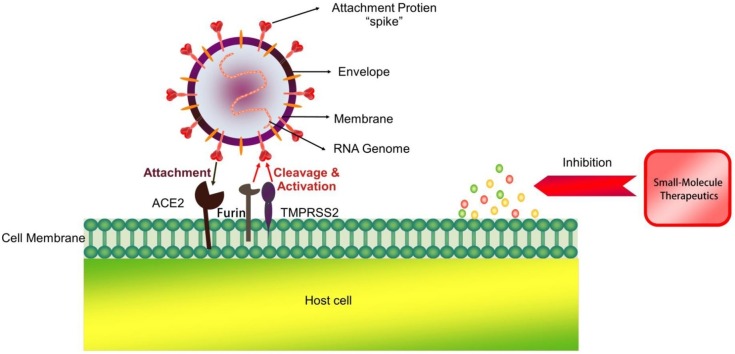
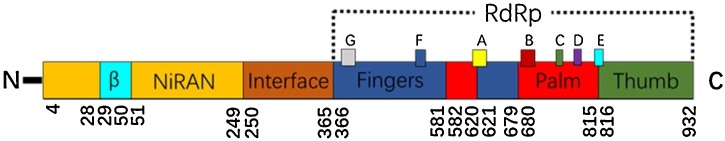
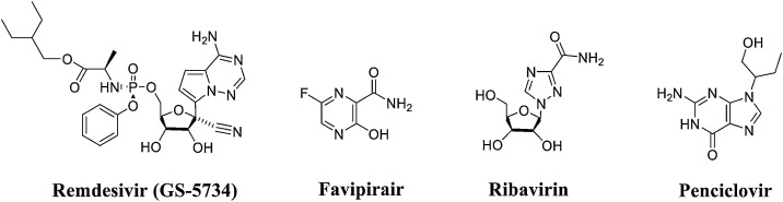
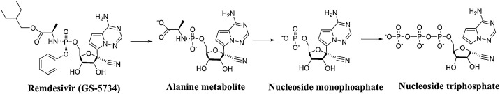
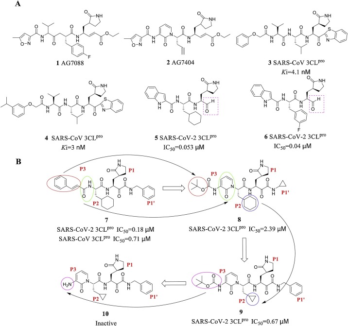
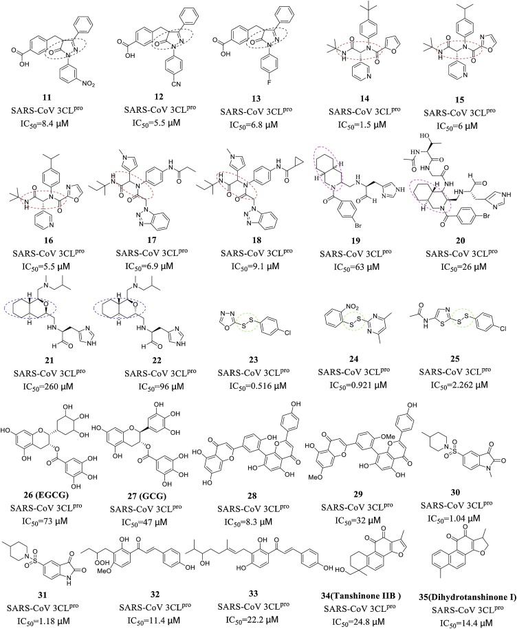
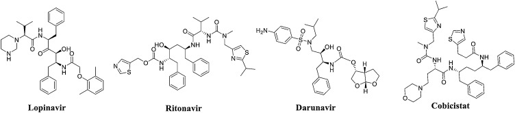
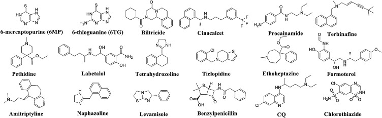
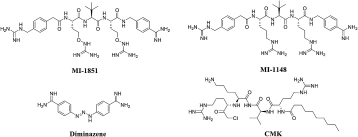
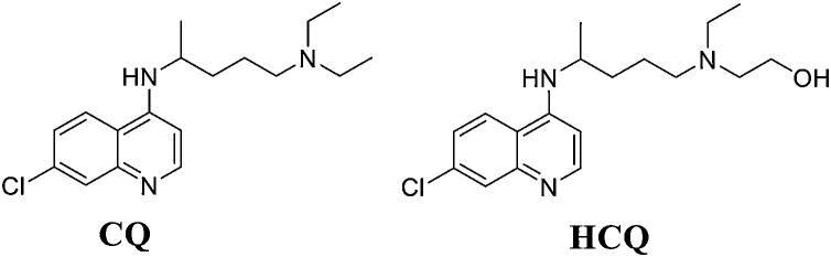
The SARS-CoV-2 outbreak and pandemic that began near the end of 2019 has posed a challenge to global health. At present, many candidate small-molecule therapeutics have been developed that can inhibit both the infection and replication of SARS-CoV-2 and even potentially relieve cytokine storms and other related complications. Meanwhile, host-targeted drugs that inhibit cellular transmembrane serine protease (TMPRSS2) can prevent SARS-CoV-2 from entering cells, and its combination with chloroquine and dihydroorotate dehydrogenase (DHODH) inhibitors can limit the spread of SARS-CoV-2 and reduce the morbidity and mortality of patients with COVID-19. The present article provides an overview of these small-molecule therapeutics based on insights from medicinal chemistry research and focuses on RNA-dependent RNA polymerase (RdRp) inhibitors, such as the nucleoside analogues remdesivir, favipiravir and ribavirin. This review also covers inhibitors of 3C-like protease (3CLpro), papain-like protease (PLpro) and other potentially innovative active ingredient molecules, describing their potential targets, activities, clinical status and side effects.

Keywords: 3C-like protease (3CL(pro)); Coronaviruses; Papain-like protease (PL(pro)); RNA-dependent RNA polymerase (RdRp); SARS-CoV-2; Transmembrane serine protease (TMPRSS2).


Conflict of interest statement

The authors declared that they have no conflicts of interest to this work.

Figures

None
Graphical abstract
Fig. 1
Fig. 1
Genome of RdRp.
Fig. 2
Fig. 2
Structures of representative nucleoside RdRp inhibitors.
Fig. 3
Fig. 3
Process of remdesivir-metabolized transformation to adenosine triphosphate.
Fig. 4
Fig. 4
Model of the incorporation of remdesivir into SARS-CoV-2 nsp12.
Fig. 5
Fig. 5
Mechanism of favipiravir-mediated inhibition of coronavirus replication in host cells (the purple wireframe and arrows represent the intracellular triphosphorylation of favipiravir and the inhibition of RdRp to terminate viral RNA replication). (For interpretation of the references to colour in this figure legend, the reader is referred to the web version of this article).
Fig. 6
Fig. 6
Mechanism of ribavirin against RNA viruses. The mechanism includes the following: a) induction of a shift from a Th2 to a Th1 immune response, b) inhibition of IMPDH to induce GTP depletion, c) direct inhibition of RdRp, and (d) induction of mutagenesis to trigger the production of defective viral particles.
Fig. 7
Fig. 7
The crystal structure of SARS-CoV-2 3CLpro (PDB code: 6LU7).
Fig. 8
Fig. 8
(A) Representative peptidomimetic 3CLpro inhibitors. (B) Chemical structures of α-ketoamide inhibitors 7-10. The coloured circles and arrows show the specific modifications at each development step.
Fig. 9
Fig. 9
Structure of representative nonpeptidic inhibitors.
Fig. 10
Fig. 10
Structure of HIV protease inhibitors with inhibitory activity against SARS-CoV-2 3CLpro.
Fig. 11
Fig. 11
Structure of representative PLpro inhibitors.
Fig. 12
Fig. 12
(A) Structures of the tetrapeptide substrate. (B) X-ray crystal structure of VIR250 and VIR251 bound to SARS-CoV-2 PLpro. The residues are shown as sticks; the red, blue and yellow colours indicate oxygen, nitrogen, and sulphur atoms, respectively, and hydrogen bonds are indicated by dashed lines (left: VI2R50, PDB code: 6WUU; right: VIR251, PDB code: 6WX4). (C) VIR250 and VIR251 bound to the binding pocket of SARS-CoV PLpro. VIR250 and VIR251 are shown as sticks, and the P2-P4 positions are labelled. (For interpretation of the references to colour in this figure legend, the reader is referred to the web version of this article).
Fig. 13
Fig. 13
Structures of S312 and S416.
Fig. 14
Fig. 14
Structures of TMPRSS2 inhibitors.
Fig. 15
Fig. 15
Sequence analysis of the SARS-CoV-2 S protein. The amino acid sequence positions of each domain are underneath. The cleavage sites of the SARS-CoV and SARS-CoV-2 S proteins are marked with diamonds.
Fig. 16
Fig. 16
Structures of furin inhibitors.
Fig. 17
Fig. 17
Structure of CQ and HCQ.

References

    1. Kaul D. An overview of coronaviruses including the SARS-2 coronavirus - Molecular biology, epidemiology and clinical implications. Curr. Med. Res. Pract. 2020;10(2):54–64. - PMC - PubMed
    1. Cui J., Li F., Shi Z.L. Origin and evolution of pathogenic coronaviruses. Nature Rev. Microbiol. 2019;17(3):181–192. - PMC - PubMed
    1. Marra M.A., Jones S.J., Astell C.R., Holt R.A., Brooks-Wilson A., Butterfield Y.S., et al. The Genome sequence of the SARS-associated coronavirus. Science. 2003;300(5624):1399–1404. - PubMed
    1. Ruan Y.J., Wei C.L., Ee A.L., Vega V.B., Thoreau H., Su S.T., Chia J.M., Ng P., Chiu K.P., Lim L., Zhang T., Peng C.K., Lin E.O., Lee N.M., Yee S.L., Ng L.F., Chee R.E., Stanton L.W., Long P.M., Liu E.T. Comparative full-length genome sequence analysis of 14 SARS coronavirus isolates and common mutations associated with putative origins of infection. Lancet (London, England) 2003;361(9371):1779–1785. - PMC - PubMed
    1. Vankadari N. Structure of furin protease binding to SARS-CoV-2 spike glycoprotein and implications for potential targets and virulence. J. Phys. Chem. Lett. 2020;11(16):6655–6663. - PubMed

MeSH terms