. 2021 Feb 4;13(4):619.
doi: 10.3390/cancers13040619.

The Potential of Raman Spectroscopy in the Diagnosis of Dysplastic and Malignant Oral Lesions

Affiliations

The Potential of Raman Spectroscopy in the Diagnosis of Dysplastic and Malignant Oral Lesions

Ola Ibrahim et al. Cancers (Basel). .

Abstract

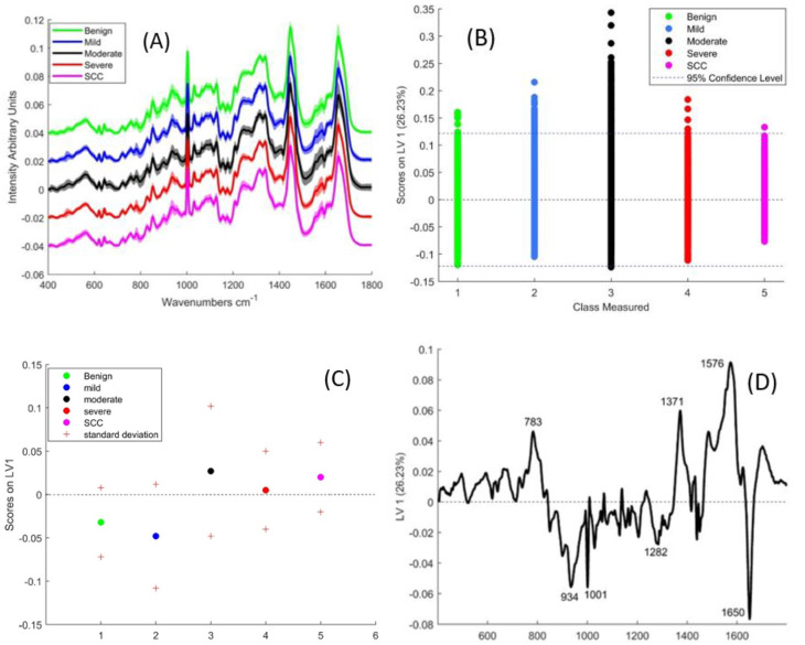
Early diagnosis, treatment and/or surveillance of oral premalignant lesions are important in preventing progression to oral squamous cell carcinoma (OSCC). The current gold standard is through histopathological diagnosis, which is limited by inter- and intra-observer errors and sampling errors. The objective of this work was to use Raman spectroscopy to discriminate between benign, mild, moderate and severe dysplasia and OSCC in formalin fixed paraffin preserved (FFPP) tissues. The study included 72 different pathologies from which 17 were benign lesions, 20 mildly dysplastic, 20 moderately dysplastic, 10 severely dysplastic and 5 invasive OSCC. The glass substrate and paraffin wax background were digitally removed and PLSDA with LOPO cross-validation was used to differentiate the pathologies. OSCC could be differentiated from the other pathologies with an accuracy of 70%, while the accuracy of the classifier for benign, moderate and severe dysplasia was ~60%. The accuracy of the classifier was lowest for mild dysplasia (~46%). The main discriminating features were increased nucleic acid contributions and decreased protein and lipid contributions in the epithelium and decreased collagen contributions in the connective tissue. Smoking and the presence of inflammation were found to significantly influence the Raman classification with respective accuracies of 76% and 94%.

Keywords: Raman spectroscopy; oral cancer; oral dysplasia; oral pre-cancer; potentially malignant lesions; premalignant lesions.


Conflict of interest statement

The authors declare no conflict of interest. The funders had no role in the design of the study; in the collection, analyses, or interpretation of data; in the writing of the manuscript, or in the decision to publish the results.

Figures

Figure 1
Figure 1
(A) Mean Raman spectra of benign, mild, moderate and severely dysplastic epithelial tissue. The spectra have been offset for clarity and shading denotes standard deviation (B) A plot of the PLSDA scores according to LV-1 (C) Mean and standard deviation of PLSDA scores of LV-1 (D) LV-1 of the PLSDA model for Epithelial tissue, including all the classes.
Figure 2
Figure 2
(A) Mean Raman spectra of benign, mild, moderate severely dysplastic and SCC connective tissue. The spectra have been offset for clarity and shading denotes standard deviation (B) A plot of the PLSDA scores of LV-1 (C) Mean and standard deviation of PLSDA scores of LV-1 (D) Loading of LV-1 of the PLSDA model which included all the classes.
Figure 2
Figure 2
(A) Mean Raman spectra of benign, mild, moderate severely dysplastic and SCC connective tissue. The spectra have been offset for clarity and shading denotes standard deviation (B) A plot of the PLSDA scores of LV-1 (C) Mean and standard deviation of PLSDA scores of LV-1 (D) Loading of LV-1 of the PLSDA model which included all the classes.
Figure 3
Figure 3
(A) Scores of smokers and non-smoker/ex-smokers on the latent variables from the PLSDA model. (B) Loading of LV-1 from PLSDA of smokers vs. non-smoker and ex-smokers in epithelial tissue.
Figure 4
Figure 4
(A) Scores of inflamed and non-inflamed moderately dysplastic connective tissue on the latent variables from the PLSDA model. (B) Loading of LV-1 from the PLSDA model of inflamed vs. non-inflamed connective tissue.
Figure 5
Figure 5
Representative H & E images showing (A) the regions of dysplasia marked by the pathologist. (B) A magnified region of moderate dysplasia (C) A magnified region of severe dysplasia. Scale bar: 200 µm.
Figure 6
Figure 6
A schematic of a Raman microspectrometer based on the Horiba Jobin Yvon LabRAM HR 800.
Figure 7
Figure 7
Spectral processing steps (A) Raw spectra. (B) Spectra after first quality control step, smoothing, baseline correction and normalization. (C) Spectra after k-means grouping; the spectra in red have high wax and low biological content while those in blue have higher biological content and less wax. (D) Spectra after glass and wax subtraction.

References

    1. Bray F., Ferlay J., Soerjomataram I., Siegel R.L., Torre L.A., Jemal A. Global cancer statistics 2018: GLOBOCAN estimates of incidence and mortality worldwide for 36 cancers in 185 countries. CA A Cancer J. Clin. 2018;68:394–424. doi: 10.3322/caac.21492. - DOI - PubMed
    1. Pfeifer G.P., Denissenko M.F., Olivier M., Tretyakova N., Hecht S.S., Hainaut P. Tobacco smoke carcinogens, DNA damage and p53 mutations in smoking-associated cancers. Oncogene. 2002;21:7435–7451. doi: 10.1038/sj.onc.1205803. - DOI - PubMed
    1. Boffetta P., Hashibe M. Alcohol and cancer. Lancet Oncol. 2006;7:149–156. doi: 10.1016/S1470-2045(06)70577-0. - DOI - PubMed
    1. Hashibe M., Brennan P., Chuang S.C., Boccia S., Castellsague X., Chen C., Curado M.P., Dal Maso L., Daudt A.W., Fabianova E., et al. Interaction between Tobacco and Alcohol Use and the Risk of Head and Neck Cancer: Pooled Analysis in the International Head and Neck Cancer Epidemiology Consortium. Cancer Epidemiol. Biomark. Prev. 2009;18:541–550. doi: 10.1158/1055-9965.EPI-08-0347. - DOI - PMC - PubMed
    1. van der Waal I. Potentially malignant disorders of the oral and oropharyngeal mucosa; terminology, classification and present concepts of management. Oral Oncol. 2009;45:317–323. doi: 10.1016/j.oraloncology.2008.05.016. - DOI - PubMed

LinkOut - more resources