. 2021 Feb 4;22(4):1590.
doi: 10.3390/ijms22041590.

Accelerated Wound Closure of Deep Partial Thickness Burns with Acellular Fish Skin Graft

Affiliations

Accelerated Wound Closure of Deep Partial Thickness Burns with Acellular Fish Skin Graft

Randolph Stone 2nd et al. Int J Mol Sci. .

Abstract

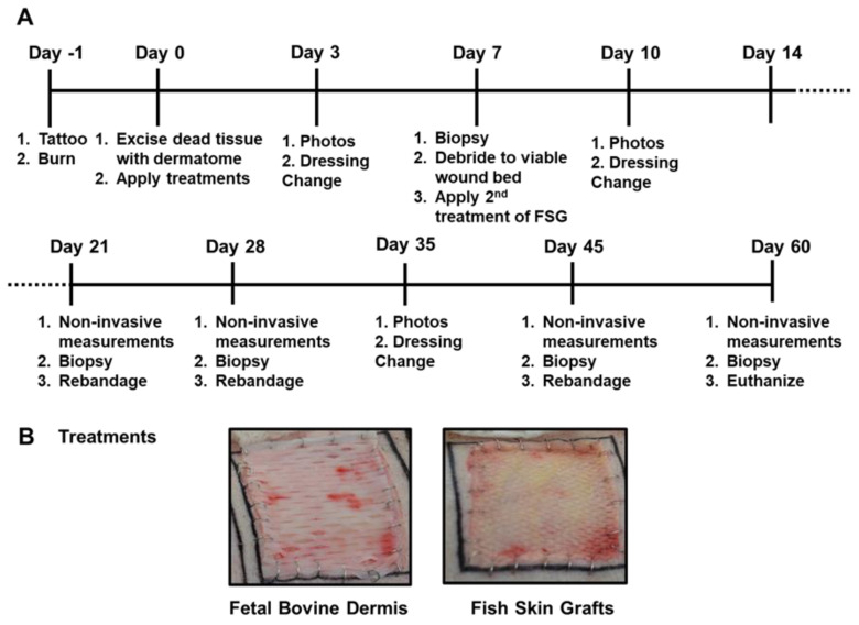
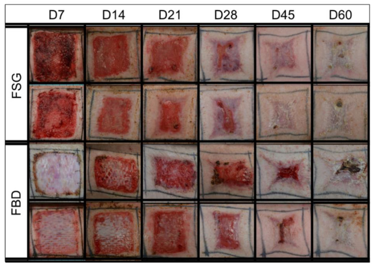
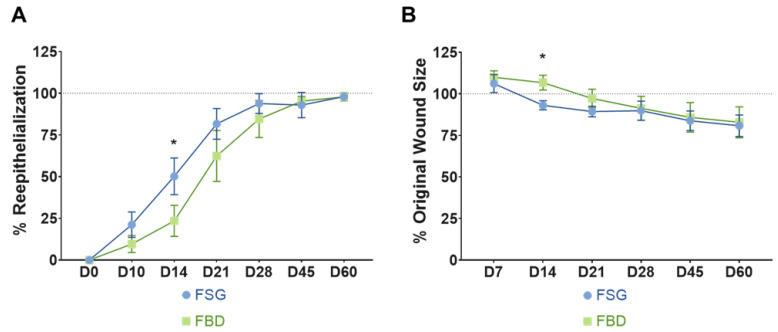
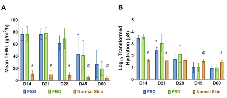
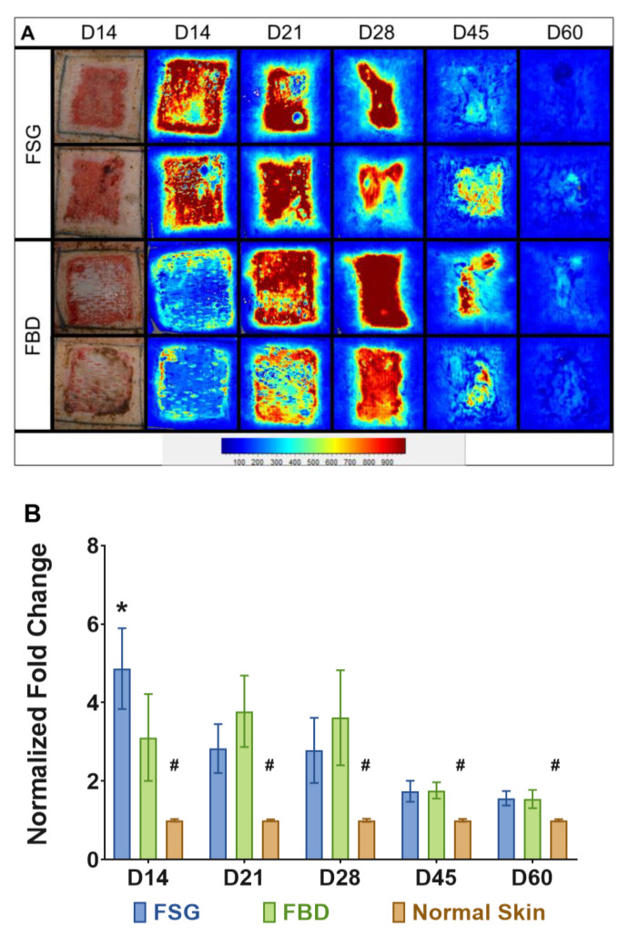
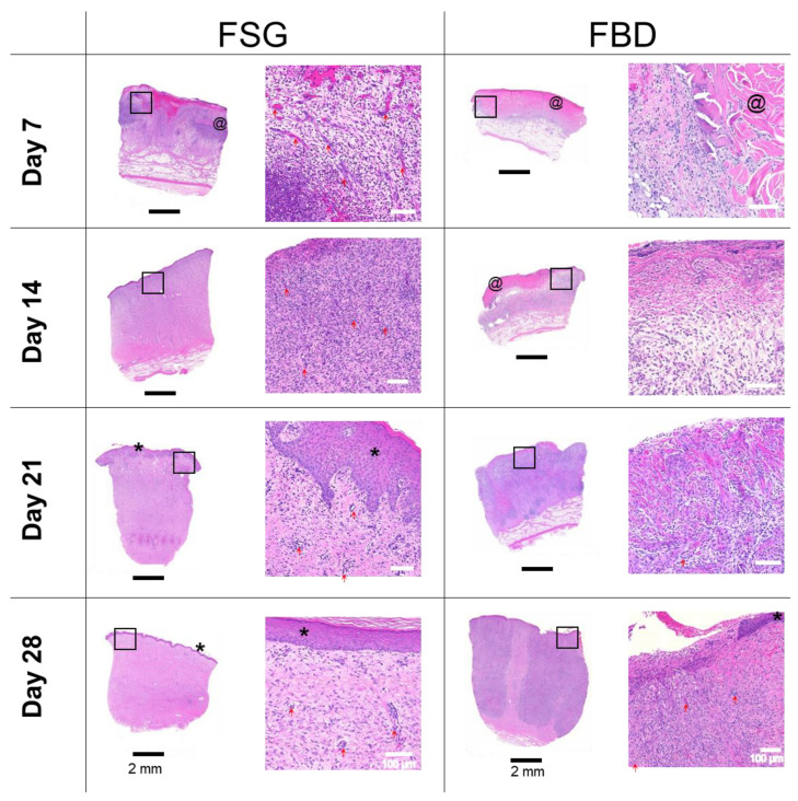
Thermal injuries are caused by exposure to a variety of sources, and split thickness skin grafts are the gold standard treatment for severe burns; however, they may be impossible when there is no donor skin available. Large total body surface area burns leave patients with limited donor site availability and create a need for treatments capable of achieving early and complete coverage that can also retain normal skin function. In this preclinical trial, two cellular and tissue based products (CTPs) are evaluated on twenty-four 5 × 5 deep partial thickness (DPT) burn wounds. Using appropriate pain control methods, DPT burn wounds were created on six anesthetized Yorkshire pigs. Wounds were excised one day post-burn and the bleeding wound beds were subsequently treated with omega-3-rich acellular fish skin graft (FSG) or fetal bovine dermis (FBD). FSG was reapplied after 7 days and wounds healed via secondary intentions. Digital images, non-invasive measurements, and punch biopsies were acquired during rechecks performed on days 7, 14, 21, 28, 45, and 60. Multiple qualitative measurements were also employed, including re-epithelialization, contraction rates, hydration, laser speckle, and trans-epidermal water loss (TEWL). Each treatment produced granulated tissue (GT) that would be receptive to skin grafts, if desired; however, the FSG induced GT 7 days earlier. FSG treatment resulted in faster re-epithelialization and reduced wound size at day 14 compared to FBD (50.2% vs. 23.5% and 93.1% vs. 106.7%, p < 0.005, respectively). No differences in TEWL measurements were observed. The FSG integrated into the wound bed quicker as evidenced by lower hydration values at day 21 (309.7 vs. 2500.4 µS, p < 0.05) and higher blood flow at day 14 (4.9 vs. 3.1 fold change increase over normal skin, p < 0.005). Here we show that FSG integrated faster without increased contraction, resulting in quicker wound closure without skin graft application which suggests FSG improved burn wound healing over FBD.

Keywords: burn; cellular and tissue-based product; contraction; epithelialization; fetal bovine dermis; fish skin graft; swine.


Conflict of interest statement

S.M and H.K. are employees of Kerecis®, Reykjavik, Iceland. For the remaining authors none are declared.

Figures

Figure 1
Figure 1
Porcine Burn Wound Experimental Timeline and Treatments. (A) The timeline is depicted for the 62 day study with the associated procedures performed throughout the study. Non-invasive measurements included digital and laser speckle imaging, TransEpidermal Water Loss (TEWL), and hydration readings. (B) Representative digital images of both fetal bovine dermis (FBD) and fish skin graft (FSG) after application on Day 0.
Figure 2
Figure 2
Representative Digital Images. Digital images shown are examples from 2 of the animals in the study. These were captured of all wounds during the 62 day study and were utilized to calculate the re-epithelialization and contraction rates. Biopsy punches harvested on previous time points are visible as small scabs in some wounds. Fetal bovine dermis (FBD); Fish skin graft (FSG).
Figure 3
Figure 3
Wound Closure rates. (A) Re-epithelialization was calculated by tracing the leading edge of the epidermis and comparing to total wound size. The dotted line represents 100% wound re-epithelialization. No day 7 results could be determined for re-ep due to the reapplication of FSG to the wounds which made observing the wound edge not possible. (B) Wound contraction was calculated by tracing the tattoos, normalizing to the growth of each animal, and comparing to the initial wound size. The dotted line represents the original wound size. Any reduction below that line is the result of the wounds contracting as they heal. For both A and B) * = p < 0.05 comparing treatments via a 2-way repeated measures ANOVA with Sidak’s post-hoc test (n = 12). Fetal bovine dermis (FBD); Fish skin graft (FSG).
Figure 4
Figure 4
Skin Barrier Function Measurements. (A) TransEpidermal Water Loss (TEWL) measures the barrier properties of the epidermis. At each time point, three TEWL measurements were taken for each of the twenty four wounds and then averaged. (# = p < 0.0001; @ = p < 0.05 for normal skin vs. treatment groups). (B) Hydration levels of wounds on follow-up days represented in Log scale (a log transformation was performed on hydration data because measurements ranged from 10 to 10,000). The hydration measures the water content of the wounds. Five measurements were obtained for each wound at each time point and averaged. (# = p < 0.001 and @ = p < 0.05 for normal skin vs. CTPs; * = p < 0.05 for FBD vs. FSG). For A and B, significance was detected using a 2-way ANOVA mixed effects model and a Tukey post-hoc test. Fetal bovine dermis (FBD); Fish skin graft (FSG).
Figure 5
Figure 5
Laser Speckle Imaging. (A) Two representative wounds are shown for both treatment groups. Digital images at day 14 and the corresponding laser speckle images (LSI) throughout the experiment are shown. The heatmap scale is shown below in which blue represents low perfusion and red if highly perfused. Note that the perfusion in uninjured skin is a sky blue and as the wounds heal by day 60, the borders of the wound are no longer obvious. (B) Quantitation of LSI measurements represented as a fold change above the normal perfusion around each wound and normalized to the growth controls on each animal at the designated time point (# = p < 0.005 for normal skin vs. treatment groups; * = p < 0.05 for FSG vs. FBD as determined by 2-way repeated measures ANOVA with Tukey post-hoc test, n = 12). Fetal bovine dermis (FBD); Fish skin graft (FSG).
Figure 6
Figure 6
H&E Histology. Representative H&E images of biopsy punches harvested at the indicated time points. Images on left are the entire biopsy punch with black scale bars = 2 mm while the area indicated by the black box is the enlarged image on the right with white scale bars = 100 um. * = newly formed epidermis; @ = residual pieces of treatment in wound bed; red arrows are pointing out some of the newly formed blood vessels. Fetal bovine dermis (FBD); Fish skin graft (FSG).
Figure 7
Figure 7
Immunohistochemistry with alpha-smooth muscle actin. Representative α-SMA images of biopsy punches harvested at the indicated time points. Positive brown staining is outlining the newly formed blood vessels. Scale bars = 100 um. Fetal bovine dermis (FBD); Fish skin graft (FSG).
Figure 8
Figure 8
Pathology Scores for Re-epithelialization. Sections were assessed by a Board-Certified Veterinarian Pathologist. (A,B) The number of slides receiving the designated score are indicated in (#) with the calculated percentages out of the 12 wounds for each treatment at each time point. Path Scores: 3 = Normal epidermis across entire wound bed; 2 = regenerating or hyperplastic with 100% coverage; 1 = partial epidermis; 0 = no epidermis. (C,D) Day 14 example of hematoxylin and eosin stained sections with a path score of “1” with the upper left image showing the entire biopsy punch. The area outlined with a green box is magnified and shown in the image to the lower right with scale bar = 250 µm. * = newly formed epidermis; @ = residual pieces of treatment in wound bed. Fetal bovine dermis (FBD); Fish skin graft (FSG).

References

    1. American Burn Association; 2016. [(accessed on 15 October 2020)]. Burn Incidence and Treatment in the United States: 2016 Fact. Sheet. Available online: http://ameriburn.org/who-we-are/media/burn-incidence-fact-sheet/
    1. Mosier M.J., Gibran N.S. Surgical excision of the burn wound. Clin. Plast. Surg. 2009;36:617–625. doi: 10.1016/j.cps.2009.05.006. - DOI - PubMed
    1. Fletcher J.L., Cancio L.C., Sinha I., Leung K.P., Renz E.M., Chan R.K. Inability to determine tissue health is main indication of allograft use in intermediate extent burns. Burns. 2015;41:1862–1867. doi: 10.1016/j.burns.2015.09.006. - DOI - PubMed
    1. Tam J., Wang Y., Farinelli W.A., Jiménez-Lozano J., Franco W., Sakamoto F.H., Cheung E.J., Purschke M., Doukas A.G., Anderson R.R. Fractional Skin Harvesting: Autologous Skin Grafting without Donor-site Morbidity. Plast. Reconstr. Surg. Glob. Open. 2013;1:e47. doi: 10.1097/GOX.0b013e3182a85a36. - DOI - PMC - PubMed
    1. Garg K., Kaszubski P.A., Moridzadeh R., Rockman C.B., Adelman M.A., Maldonado T.S., Veith F.J., Mussa F.F. Endovascular-first approach is not associated with worse amputation-free survival in appropriately selected patients with critical limb ischemia. J. Vasc Surg. 2014;59:392–399. doi: 10.1016/j.jvs.2013.09.001. - DOI - PubMed

LinkOut - more resources