. 2021 Nov;36(13-14):1162-1168.
doi: 10.1177/0883073821991295. Epub 2021 Feb 9.

Magnetic Resonance Imaging (MRI) and Spectroscopy in Succinic Semialdehyde Dehydrogenase Deficiency

Affiliations

Magnetic Resonance Imaging (MRI) and Spectroscopy in Succinic Semialdehyde Dehydrogenase Deficiency

Onur Afacan et al. J Child Neurol. 2021 Nov.

Abstract

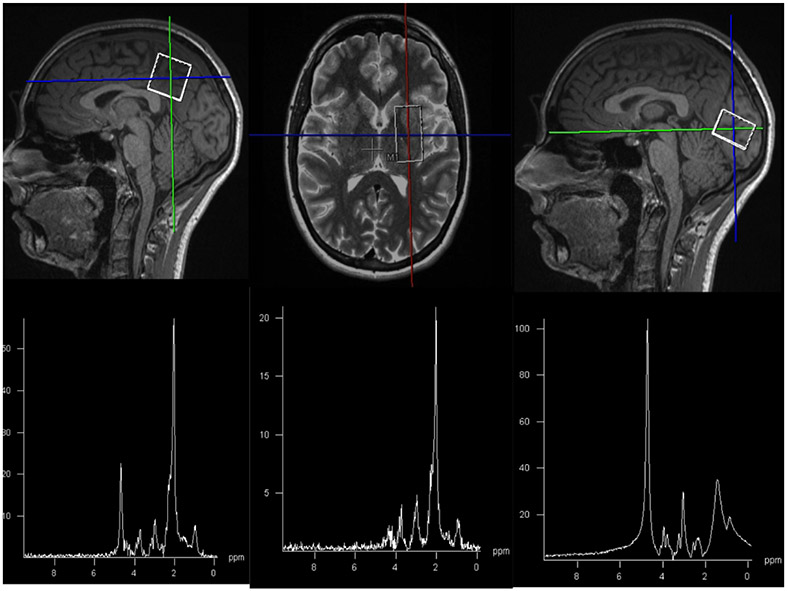
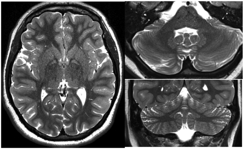
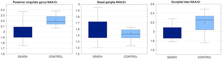
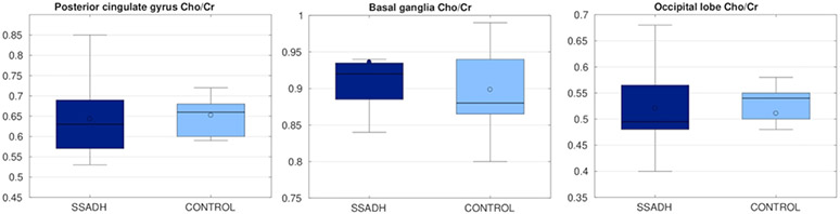
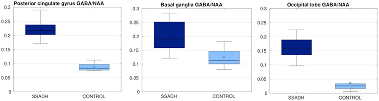
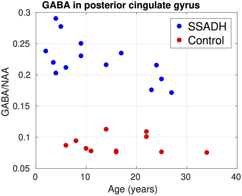
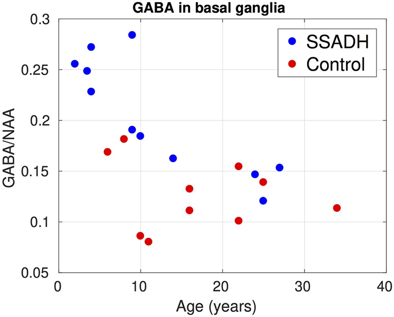
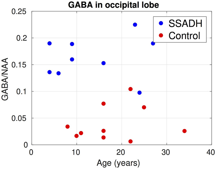
Succinic semialdehyde dehydrogenase (SSADH) deficiency is an autosomal recessive disorder of γ-aminobutyric acid (GABA) degradation, resulting in elevations of brain GABA and γ-hydroxybutyric acid (GHB). Previous magnetic resonance (MR) spectroscopy studies have shown increased levels of Glx in SSADH deficiency patients. Here in this work, we measure brain GABA in a large cohort of SSADH deficiency patients using advanced MR spectroscopy techniques that allow separation of GABA from overlapping metabolite peaks. We observed significant increases in GABA concentrations in SSADH deficiency patients for all 3 brain regions that were evaluated. Although GABA levels were higher in all 3 regions, each region had different patterns in terms of GABA changes with respect to age. We also report results from structural magnetic resonance imaging (MRI) of the same cohort compared with age-matched controls. We consistently observed signal hyperintensities in globus pallidus and cerebellar dentate nucleus.

Keywords: GABA; MEGAPRESS; MRI; SSADH; spectroscopy.


Conflict of interest statement

Disclosure of Conflict of Interests:

The authors have no conflicts of interest.

Figures

Figure 1:
Figure 1:
Sample images showing voxel placement and corresponding spectra in posterior cingulate gyrus (left), basal ganglia (middle), and occipital lobe (right).
Figure 2:
Figure 2:
Representative T2 weighted imaging from SSADH patients, highlighting the hyperintense signal in globus pallidus (left) and dentate nuclei (right).
Figure 3:
Figure 3:
NAA/Cr ratio in three different regions, compared between SSADH patients and control subjects. Control group had a significantly higher NAA/Cr ratio.
Figure 4:
Figure 4:
Cho/Cr ratio in three different regions, compared between SSADH patients and control subjects. There was no significant difference between two groups.
Figure 5:
Figure 5:
GABA/NAA ratio in three different regions, compared between SSADH patients and control subjects. In all three regions GABA/NAA was significantly higher in SSADH patients.
Figure 6:
Figure 6:
Change of GABA/NAA in the posterior cingulate gyrus with respect to subject age. Two groups were completely separated. In the controls GABA/NAA was mostly constant over age whereas in SSADH patients there was a small decline with respect to age.
Figure 7:
Figure 7:
Change of GABA/NAA in the basal ganglia with respect to subject age. Although the GABA/NAA was higher in younger SSADH subjects, in the older subjects there was no significant difference between the two groups.
Figure 8:
Figure 8:
Change of GABA/NAA in the occipital lobe with respect to subject age. GABA/NAA was significantly higher in SSADH deficiency patients.

References

    1. Gibson KM, Gupta M, Pearl PL, et al. Significant behavioral disturbances in succinic semialdehyde dehydrogenase (SSADH) deficiency (gamma-hydroxybutyric aciduria). Biol Psychiatry. 2003. doi:10.1016/S0006-3223(03)00113-6 - DOI - PubMed
    1. Kim KJ, Pearl PL, Jensen K, et al. Succinic semialdehyde dehydrogenase: Biochemical-molecular-clinical disease mechanisms, redox regulation, and functional significance. Antioxidants Redox Signal. 2011. doi:10.1089/ars.2010.3470 - DOI - PMC - PubMed
    1. Pearl PL, Wiwattanadittakul N, Roullet J-B, Gibson KM. Succinic semialdehyde dehydrogenase deficiency. In: GeneReviews®[Internet]. University of Washington, Seattle; 2016. - PubMed
    1. Pearl PL, Novomy EJ, Acosta MT, Jakobs C, Gibson KM. Succinic semialdehyde dehydrogenase deficiency in children and adults. In: Annals of Neurology. ; 2003. doi:10.1002/ana.10629 - DOI - PubMed
    1. Acosta MT, Munasinghe J, Pearl PL, et al. Cerebellar atrophy in human and murine succinic semialdehyde dehydrogenase deficiency. J Child Neurol. 2010. doi:10.1177/0883073810368137 - DOI - PMC - PubMed

Publication types

MeSH terms

Substances

Supplementary concepts