. 2021 Apr 15;81(8):2171-2183.
doi: 10.1158/0008-5472.CAN-20-1458. Epub 2021 Feb 8.

Ubiquitin-Specific Protease 6 Functions as a Tumor Suppressor in Ewing Sarcoma through Immune Activation

Affiliations

Ubiquitin-Specific Protease 6 Functions as a Tumor Suppressor in Ewing Sarcoma through Immune Activation

Ian C Henrich et al. Cancer Res. .

Abstract

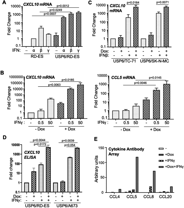
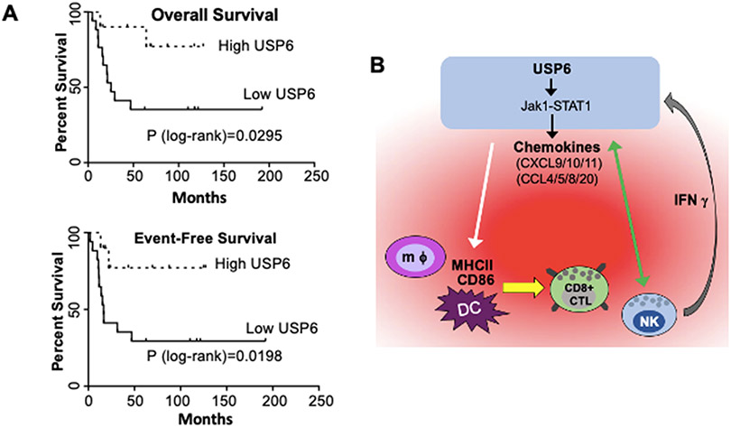
Ewing sarcoma is the second most common pediatric bone cancer, with a 5-year survival rate for metastatic disease of only 20%. Recent work indicates that survival is strongly correlated with high levels of tumor-infiltrating lymphocytes (TIL), whose abundance is associated with IFN-inducible chemokines CXCL10 and CCL5. However, the tumor-intrinsic factors that drive chemokine production and TIL recruitment have not been fully elucidated. We previously showed that ubiquitin-specific protease 6 (USP6) directly deubiquitinates and stabilizes Jak1, thereby inducing an IFN signature in Ewing sarcoma cells. Here, we show that this gene set comprises chemokines associated with immunostimulatory, antitumorigenic functions, including CXCL10 and CCL5. USP6 synergistically enhanced chemokine production in response to exogenous IFN by inducing surface upregulation of IFNAR1 and IFNGR1. USP6-expressing Ewing sarcoma cells stimulated migration of primary human monocytes and T lymphocytes and triggered activation of natural killer (NK) cells in vitro. USP6 inhibited Ewing sarcoma xenograft growth in nude but not NSG mice and was accompanied by increased intratumoral chemokine production and infiltration and activation of NK cells, dendritic cells, and macrophages, consistent with a requirement for innate immune cells in mediating the antitumorigenic effects of USP6. High USP6 expression in patients with Ewing sarcoma was associated with chemokine production, immune infiltration, and improved survival. This work reveals a previously unrecognized tumor-suppressive function for USP6, which engenders an immunostimulatory microenvironment through pleiotropic effects on multiple immune lineages. This further raises the possibility that USP6 activity may be harnessed to create a "hot" tumor microenvironment in immunotherapy. SIGNIFICANCE: This study reveals a novel tumor-suppressive function for USP6 by inducing an immunostimulatory microenvironment, suggesting that USP6 activity may be exploited to enhance immunotherapy regimens.


Conflict of interest statement

Conflict of Interest: This manuscript presents original research that has not been or is in the process of being published elsewhere. The Children’s Hospital of Philadelphia has ownership of PCT/US2019/063594 “Modulation of USP6 Expression as a Cancer Therapeutic” which lists Drs. Henrich and Chou as co-inventors.

Figures

Figure 1:
Figure 1:. USP6 elicits synergistic induction of IFN-regulated chemokines in ES cells
A-C) RT-qPCR was performed for cells treated as indicated. A) Parental RD-ES or USP6(Pool)/RD-ES were treated with dox and the indicated IFN overnight. B) USP6(Pool)/RD-ES were treated with dox and IFNγ overnight. C) ES cell lines TC71 and SK-N-MC were treated as indicated. D) Cells were treated with dox and IFNγ (5 ng/ml) as shown, and CM subjected to CXCL10 ELISA (RD-ES n=3, A673 n=2). See Supplemental Figure 2C for absolute concentrations. E) CM from USP6/RD-ES treated as indicated was used to probe a RayBiotech chemokine antibody array, followed by LI-COR detection. Results represent data from probing a single set of cytokine arrays (n=1).
Figure 2:
Figure 2:. USP6-regulated chemokines are induced by Jak-STAT1/NF-κB and can stimulate immune cell migration
A) USP6/RD-ES with the indicated CRISPR deletions were treated with dox and IFNγ (50 ng/ml), and CXCL9/10 expression was quantified. B) USP6(Pool)/RD-ES were treated with dox and IFNγ overnight, in the presence of PS1145 (15 μM) and Jak inhibitor (1 μM) as indicated, and CXCL9/10 expression quantified. C) Firefly (FF) luciferase driven by the CXCL10 promoter (WT or mutants in the indicated response elements) were co-transfected with renilla luciferase and USP6-encoding vectors. Cells were treated overnight with IFNγ, then subjected to dual luciferase assays. FF/renilla ratios relative to WT (black bar) were calculated; all p values are in comparison to this sample. Transwell migration assays were performed on THP-1 (D), and primary monocytes (E). Migration was monitored after 2 hr in response to serum-free conditioned medium (CM) from USP6/RD-ES cells treated as indicated, control serum-free medium, or recombinant CCL5 (10 ng/ml), CXCL10 (250 ng/ml), or 1% fetal bovine serum (FBS).
Figure 3:
Figure 3:. USP6 increases IFNGR1 surface expression
A) Top, cells were treated with dox and IFNg as shown, lysed, then subjected to western for IFNGR1 and b-catenin (as a loading control). Bottom, quantification of IFNGR1 protein levels, normalized against b-catenin, using LI-COR. B) RT-qPCR to quantify IFNGR1 mRNA. C) Surface IFNGR1 was quantified by flow cytometry in cells treated with or without dox. USP6/A673 and USP6/RD-ES are pooled cell lines that express heterogenous levels of HA-USP6; in dox-treated samples, IFNGR1 levels were assessed in the HA+ vs. HA populations. IFNGR1 CRISPR knockout cells were used as an antibody specificity control. D) Quantification of IFNGR1 flow results in C.
Figure 4:
Figure 4:. USP6 inhibits growth of RD-ES xenografts in nude mice coincident with enhanced chemokine production and immune cell infiltration in vivo
A) Nude mice were subcutaneously injected with clonal RD-ES cell lines expressing high or medium levels of USP6 (USP6(high) and USP6(med), respectively (17)), and maintained on dox or control water. Tumors were harvested after approximately 4 weeks; terminal masses are indicated for each mouse. Though dox treatment yielded a clear inhibitory trend in nude mice, this difference did not reach statistical significance. B) USP6/RD-ES were subcutaneously xenografted into NSG mice and terminal mass determined after approximately 4 weeks; differences in terminal mass were not statistically different. C) RT-qPCR was performed on tumors, normalizing against GAPDH. D) USP6(med) tumors were digested and surface flow cytometry performed using antibodies against: CD45 (pan-leukocyte), CD11b (pan-myeloid), CD11c (dendritic cell), F4/80 (macrophage), Ly6c (monocyte), and Ly6g (granulocyte). The percent of cells positive for the indicated marker relative to the total live cells in the bulk tumor is shown. See Supplemental Figures 8/9 for gating strategy.
Figure 5:
Figure 5:. USP6 stimulates intra-tumoral infiltration and activation of myeloid lineages
A) Nude mice were subcutaneously injected with USP6/A673, and maintained on dox or control water. Tumors were measured 3 times weekly until a terminal volume of 2000 mm3 was reached. Asterisks, mice whose tumors failed to reach terminal volume ("strong responders"). B) Time to terminal tumor volume was determined (presented as percent of mice whose tumors have yet to reach terminal volume). C-F) USP6/A673 tumors were digested and flow cytometry was performed using the indicated antibodies. Cell viability was assessed by Zombie-UV exclusion (C). Gray dots correspond to the strong responders (i.e. the mice labeled with asterisks in A). The percent of cells positive for the indicated marker relative to the total live cells in the bulk tumor is shown (see Supplemental Figures 8/9 for gating strategy). For p-values in gray, strong responders were included in the calculation; for p-values in black, they were excluded].
Figure 6:
Figure 6:. USP6 promotes NK activation in vivo, and directly induces NK activation in vitro
A) Digested USP6/A673 tumors were subjected to flow cytometry. The percent of cells positive for NK1.1+ relative to total live cells in the digested tumor was quantified, as was CD69 and CD25 in the NK1.1+/CD45+ live population. See Supplemental Figures 12/13 for gating strategy. B) NK92 (effector, E) cells were co-incubated with USP6/RD-ES (GFP+) (target, T) cells at various effector E:T ratios overnight, in the absence or presence of dox. Percent survival of the USP6/RD-ES (relative to sample without NK92) was quantified by monitoring the GFP+/propidium iodide-excluded population. C/D) USP6/RDES and NK92 were co-cultured overnight with or without dox, then subjected to (C) flow cytometry to monitor surface CD69 and CD107a on the NK92 (CD45+/GFP) cells, or (D) RT-qPCR to quantify IFNγ, CXCL9, and CXCL10 expression.
Figure 7:
Figure 7:. USP6 Expression is Associated with Increased Immune Cell Infiltration and Prolonged Survival in ES Patients
A) Primary untreated ES patient samples (GSE17679) (37) were ranked by USP6 expression, and overall and event-free survival was plotted. USP6high, top ~1/3; USP6low, bottom ~2/3; total patients in dataset n=27. B) Model for establishment of an immunostimulatory, anti-tumorigenic TME by USP6 to drive cancer cell clearance. USP6, via Jak1/STAT1, induces low level production of multiple chemokines (red cloud), leading to recruitment of multiple immune lineages (including DCs, macrophages (mφ), NK cells, and CD8+ cytotoxic T lymphocytes) into the TME. USP6 expression activates NK cells, leading to enhanced killing of USP6+ ES cells (green arrow), and IFNγ production. IFNγ feeds back on USP6-expressing ES cells, which synergistically induce production of chemokines (due to upregulation of IFNAR1, IFNGR, and Jak1 in these cells), and amplifying immune cell recruitment and activation. USP6 also induces activation of DCs/macrophages by stimulating their expression of CD86 and MHCII (white arrow), which we speculate would enhance their ability to activate recruited CD8+ CTLs.

References

    1. Lawlor ER, Sorensen PH. Twenty Years On – What Do We Really Know About Ewing Sarcoma And What Is The Path Forward? Crit Rev Oncog 2015;20:155–71. - PMC - PubMed
    1. Grohar PJ, Helman LJ. Prospects and challenges for the development of new therapies for Ewing sarcoma. Pharmacol Ther 2013;137:216–24. - PMC - PubMed
    1. Arnaldez FI, Helman LJ. New strategies in ewing sarcoma: lost in translation? Clin Cancer Res 2014;20:3050–6. - PMC - PubMed
    1. Pishas KI, Lessnick SL. Recent advances in targeted therapy for Ewing sarcoma. F1000Res 2016;5:1–11. - PMC - PubMed
    1. Hutzen B, Ghonime M, Lee J, Mardis ER, Wang R, Lee DA, et al. Immunotherapeutic Challenges for Pediatric Cancers. Mol Ther Oncolytics 2019;15:38–48. - PMC - PubMed

Publication types

MeSH terms