. 2021 Mar 15;206(6):1348-1360.
doi: 10.4049/jimmunol.2001228. Epub 2021 Feb 8.

AT-RvD1 Mitigates Secondhand Smoke-Exacerbated Pulmonary Inflammation and Restores Secondhand Smoke-Suppressed Antibacterial Immunity

Affiliations

AT-RvD1 Mitigates Secondhand Smoke-Exacerbated Pulmonary Inflammation and Restores Secondhand Smoke-Suppressed Antibacterial Immunity

Tariq A Bhat et al. J Immunol. .

Abstract

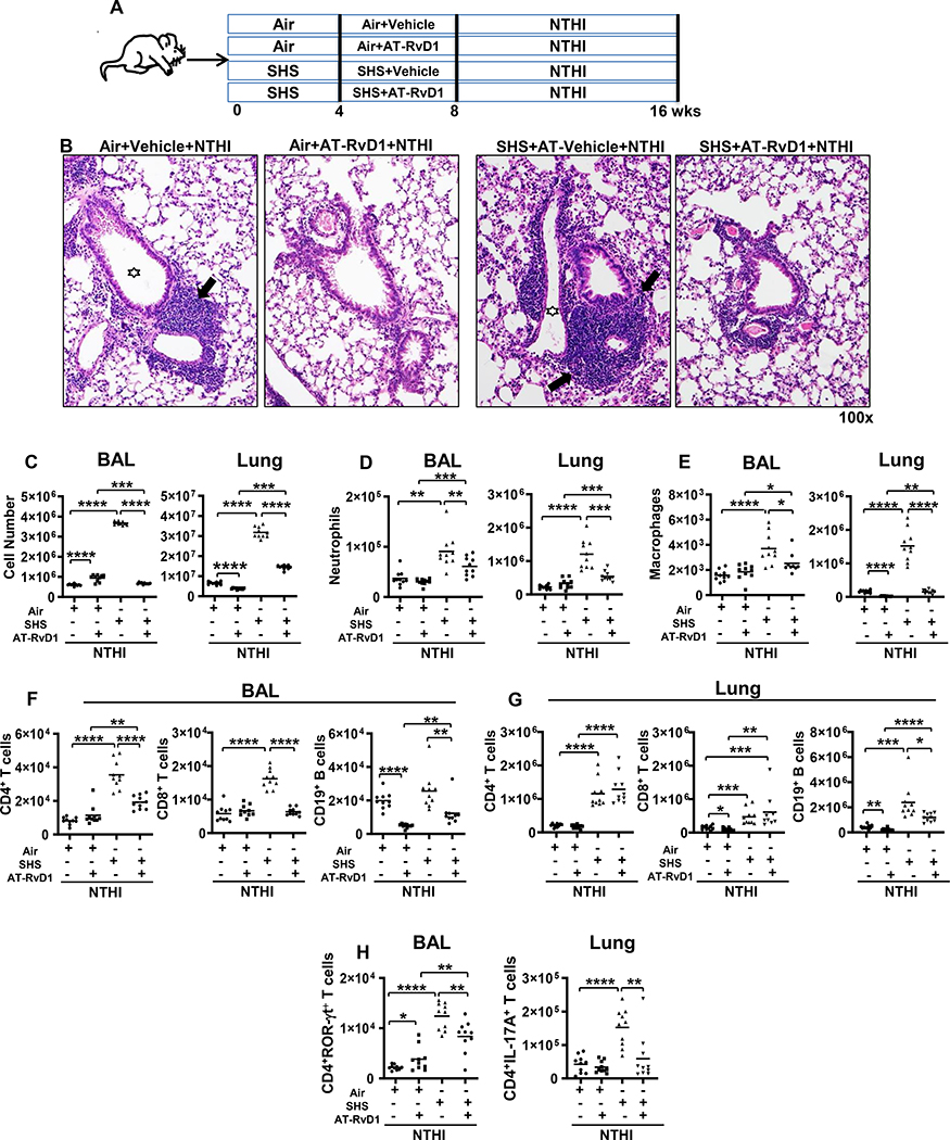
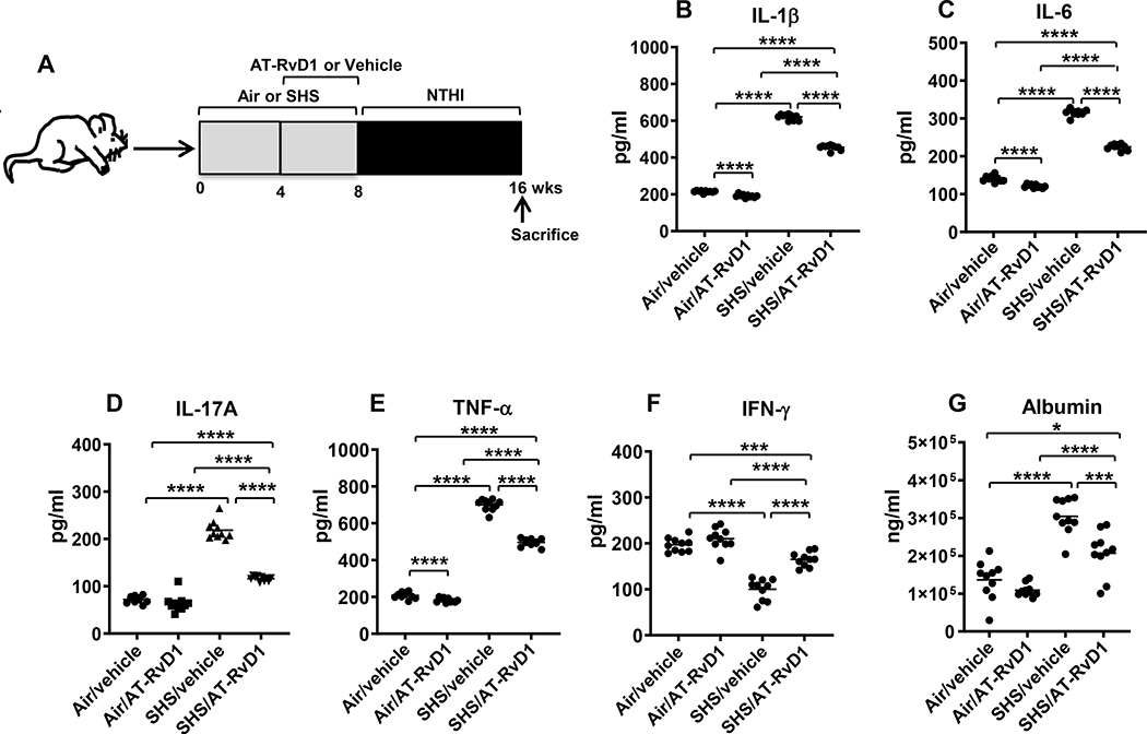
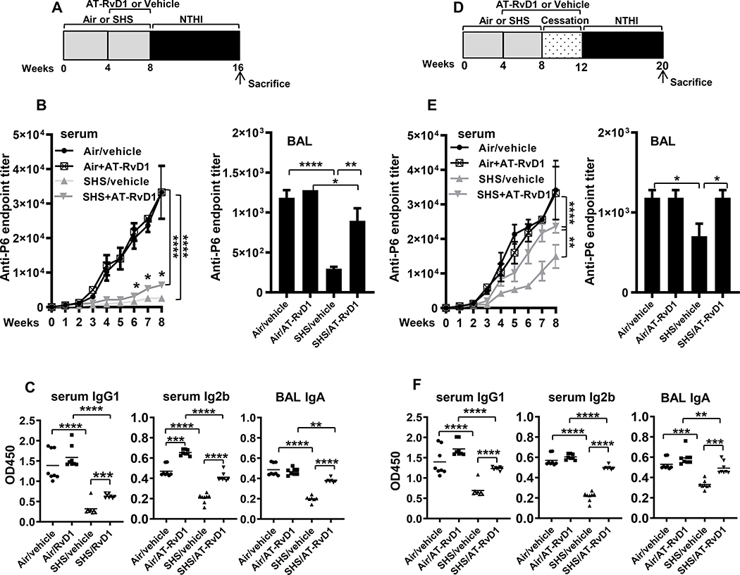
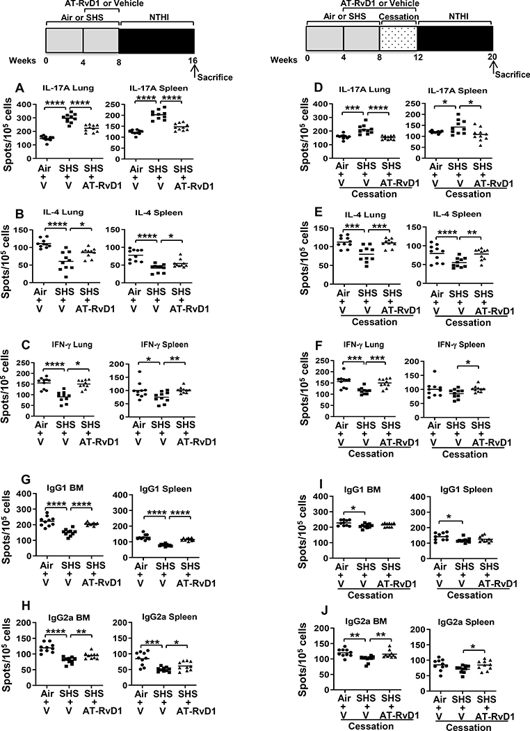
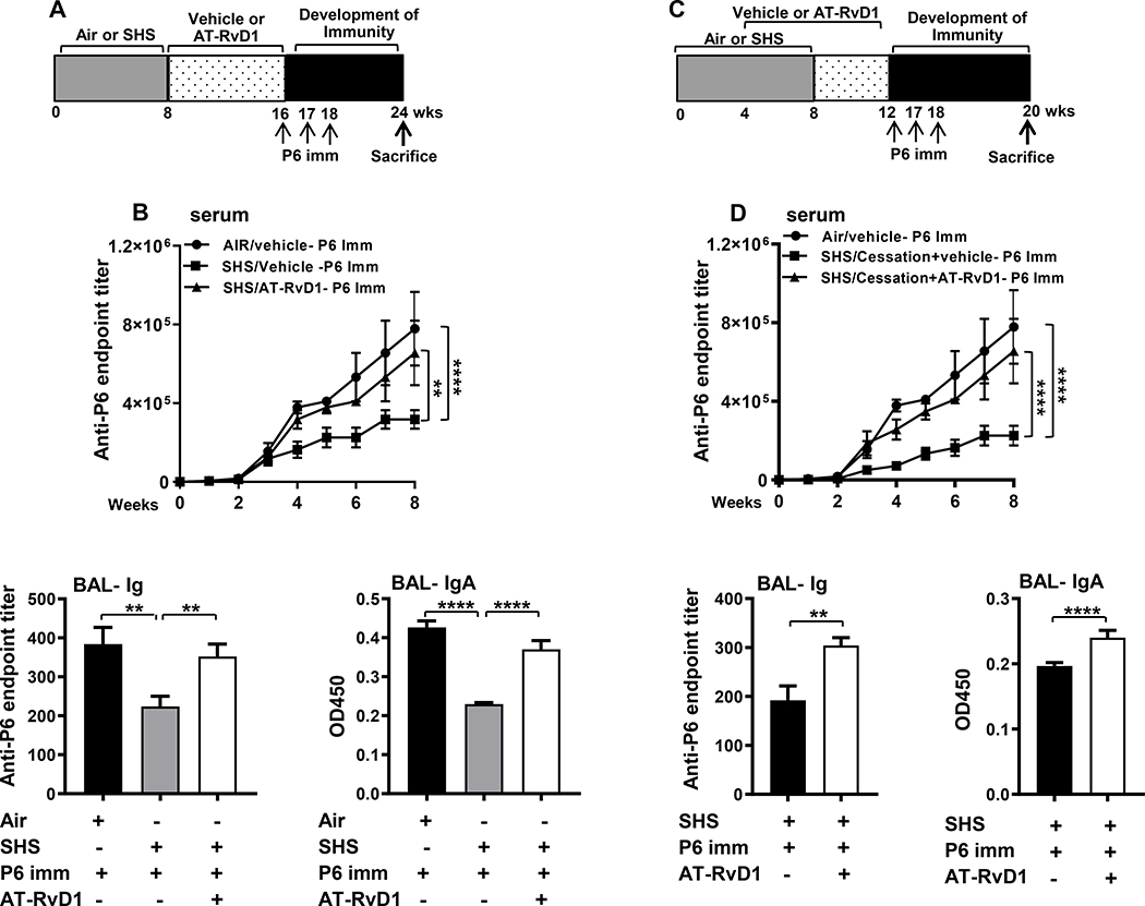
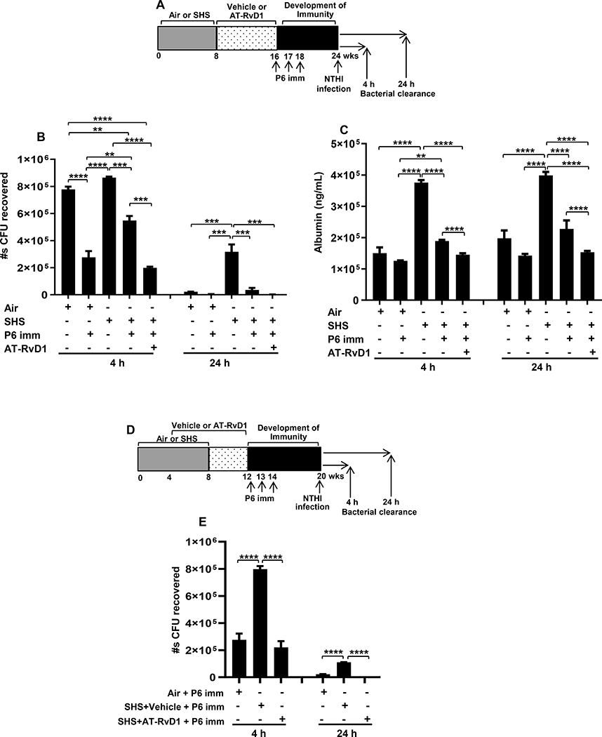
Cigarette smoke is a potent proinflammatory trigger contributing to acute lung injury and the development of chronic lung diseases via mechanisms that include the impairment of inflammation resolution. We have previously demonstrated that secondhand smoke (SHS) exposure exacerbates bacterial infection-induced pulmonary inflammation and suppresses immune responses. It is now recognized that resolution of inflammation is a bioactive process mediated by lipid-derived specialized proresolving mediators that counterregulate proinflammatory signaling and promote resolution pathways. We therefore hypothesized that proresolving mediators could reduce the burden of inflammation due to chronic lung infection following SHS exposure and restore normal immune responses to respiratory pathogens. To address this question, we exposed mice to SHS followed by chronic infection with nontypeable Haemophilus influenzae (NTHI). Some groups of mice were treated with aspirin-triggered resolvin D1 (AT-RvD1) during the latter half of the smoke exposure period or during a period of smoking cessation and before infection. Treatment with AT-RvD1 markedly reduced the recruitment of neutrophils, macrophages, and T cells in lung tissue and bronchoalveolar lavage and levels of proinflammatory cytokines in the bronchoalveolar lavage. Additionally, treatment with AT-RvD1 improved Ab titers against the NTHI outer membrane lipoprotein Ag P6 following infection. Furthermore, treatment with AT-RvD1 prior to classically adjuvanted immunization with P6 increased Ag-specific Ab titers, resulting in rapid clearance of NTHI from the lungs after acute challenge. Collectively, we have demonstrated that AT-RvD1 potently reverses the detrimental effects of SHS on pulmonary inflammation and immunity and thus could be beneficial in reducing lung injury associated with smoke exposure and infection.


Conflict of interest statement

Disclosures

The authors have no financial conflicts of interest.

Figures

FIGURE 1.
FIGURE 1.. AT-RvD1 treatment reduces SHS-exacerbated, NTHI-mediated pulmonary inflammation.
(A) C57BL/6J mice were exposed to air or SHS for 8 wks (5 days/wk; 5 h/day) with AT-RvD1 or vehicle given during the last 4 wks of exposure, and were then chronically infected by intratracheal (i.t.) NTHI instillation for an additional 8 wks. Animals were euthanized 48 h after the final instillation. (B) Representative H&E stained histological sections of lung from each group showing resolution of SHS-exacerbated pulmonary inflammation as a consequence of AT-RvD1 treatment. Large areas of lymphocytic inflammation around bronchovascular bundles are depicted by arrows and airway lumen are shown with stars. Original magnification x100. (C) Count of total white blood cells in the BAL (left) and lungs (right) of air or SHS-exposed mice that were treated with vehicle or AT-RvD1 and then infected with NTHI by i.t. administration for 8 wks (n = 10 per group). Innate (D-E), adaptive (F-G) and inflammatory (H) immune cell composition in the BAL and lungs of treated mice were evaluated by flow cytometry using cell type-specific markers as described in materials and methods. Our gating strategy is shown in Supplemental Figure 2. Line represents mean value for the group. All treatment groups were performed at the same time, and data represent results generated from a single experiment using a total of n=10 mice/group. The results are depicted as mean ± SE. Statistical significance between the treatment groups was determined using two-tailed unpaired students t test, *p<0.05, **p<0.01, ***p<0.001, ****p<0.0001.
FIGURE 2.
FIGURE 2.. AT-RvD1 treatment markedly diminishes SHS-augmented, NTHI-induced proinflammatory cytokine production, reducing SHS-enhanced lung epithelial damage.
(A) C57BL/6J mice were exposed to air or SHS for a total of 8 wks, with or without AT-RvD1 treatment given in the last 4 wks of exposure, and then subjected to chronic infection for an additional 8 wks, as described in material and methods. (B-F) After mice were euthanized, lungs were lavaged and cytokine levels in the BAL were determined by ELISA (n = 10 mice per group). (G) The levels of mouse albumin in the BAL were determined by ELISA as described in methods. Line represents mean value for the group. All treatment groups were performed at the same time, and data show results generated from a single experiment using a total of n=10 mice/group. The results are shown as mean ± SE. Statistical significance between the treatment groups was determined by two-tailed unpaired students t test, *p<0.05, ***p<0.001, ****p<0.0001.
FIGURE 3.
FIGURE 3.. AT-RvD1 treatment of SHS-exposed mice enhances antigen-specific antibody responses, which further are augmented by a period of cessation.
(A) C57BL/6J mice were exposed to air or SHS for 8 wks, with or without AT-RvD1 treatment given in the last 4 wks of exposure, and then subjected to the chronic infection model for 8 wks, as described. (B) Total anti-P6 IgG antibodies were measured by ELISA in serum (left panel) collected weekly during the period of chronic NTHI infection and in end-point BAL (right panel) collected at the time of euthanasia. (C) Levels of antigen-specific Ab subclasses IgG1 and IgG2b in end-point (wk 8) serum (left and middle panels) and IgA in BAL collected at euthanasia (right panel) were measured by determining OD450 values at dilutions of 1:1600 for serum and 1:400 for the BAL by ELISA assays as described in methods. (D) C57BL/6J mice were exposed to air or SHS for 8 wks, followed by a 4-wk period of cessation. Mice were treated with AT-RvD1 or vehicle between wks 4–12, encompassing part of the period of SHS exposure and period of cessation. All mice were chronically infected with NTHI by i.t. instillations starting at wk 12 as described in methods. (E) Total anti-P6 IgG antibodies were measured in weekly serum (left panel) and end-point BAL (right panel) by ELISA. (F) Levels of antigen-specific Ab subclasses IgG1 and IgG2b in end-point serum (left and middle panels) and IgA in end-point BAL (right panel) were determined by measuring the OD450 values for serum at 1:1600 dilution and for the BAL at 1:400 dilution by ELISA assays as described in methods. Line represents mean value for the group. All treatment groups were performed at the same time, and data represent results generated from a single experiment using a total of n=7–10 mice/group. The results are shown as mean ± SE. Statistical significance was determined by either two-way ANOVA with Tukey’s posttest for multiple comparisons (B and E, left panels) or by two-tailed unpaired students t test (all other figures), **p<0.01, ***p<0.001, ****p<0.0001.
FIGURE 4.
FIGURE 4.. AT-RvD1 improves SHS-impaired frequency of antigen-specific cytokine-secreting T cells, thereby augmenting antigen-specific antibody-secreting B cell frequency.
(A-F) Lung lymphocytes (Left panel) and splenocytes (Right panel) were isolated from air- or SHS-exposed, AT-RvD1 or vehicle treated mice after 8 wks of chronic NTHI infection. Cells were incubated with P641–55 peptide-pulsed APCs to determine the frequency of IL-17A-, IL-4- and IFN-γ-cytokine-secreting T cells by ELISPOT assays as described in methods. (G-J) B cell ELISPOTs were performed to quantify the frequency of P6-specific IgG1-secreting (upper panels) and IgG2a-secreting (bottom panels) B cells, from BM (left panels) and spleens (right panels) of air- or SHS-exposed, AT-RvD1 or vehicle treated mice. Spots were enumerated using a CTL ELISPOT reader. All treatment groups were performed at the same time, and data depict results generated from a single experiment using a total of n=10 mice/group. The results are depicted as mean ± SE. Line represents mean value for the group. Two-tailed unpaired students t test was utilized to determine the statistical significance between the treatment groups. *p≤0.05, **p≤0.01, ***p≤0.005, ****p≤0.0001,
FIGURE 5.
FIGURE 5.. AT-RvD1 treatment of SHS-exposed mice augments the efficacy of P6 vaccination.
(A) Mice exposed to air or SHS for 8 wks and then treated with AT-RvD1 or vehicle for an additional 8 wks were vaccinated with purified P6 lipoprotein as described in methods. (B) Total P6-specific IgG Ab titers were quantified by ELISA. in weekly serum (upper panel) collected after the start of vaccination and in end-point BAL (lower left panel) collected at the time of euthanasia Levels of mucosal anti-P6 IgA (lower right panel) were quantified by measuring OD450 values in the BAL at dilutions of 1:400 in ELISA assays as described in methods. (C) Mice were exposed to air or SHS for a total of 8 wks, with or without AT-RvD1 treatment between wks 4–12, and then vaccinated with NTHI P6 antigen as described in methods. (D) Total P6-specific IgG Ab titers were measured by ELISA in serum (upper panel) collected weekly after P6 vaccination and in end-point BAL (lower left panel). Mucosal anti-P6 specific IgA levels (lower right panel) were determined by measuring OD450 values in the BAL at 1:400 dilutions by ELISA as described in methods. All treatment groups were performed at the same time, and data represent results generated from a single experiment using a total of n=10 mice/group at each time point. The results are depicted as mean ± SE. Statistical significance was determined either by two-way ANOVA with Tukey’s posttest for multiple comparisons (B & D) or two-tailed unpaired students t test (**p<0.01, ****p<0.0001).
FIGURE 6.
FIGURE 6.. AT-RvD1 treatment-enhanced vaccination efficacy translates into marked decrease in SHS-augmented pulmonary bacterial burden and lung epithelial-cell damage following acute infection.
(A) Mice exposed to air or SHS for 8 wks were then treated with AT-RvD1 or vehicle for another 8 wks. All mice were vaccinated i.p. with 40 μg purified native P6 emulsified in CFA at wk16 and boosted 1 wk later with Ag in IFA and 2 wks later with Ag alone and bacterial challenge was done at wk24 as described in methods. Bacterial clearance was measured at 4 and 24 h following bacterial challenge. (B) Bacterial burden in the lungs of mice was measured by bacterial colony-plating assay and data are represented as total number of CFUs recovered from the lungs. (C) The levels of albumin in the BAL were measured by ELISA. (D-E) A group of SHS-exposed mice were given AT-RvD1- or vehicle-treatment that started immediately at week 4 and continued till week 12. Treated mice were vaccinated i.p. with 40 μg purified native P6 emulsified in CFA at wk12 and boosted 1 wk later with Ag in IFA and 2 wks later with Ag alone and then challenged with acute bacterial infection 8 wks after immunization at wk20. Bacterial burden in the lungs of this group of mice was measured at 4 and 24 h after bacterial challenge and data represented as total number of CFUs recovered from the lungs. All treatment groups were performed at the same time, and data represent results generated from a single experiment using a total of n=3 mice/group (B & E) or n=5 mice/group (C) at each time point. The results are depicted as mean ± SE. Statistical significance was determined by two-way ANOVA with Tukey’s posttest for multiple comparisons (**p≤0.01; ***p≤0.001; ****p≤0.0001).

References

    1. World Health Organization. 2016. Tobacco – fact sheet. Available at: http://www.who.int/mediacentre/factsheets/fs339/en/. Accessed: July 26, 2019.
    1. U.S. Department of Health and Human Services. 2014. The Health Consequences of Smoking-50 Years of Progress: A Report of the Surgeon General. U.S. Department of Health and Human Services, Centers for Disease Control and Prevention, National Center for Chronic Disease Prevention and Health Promotion, Office on Smoking and Health, Atlanta, GA.
    1. Eisner MD, Balmes J, Katz PP, Trupin L, Yelin EH, and Blanc PD. 2005. Lifetime environmental tobacco smoke exposure and the risk of chronic obstructive pulmonary disease. Environ. Health 4: 7. - PMC - PubMed
    1. Stampfli MR, and Anderson GP. 2009. How cigarette smoke skews immune responses to promote infection, lung disease and cancer. Nat. Rev. Immunol. 9: 377–384. - PubMed
    1. Oberg M, Jaakkola MS, Woodward A, Peruga A, and Pruss-Ustun A. 2011. Worldwide burden of disease from exposure to second-hand smoke: a retrospective analysis of data from 192 countries. Lancet. 377: 139–146. - PubMed

Publication types

MeSH terms