

Characterisation of Prostate Lesions Using Transrectal Shear Wave Elastography (SWE) Ultrasound Imaging: A Systematic Review
- PMID: 33558449
- PMCID: PMC7795187
- DOI: 10.3390/cancers13010122
Characterisation of Prostate Lesions Using Transrectal Shear Wave Elastography (SWE) Ultrasound Imaging: A Systematic Review
Abstract
Background: ultrasound-based shear wave elastography (SWE) can non-invasively assess prostate tissue stiffness. This systematic review aims to evaluate SWE for the detection of prostate cancer (PCa) and compare diagnostic estimates between studies reporting the detection of all PCa and clinically significant PCa (csPCa).
Methods: a literature search was performed using the MEDLINE, EMBASE, Cochrane Library, ClinicalTrials.gov, and CINAHL databases. Studies evaluating SWE for the detection of PCa using histopathology as reference standard were included.
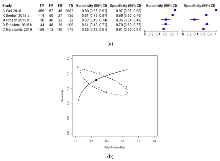
Results: 16 studies including 2277 patients were included for review. Nine studies evaluated SWE for the detection of PCa using systematic biopsy as a reference standard at the per-sample level, with a pooled sensitivity and specificity of 0.85 (95% CI = 0.74-0.92) and 0.85 (95% CI = 0.75-0.91), respectively. Five studies evaluated SWE for the detection of PCa using histopathology of radical prostatectomy (RP) specimens as the reference standard, with a pooled sensitivity and specificity of 0.71 (95% CI = 0.55-0.83) and 0.74 (95% CI = 0.42-0.92), respectively. Sub-group analysis revealed a higher pooled sensitivity (0.77 vs. 0.62) and specificity (0.84 vs. 0.53) for detection of csPCa compared to all PCa among studies using RP specimens as the reference standard.
Conclusion: SWE is an attractive imaging modality for the detection of PCa.
Keywords: prostate cancer; shear wave elastography; ultrasound.
Conflict of interest statement
The authors declare no conflict of interest.
Figures


References
-
- Abraham N.E., Mendhiratta N., Taneja S.S. Patterns of repeat prostate biopsy in contemporary clinical practice. [(accessed on 5 August 2020)];J. Urol. 2015 193:1178–1184. doi: 10.1016/j.juro.2014.10.084. Available online: http://www.ncbi.nlm.nih.gov/pubmed/25444971. - DOI - PubMed
-
- Ahmed H.U., El-Shater Bosaily A., Brown L.C., Gabe R., Kaplan R., Parmar M.K., Collaco-Moraes Y., Ward K., Hindley R.G., Freeman A., et al. Diagnostic accuracy of multi-parametric MRI and TRUS biopsy in prostate cancer (PROMIS): A paired validating confirmatory study. Lancet. 2017;389:815–822. doi: 10.1016/S0140-6736(16)32401-1. - DOI - PubMed
-
- Serefoglu E.C., Altinova S., Ugras N.S., Akincioglu E., Asil E., Balbay M.D. How reliable is 12-core prostate biopsy procedure in the detection of prostate cancer? [(accessed on 5 August 2020)];J. Can. Urol. Assoc. 2013 7:E293–E298. doi: 10.5489/cuaj.1248. Available online: http://www.ncbi.nlm.nih.gov/pubmed/22398204. - DOI - PMC - PubMed
-
- EAU Guidelines: Prostate Cancer|Uroweb. [(accessed on 5 August 2020)]; Available online: https://uroweb.org/guideline/prostate-cancer/#note_193.
-
- Richenberg J., Løgager V., Panebianco V., Rouviere O., Villeirs G., Schoots I.G. The primacy of multiparametric MRI in men with suspected prostate cancer. [(accessed on 5 August 2020)];Eur. Radiol. 2019 29:6940–6952. doi: 10.1007/s00330-019-06166-z. Available online: http://www.ncbi.nlm.nih.gov/pubmed/31172275. - DOI - PMC - PubMed
Publication types
Grants and funding
LinkOut - more resources
Full Text Sources
Other Literature Sources
