Review
. 2021 Feb 8;6(1):53.
doi: 10.1038/s41392-021-00487-6.

Viral vector platforms within the gene therapy landscape

Affiliations
Review

Viral vector platforms within the gene therapy landscape

Jote T Bulcha et al. Signal Transduct Target Ther. .

Abstract

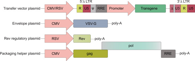
Throughout its 40-year history, the field of gene therapy has been marked by many transitions. It has seen great strides in combating human disease, has given hope to patients and families with limited treatment options, but has also been subject to many setbacks. Treatment of patients with this class of investigational drugs has resulted in severe adverse effects and, even in rare cases, death. At the heart of this dichotomous field are the viral-based vectors, the delivery vehicles that have allowed researchers and clinicians to develop powerful drug platforms, and have radically changed the face of medicine. Within the past 5 years, the gene therapy field has seen a wave of drugs based on viral vectors that have gained regulatory approval that come in a variety of designs and purposes. These modalities range from vector-based cancer therapies, to treating monogenic diseases with life-altering outcomes. At present, the three key vector strategies are based on adenoviruses, adeno-associated viruses, and lentiviruses. They have led the way in preclinical and clinical successes in the past two decades. However, despite these successes, many challenges still limit these approaches from attaining their full potential. To review the viral vector-based gene therapy landscape, we focus on these three highly regarded vector platforms and describe mechanisms of action and their roles in treating human disease.


Conflict of interest statement

G.G. is a scientific co-founder of Voyager Therapeutics, Adrenas Therapeutics, and Aspa Therapeutics, and holds equity in these companies. G.G. is an inventor on patents with potential royalties licensed to Voyager Therapeutics, Aspa Therapeutics, and other biopharmaceutical companies. The remaining authors declare no competing interests.

Figures

Fig. 1
Fig. 1. Summary of viral gene therapy modalities.
In vivo gene therapy entails the direct administration of vector carrying a therapeutic transgene into the patient. Ex vivo gene therapy involves the extraction of a patient’s cells or from an allogenic source, genetic modification by a vector carrying a therapeutic transgene, selection and expansion in culture, and infusion to re-introduce the engineered cells back into the patient
Fig. 2
Fig. 2. Summary of viral vectors used in clinical trials.
a Pie chart showing the percentage of adenovirus, adeno-associated virus, or lentivirus vectors in use. b A table of the current number of clinical trials employing the different viral vectors. Data source: Wiley database on Gene Therapy Trials Worldwide. http://www.abedia.com/wiley/vectors.php
Fig. 3
Fig. 3. Schematic of the wild-type adenovirus type 5 (Ad5) genome and the genetic modifications of common Ad5-based vectors.
a Diagram of the wild-type Ad5 genome structure. The 36 kb genome consists of four early transcription elements (E1, E2, E3, and E4), five late expression genes (L1–L5), cis-packaging elements (ψ) and two inverted terminal repeat sequences (ITR). The E1A (red arrow) gene contains four conserved domains (CR1-4), each of which interacts with special cellular proteins. The E1B gene encodes for two distinct tumor antigens, the 19 kDa (19K) and 55 kDa (55K) proteins. The E2 gene encodes DNA-binding protein (DBP), terminal protein (pTP), IVa2, and DNA polymerase (Pol). The E4 gene encodes 1–7 open reading frames. The major late messenger RNAs (L1–L5) mainly encode for virion structural proteins and are derived from a pre-mRNA, which is driven by a major late promoter (MLP) via alternative splicing and polyadenylation. L1 encodes for IIIa and 52K. L2 encodes for the penton base gene (capsid protein III) and the core proteins V, pVII, and pX. L3 encodes for the hexon (capsid protein II), capsid protein precursor (pVI), and protease (Pr) genes. L4 encodes for 100K, 33K, 22K, and pVIII. L5 encodes for the fiber gene (capsid protein IV). be Diagrams of rAd vectors. b Replication-defective (RD) vector. The E1A and E1B regions are deleted and replaced with an expression cassette containing an exogenous promoter and a transgene of interest (indicated by the solid red X and yellow arrow). The E3 and E4 regions are usually deleted to accommodate larger insertions and eliminates leaky expression of other viral genes. c E1B-55K replication-competent vector. The E1B-55K region is deleted (solid red X and dashed blue arrow), whereas in some vectors, the E3 region is deleted and replaced with an expression cassette (dashed red X and dashed blue arrow). d E1A-Δ24 (Δ24) replication-competent vector. The CR2 region in E1A is deleted (solid red X and dashed red arrow) and the E1A promoter can be replaced with various tumor-specific promoters to drive CR2-deleted E1A expression. In some vector designs, the E3 region is deleted and replaced with an expression cassette (dashed red X and dashed blue arrow). e Helper-dependent Ad vectors (HDAds). Most or all of the Ad genomic elements are replaced with a therapeutic expressing cassette (yellow arrow), save for the cis-packaging sequences (ψ) and ITRs. These vectors are propagated in the presence of an Ad helper vector
Fig. 4
Fig. 4. Schematic of the AAV genome and sites used for PCR screening.
The AAV genome comprised four known open reading frames, rep (green), cap (salmon), MAAP (orange), and AAP (yellow). The rep and cap ORFs encode four and three isoforms, respectively. Transcription is driven by the viral P5, P19, and P40 promoters (arrows). The genome is flanked by inverted terminal repeat (ITR, cyan) sequences
Fig. 5
Fig. 5. Third-generation HIV-1-based lentiviral vector design.
The third generation of lentiviral vectors are produced using four plasmids. The first plasmid has a construct carrying the gene of interest driven by a desired promoter flanked by long terminal repeats (LTRs). Both 5′ and 3′ LTRs in wild-type HIV-1 is composed of U3, R, and U5 sequences. The U3 sequence constitutes promoter/enhancer elements. Part of the U3 sequence in the 3′-LTR is deleted, and the entire U3 sequence within the 5′-LTR is replaced by a strong viral promoter, such as CMV. The psi (ψ) packaging signal is followed by the rev response element (RRE). The envelope glycoprotein is encoded by VSV-G (vesicular stomatitis virus) and is expressed under a strong promoter, such as CMV. The rev gene is split from the packaging plasmid and is provided on a separate plasmid construct. The packaging plasmid harbors the viral gag and pol genes, and notably lacks the tat regulatory gene

References

    1. Terheggen HG, et al. Unsuccessful trial of gene replacement in arginase deficiency. Z. Kinderheilkd. 1975;119:1–3. doi: 10.1007/BF00443566. - DOI - PubMed
    1. Cline MJ. Perspectives for gene therapy: inserting new genetic information into mammalian cells by physical techniques and viral vectors. Pharm. Ther. 1985;29:69–92. doi: 10.1016/0163-7258(85)90017-8. - DOI - PubMed
    1. Anderson WF. Human gene therapy. Science. 1992;256:808–813. doi: 10.1126/science.1589762. - DOI - PubMed
    1. Kohn DB, et al. Establishment and characterization of adenosine deaminase-deficient human T cell lines. J. Immunol. 1989;142:3971–3977. - PubMed
    1. Arrand JR, Roberts RJ. The nucleotide sequences at the termini of adenovirus-2 DNA. J. Mol. Biol. 1979;128:577–594. doi: 10.1016/0022-2836(79)90294-8. - DOI - PubMed

Publication types