. 2021 Feb 8;12(1):848.
doi: 10.1038/s41467-021-21118-2.

SARS-CoV-2 D614G spike mutation increases entry efficiency with enhanced ACE2-binding affinity

Affiliations

SARS-CoV-2 D614G spike mutation increases entry efficiency with enhanced ACE2-binding affinity

Seiya Ozono et al. Nat Commun. .

Abstract

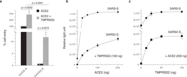
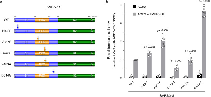
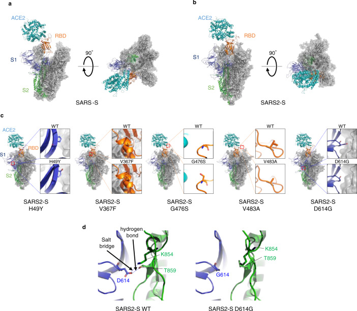
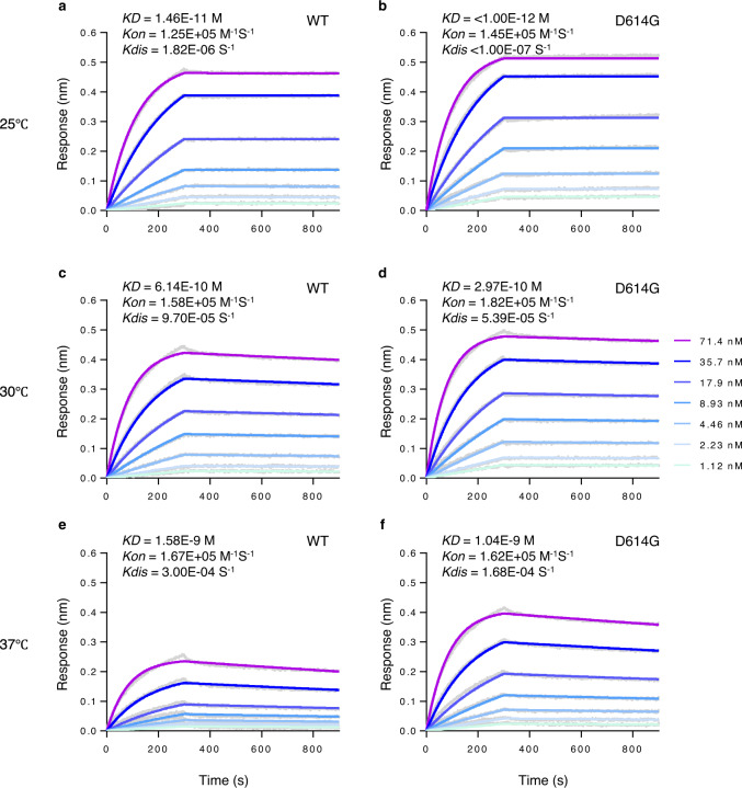
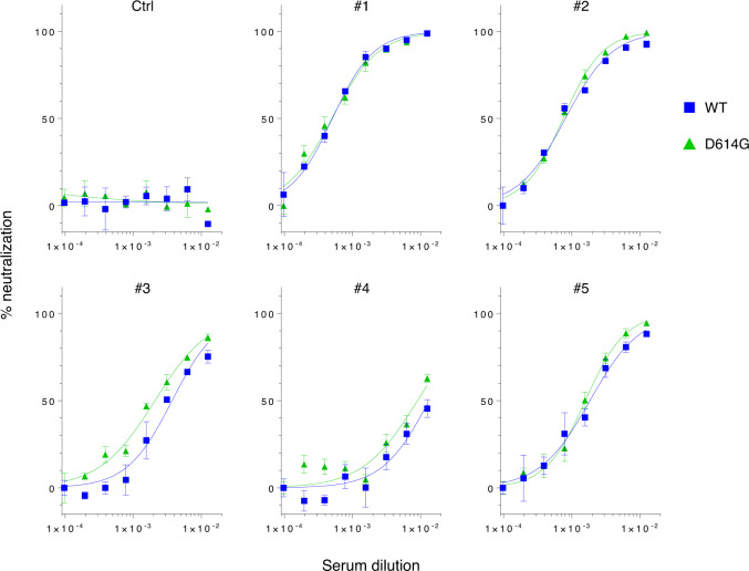
The causative agent of the COVID-19 pandemic, SARS-CoV-2, is steadily mutating during continuous transmission among humans. Such mutations can occur in the spike (S) protein that binds to the ACE2 receptor and is cleaved by TMPRSS2. However, whether S mutations affect SARS-CoV-2 cell entry remains unknown. Here, we show that naturally occurring S mutations can reduce or enhance cell entry via ACE2 and TMPRSS2. A SARS-CoV-2 S-pseudotyped lentivirus exhibits substantially lower entry than that of SARS-CoV S. Among S variants, the D614G mutant shows the highest cell entry, as supported by structural and binding analyses. Nevertheless, the D614G mutation does not affect neutralization by antisera against prototypic viruses. Taken together, we conclude that the D614G mutation increases cell entry by acquiring higher affinity to ACE2 while maintaining neutralization susceptibility. Based on these findings, further worldwide surveillance is required to understand SARS-CoV-2 transmissibility among humans.


Conflict of interest statement

The authors declare no competing interests.

Figures

Fig. 1
Fig. 1. SARS2-S-mediated cell entry is highly dependent on TMPRSS2.
a Viruses were prepared by transfection of 293T cells with the HiBiT-tagged lentiviral packaging plasmid, the firefly luciferase-reporter lentiviral transfer plasmid, and either a SARS-CoV S (SARS-S) or SARS-CoV-2 S (SARS2-S) expression plasmid. Viral supernatants were subjected to HiBiT assays, and S-pseudotyped viruses normalized by HiBiT activity were used for infection of 293T cells expressing the host receptor ACE2 alone (black) or coexpressing TMPRSS2 (gray). Cell entry was determined by firefly luciferase activity in cell lysates. Data from four experiments are shown as a percentage of cell entry of SARS-S-pseudotyped viruses into 293T cells expressing ACE2 only (mean ± s.d., n = 3 technical replicates). The p value was calculated using a two-tailed paired or unpaired Student’s t-test. b, c The effect of ACE2 or TMPRSS2 expression levels on cell entry activity. 293T cells were transfected with a high and constant level of an expression plasmid encoding ACE2 together with increasing levels of a TMPRSS2 expression plasmid (b), and vice versa (c). Transfected cells were infected with lentiviruses pseudotyped with either SARS-S (circle) or SARS2-S (square), as described in a. Data shown are representative of three independent experiments (mean ± s.d., n = 3 technical replicates). Source data are provided as a Source Data file.
Fig. 2
Fig. 2. SARS2-S D614G variant protein display highest levels of entry activity.
a Schematic illustration of the prototype (wild-type, WT) and globally spread variant SARS2-S proteins. Numbers indicate amino acid positions. The signal peptide (SP), transmembrane domain (TM), cytoplasmic tail (CT), S1 subunit (S1), S2 subunit (S2), and receptor-binding domain (RBD) are indicated. The positions of mutations are indicated by arrows. b Functional comparison of the entry activity of WT and mutant SARS2-S proteins. Different S-pseudotyped viruses were prepared as described in a and used for infection of 293T cells expressing the host receptor ACE2 alone (black) or coexpressing TMPRSS2 (gray). Cell entry was determined by firefly luciferase activity in cell lysates. Data were shown as the fold difference of cell entry relative to that of WT into 293T cells coexpressing ACE2 and TMPRSS2 (mean ± s.d. from seven independent experiments (except for that of G476S, n = 6 with three technical replicates); compared with WT using one-way ANOVA with Dunnett’s multiple comparison test. Source data for b are provided as a Source Data file.
Fig. 3
Fig. 3. SARS2-S D614G protein shows the prominent structural difference.
a, b Trimeric structure of the S proteins from SARS-CoV (a) and SARS-CoV-2 (b) that bind to the host receptor ACE2 (cyan). One protomer of the S protein is shown as a ribbon in navy (S1 subunit), orange (receptor-binding domain (RBD) in S1 subunit), and green (S2 subunit), while two other protomers are shown as gray transparent surfaces and ribbons. The structures are viewed from two different angles. c Comparison between wild-type (WT) and mutant S proteins from globally spread SARS-CoV-2 variants. (Inset) Enlargement of the region in which each amino acid is mutated, with comparison with the WT S protein. H49Y; as the histidine at position 49 is located distant from the RBD and putative cleavage sites, the effect of this mutation on S’s function is likely limited. V367F; the substitution from a valine to a phenylalanine at position 367 in the RBD introduces a larger side chain at a protomer–protomer interface, which might provide a more rigid RBD structure. G476S; the substitution at position 476 in the RBD results in a protruded surface, which appears to interfere with the ACE2–RBD interaction. V483A; both valine and alanine residues have short side chains, likely sharing similar phenotypes. D614G; details are depicted in Fig. 3d. Note that these structural bases are largely consistent with the results of cell entry activity shown in Fig. 2b. d Structural difference between WT and D614G SARS-CoV-2 S proteins. WT (left); an aspartic acid (D614) in the S1 subunit (navy) of a protomer binds to a threonine (T859) and/or a lysine (K854) in the S2 subunit (green) of the other protomer though electrostatic interaction between the pairs of these residues. D614G (right); the short nonpolar side chain of glycine (G614), which does not bind to T859 and K854, provides flexible space between the two protomers. The figures were drawn with PyMOL ver. 2.4 (https://pymol.org).
Fig. 4
Fig. 4. SARS2-S D614G protein binds ACE2 with higher affinity than WT S protein.
Binding affinity between dimeric ACE2 and trimeric SARS2-S WT (a, c, and e) or D614G variant (b, d, and f) (two-fold serially diluted from 71.4 nM to 1.12 nM) was evaluated at 25 °C (a and b), 30 °C (c and d), or 37 °C (e and f) using Octet RED96 instrument. The association rate constants (Kon) and dissociation rate constants (Kdis) were determined by global fitting of the experimental data using a 1:1 binding model. Equilibrium dissociation constants (KD) were obtained from KdisKon. Data shown are representative sensorgrams from two independent experiments at 25/37°C and from three independent experiments at 30°C using different protein preparations. The difference in KD and Kdis at 30°C was statistically significant (p = 0.0042 and 0.0129, respectively) by a two-tailed unpaired Student’s t-test, as shown in Supplementary Fig. 5. Source data are provided as a Source Data file.
Fig. 5
Fig. 5. SARS-2 S WT and D614G proteins are similarly neutralized by patient sera.
Lentiviruses pseudotyped with either the WT or D614G mutant SARS2-S were preincubated with two-fold serially diluted human sera (80-fold to 10, 240-fold) obtained from a healthy donor (Ctrl) or collected at 15–30 days post-symptom onset from confirmed case patients (#1–#5) infected with the prototypic viruses. The mixture was used for infection of 293T cells coexpressing ACE2 and TMPRSS2, and cell entry levels of pseudoviruses in the presence of diluted patient sera were determined by luciferase assays. Representative data from two independent experiments are shown as percent neutralization (mean ± s.d., n = 3 technical replicates). Source data are provided as a Source Data file.

Similar articles

Cited by

References

    1. Wu F, et al. A new coronavirus associated with human respiratory disease in China. Nature. 2020;579:265–269. doi: 10.1038/s41586-020-2008-3. - DOI - PMC - PubMed
    1. Li Q, et al. Early transmission dynamics in Wuhan, China, of novel coronavirus–infected pneumonia. N. Engl. J. Med. 2020;382:1199–1207. doi: 10.1056/NEJMoa2001316. - DOI - PMC - PubMed
    1. Huang C, et al. Clinical features of patients infected with 2019 novel coronavirus in Wuhan, China. Lancet. 2020;395:497–506. doi: 10.1016/S0140-6736(20)30183-5. - DOI - PMC - PubMed
    1. Zhou P, et al. A pneumonia outbreak associated with a new coronavirus of probable bat origin. Nature. 2020;579:270–273. doi: 10.1038/s41586-020-2012-7. - DOI - PMC - PubMed
    1. Hoffmann M, et al. SARS-CoV-2 cell entry depends on ACE2 and TMPRSS2 and is blocked by a clinically proven protease inhibitor. Cell. 2020;181:271–280.e278. doi: 10.1016/j.cell.2020.02.052. - DOI - PMC - PubMed

Publication types

MeSH terms