. 2021 Feb 8;12(2):159.
doi: 10.1038/s41419-021-03454-9.

Tetraarsenic hexoxide enhances generation of mitochondrial ROS to promote pyroptosis by inducing the activation of caspase-3/GSDME in triple-negative breast cancer cells

Affiliations

Tetraarsenic hexoxide enhances generation of mitochondrial ROS to promote pyroptosis by inducing the activation of caspase-3/GSDME in triple-negative breast cancer cells

Haein An et al. Cell Death Dis. .

Abstract

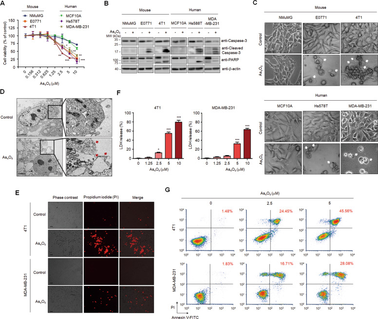
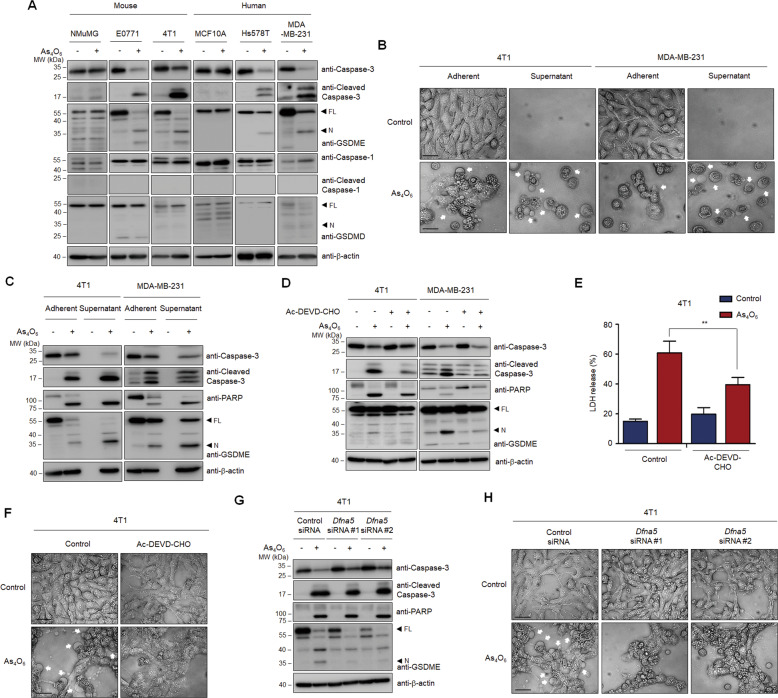
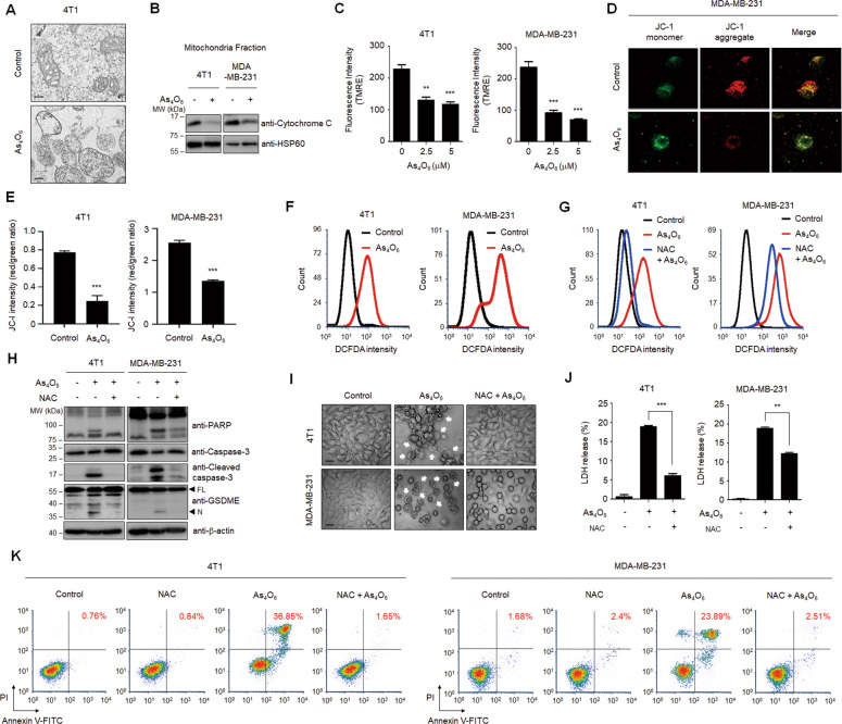
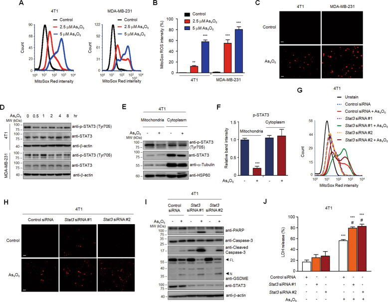
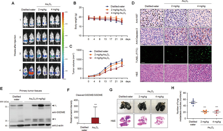
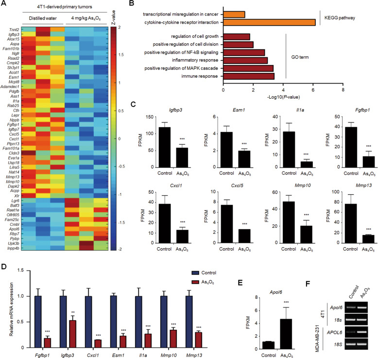
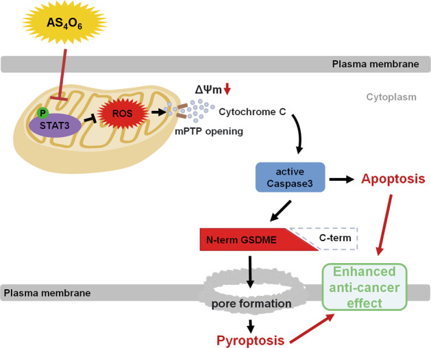
Although tetraarsenic hexoxide is known to exert an anti-tumor effect by inducing apoptosis in various cancer cells, its effect on other forms of regulated cell death remains unclear. Here, we show that tetraarsenic hexoxide induces the pyroptotic cell death through activation of mitochondrial reactive oxygen species (ROS)-mediated caspase-3/gasdermin E (GSDME) pathway, thereby suppressing tumor growth and metastasis of triple-negative breast cancer (TNBC) cells. Interestingly, tetraarsenic hexoxide-treated TNBC cells exhibited specific pyroptotic characteristics, including cell swelling, balloon-like bubbling, and LDH releases through pore formation in the plasma membrane, eventually suppressing tumor formation and lung metastasis of TNBC cells. Mechanistically, tetraarsenic hexoxide markedly enhanced the production of mitochondrial ROS by inhibiting phosphorylation of mitochondrial STAT3, subsequently inducing caspase-3-dependent cleavage of GSDME, which consequently promoted pyroptotic cell death in TNBC cells. Collectively, our findings highlight tetraarsenic hexoxide-induced pyroptosis as a new therapeutic strategy that may inhibit cancer progression of TNBC cells.


Conflict of interest statement

I.B. has personal financial interests as shareholders in CHEMAS Co., Ltd. No potential conflicts of interest were disclosed by the other authors.

Figures

Fig. 1
Fig. 1. Tetraarsenic hexoxide induces pyroptotic cell death in TNBC cells.
A Cell doubling times of tetraarsenic hexoxide-treated mouse normal mammary epithelial cells (NMuMG), mouse TNBC cells (EO771, 4T1), human normal-like mammary epithelial cells (MCF10A), and human TNBC cells (Hs578T, MDA-MB-231) cells for 24 h in a dose-dependent manner. *P < 0.05, **P < 0.005, ***P < 0.0005 versus control cells. The data represent the mean ± S.D. of three independent experiments. B Representative immunoblot analysis of cleaved caspase-3 and PARP using lysates of tetraarsenic hexoxide-treated cells. β-actin was used as an internal control. Cells were treated with 5 μM tetraarsenic hexoxide for 24 h. C Phase-contrast images of tetraarsenic hexoxide-treated cells. Pyroptotic cell morphology was indicated by white arrows. Original magnification, ×200. Scale bar, 50 μm. D Representative transmission electron microscopy (TEM) images of 4T1 cells treated with 5 μM tetraarsenic hexoxide for 24 h. Red arrows indicate the large bubbles of the plasma membrane. Scale bar, 2 μm. E Fluorescent microscopy images showing propidium iodide (PI) staining in 5 μM tetraarsenic hexoxide-treated TNBC cells for 24 h. Original magnification, ×50. Scale bar, 25 μm. F Release of LDH from TNBC cells treated with tetraarsenic hexoxide for 24 h in a dose-dependent manner. *P < 0.05, **P < 0.005, ***P < 0.0005 versus control cells. The data represent the mean ± S.D. of three independent experiments. G Flow cytometry analysis of 2.5 and 5 μM tetraarsenic hexoxide-treated TNBC cells for 24 h stained by Annexin V-FITC and PI. The percentage of double-positive cells, which may indicate pyroptotic cells, was labeled in red. All P values were calculated by unpaired two-tailed Student’s t-tests (A, F).
Fig. 2
Fig. 2. Caspase-3-mediated cleavage of GSDME has involved in tetraarsenic hexoxide-induced pyroptosis in TNBC cells.
A Representative immunoblot analysis of cleaved caspase-3, PARP, GSDME, caspase-1, and GSDMD in mouse normal mammary epithelial cells (NMuMG), mouse TNBC cells (EO771, 4T1), human normal-like mammary epithelial cells (MCF10A), and human TNBC cells (Hs578T, MDA-MB-231) cells treated with 5 μM tetraarsenic hexoxide for 24 h. β-actin was used as an internal control. B, C Phase-contrast images (B) and representative immunoblot analysis showing cleaved caspase-3, PARP, and GSDME (C) from tetraarsenic hexoxide-treated adherent and supernatant TNBC cells. Cells were treated with 5 μM tetraarsenic hexoxide for 24 h, followed by separating adherent and supernatant cells, respectively. DF Representative immunoblot analysis of cleaved caspase-3, PARP, and GSDME (D) and LDH release (E) and phase-contrast images (F) from TNBC cells treated with 5 μM tetraarsenic hexoxide for 24 h in the presence or absence of 100 μM Ac-DEVD-CHO. The data represent the mean ± S.D. of three independent experiments. **P < 0.01 using unpaired two-tailed Student’s t-tests. G, H Representative immunoblot analysis (G) and phase-contrast images (H) of GSDME (encoded by DFNA5)-knockdown 4T1 cells upon 5 μM tetraarsenic hexoxide for 24 h. Pyroptotic cell morphology was indicated by white arrows. Original magnification, ×200. Scale bar, 50 μm (B, F and H). FL full length, N N-terminus.
Fig. 3
Fig. 3. Production of ROS is required for tetraarsenic hexoxide-induced pyroptosis in TNBC cells.
A Representative transmission electron microscopy images of 4T1 cells treated with 5 μM tetraarsenic hexoxide for 24 h. Scale bar, 200 nm. B Representative immunoblot analysis showing cytochrome c expression in mitochondria fractions. Cells were treated with 5 μM tetraarsenic hexoxide for 24 h and then mitochondria were fractionated. HSP60 was used as an internal control of mitochondria fractions. CE Quantification of TMRE fluorescence intensity (C) and confocal images and quantification of JC-1 dye (D, E) showing mitochondrial membrane potential in TNBC cells treated with 5 μM tetraarsenic hexoxide for 24 h. F Flow cytometry analysis showing cellular ROS levels in TNBC cells. Cells were treated with 5 μM tetraarsenic hexoxide for 24 h and then stained with DCFDA. G Cells were pretreated with or without 5 mM NAC for 2 h before treatment of 5 μM tetraarsenic hexoxide for 24 h, and then analyzed using a flow cytometer. HK Representative immunoblot analysis (H), Phase-contrast images (I), LDH release (J), and flow cytometry analysis (K) showing ROS-mediated pyroptotic characteristics induced by tetraarsenic hexoxide upon pretreatment of NAC in TNBC cells. Cells were pretreated with or without 5 mM NAC for 2 h before treatment of 5 μM tetraarsenic hexoxide for 24 h. Pyroptotic cell morphology was indicated by white arrows. Original magnification, ×200. Scale bar, 50 μm (I). The data represent the mean ± S.D. of three independent experiments. ** P < 0.01, ***P < 0.001 using unpaired two-tailed Student’s t-tests (C, E and J). The data represent the mean ± S.D. of three independent experiments. FL full length, N N-terminus.
Fig. 4
Fig. 4. Tetraarsenic hexoxide promotes the production of mitochondrial ROS by inhibiting the phosphorylation of mitochondrial STAT3.
A, B Flow cytometry analysis (A) and its quantification (B) showing the production of mitochondrial ROS in tetraarsenic hexoxide-treated TNBC cells. Cells were treated with 2.5 and 5 μM tetraarsenic hexoxide for 24 h and then were stained with MitoSox Red. **P < 0.01, ***P < 0.001 versus control cells; C Fluorescent microscopy images showing MitoSox Red staining in 5 μM tetraarsenic hexoxide-treated TNBC cells for 24 h. Original magnification, ×50. Scale bar. D Representative immunoblot analysis showing the phosphorylation of STAT3 in tetraarsenic hexoxide–treated TNBC cells for the indicated times. E, F Representative immunoblot analysis (E) and densitometric quantitation (F) showing the phosphorylation of STAT3 in mitochondria and cytoplasmic fractions. 4T1 cells were treated with 5 μM tetraarsenic hexoxide for 24 h and then mitochondria and cytoplasm were isolated. ***P < 0.0005 versus control cells. G, H Flow cytometry analysis (G) and fluorescent microscopy images (H) showing the production of mitochondrial ROS in Stat3-knockdown 4T1 cells. 4T1 cells were transiently transfected with Stat3 siRNA and then treated with 5 μM tetraarsenic hexoxide for 24 h, followed by staining with MitoSox Red. Original magnification, ×50. Scale bar. i Representative immunoblot analysis showing cleaved caspase-3, PARP, and GSDME from tetraarsenic hexoxide-treated 4T1 cells transiently transfected with Stat3 siRNA. J LDH release by tetraarsenic hexoxide in Stat3-knockdown 4T1 cells. The data represent the mean ± S.D. of three independent experiments. ***P < 0.001 versus tetraarsenic hexoxide-untreated cells; #P < 0.01 versus tetraarsenic hexoxide-treated control cells. All P values were calculated by unpaired two-tailed Student’s t-tests. The data represent the mean ± S.D. of three independent experiments. FL full length, N N-terminus.
Fig. 5
Fig. 5. Tetraarsenic hexoxide significantly decreases the primary tumor and spontaneous lung metastasis in aggressive breast cancer cells.
A Representative Bio Layer Interferometry (BLI) imaging of Balb/c mice showing primary tumors and spontaneous lung metastasis generated by 4T1-Luc cells upon administration of tetraarsenic hexoxide. B, C Body weight (B) and tumor volume (C) curves in tetraarsenic hexoxide-administrated 4T1-injected mice. D Representative IHC images showing Ki67, caspase-3, and TUNEL in primary tumor tissues from (A). Original magnification, ×100. Scale bar, 50 μm. E, F Representative immunoblot analysis (E) and densitometric quantitation (F) showing the cleavage of GSDME in primary tumor tissues from (A). G Representative whole-lung images stained with India ink (upper) and H&E (lower) from (A). H Scatter plot showing number of lung metastatic nodules from (A). *P < 0.05, ***P < 0.0005 versus tetraarsenic hexoxide-treated mice. All P values were calculated by unpaired two-tailed Student’s t tests (C, F, H). The data represent the mean ± S.D. FL full length, N N-terminus.
Fig. 6
Fig. 6. Tetraarsenic hexoxide regulates the expression of cancer progression-associated genes in TNBC cells.
A Heatmap showing the upregulated and downregulated genes in primary tumor tissue. Threshold values are as follows: corrected value P < 0.05 and absolute log2 fold-change (log2FC) > 1.0. B KEGG pathways and GO terms enriched in differentially expressed genes (DEGs) from (A). C, D FPKM values and real-time qRT-PCR showing tumor growth-related downregulated genes from (A). E, F FPKM value and RT-PCR showing expression of APOL6, a tumor suppressor gene in TNBC cells. **P < 0.01, ***P < 0.001 using unpaired two-tailed Student’s t-tests (CE). The data represent the mean ± S.D. of three independent experiments.
Fig. 7
Fig. 7. Schematic models demonstrating the anti-tumor effect of tetraarsenic hexoxide in TNBC cells.
Tetraarsenic hexoxide promotes pyroptosis through increased production of mitochondrial ROS by inhibiting the phosphorylation of mitochondrial STAT3, subsequently triggering the cleavage of caspase-3/GSDME, eventually suppressing cancer progression of TNBC cells. mPTP mitochondrial permeability transition pore.

References

    1. Foulkes WD, Smith IE, Reis-Filho JS. Triple-negative breast cancer. N. Engl. J. Med. 2010;363:1938–1948. doi: 10.1056/NEJMra1001389. - DOI - PubMed
    1. Sotiriou C, et al. Breast cancer classification and prognosis based on gene expression profiles from a population-based study. Proc. Natl Acad. Sci. USA. 2003;100:10393–10398. doi: 10.1073/pnas.1732912100. - DOI - PMC - PubMed
    1. Marra A, Viale G, Curigliano G. Recent advances in triple negative breast cancer: the immunotherapy era. BMC Med. 2019;17:90. doi: 10.1186/s12916-019-1326-5. - DOI - PMC - PubMed
    1. Anders CK, et al. Poly(ADP-Ribose) polymerase inhibition: “targeted” therapy for triple-negative breast cancer. Clin. Cancer Res. 2010;16:4702–4710. doi: 10.1158/1078-0432.CCR-10-0939. - DOI - PMC - PubMed
    1. Wahba HA, El-Hadaad HA. Current approaches in treatment of triple-negative breast cancer. Cancer Biol. Med. 2015;12:106–116. - PMC - PubMed

Publication types

MeSH terms

LinkOut - more resources