Clinical Trial
. 2021 Feb 8;11(1):3293.
doi: 10.1038/s41598-021-82716-0.

IE-Map: a novel in-vivo atlas and template of the human inner ear

Affiliations
Clinical Trial

IE-Map: a novel in-vivo atlas and template of the human inner ear

Seyed-Ahmad Ahmadi et al. Sci Rep. .

Abstract

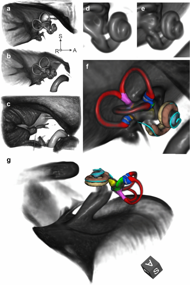
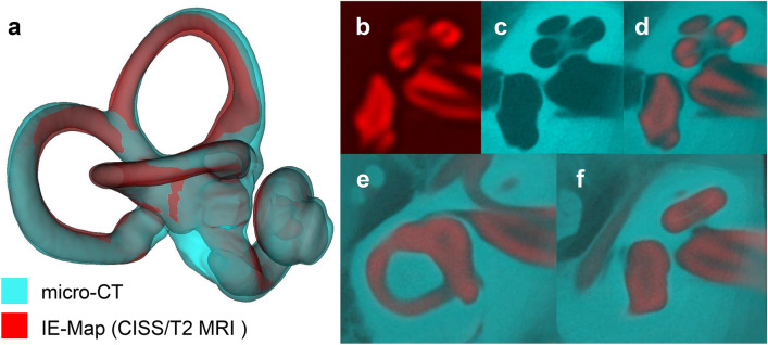
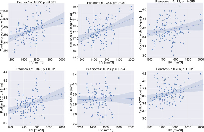
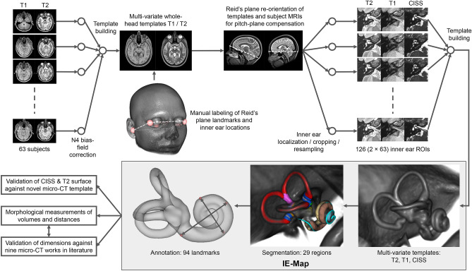
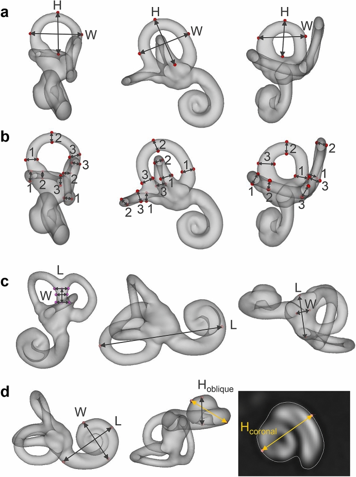
Brain atlases and templates are core tools in scientific research with increasing importance also in clinical applications. Advances in neuroimaging now allowed us to expand the atlas domain to the vestibular and auditory organ, the inner ear. In this study, we present IE-Map, an in-vivo template and atlas of the human labyrinth derived from multi-modal high-resolution magnetic resonance imaging (MRI) data, in a fully non-invasive manner without any contrast agent or radiation. We reconstructed a common template from 126 inner ears (63 normal subjects) and annotated it with 94 established landmarks and semi-automatic segmentations of all relevant macroscopic vestibular and auditory substructures. We validated the atlas by comparing MRI templates to a novel CT/micro-CT atlas, which we reconstructed from 21 publicly available post-mortem images of the bony labyrinth. Templates in MRI and micro-CT have a high overlap, and several key anatomical measures of the bony labyrinth in IE-Map are in line with micro-CT literature of the inner ear. A quantitative substructural analysis based on the new template, revealed a correlation of labyrinth parameters with total intracranial volume. No effects of gender or laterality were found. We provide the validated templates, atlas segmentations, surface meshes and landmark annotations as open-access material, to provide neuroscience researchers and clinicians in neurology, neurosurgery, and otorhinolaryngology with a widely applicable tool for computational neuro-otology.


Conflict of interest statement

The authors declare no competing interests.

Figures

Figure 1
Figure 1
Volume renderings of inner ear templates and atlas. ROI size: 4 × 4 × 3 cm; resolution: 0.2 mm isotropic; viewpoint: right-lateral view onto right inner ear. Panels: (a) T2 template; (b) CISS template; (c) T1 template; (d) Closeup of cochlea in T2 template; (e) Closeup of cochlea in CISS template, with a clear separation of scala tympani and vestibuli; (f) Atlas surface meshes on CISS template volume rendering. (g) Same as (f), with a different perspective focussing on the vestibule. Colours and regions: red: semicircular canals; blue: ampullae; pink: common crus; grey: cupula walls; green: utricle; yellow: saccule; brown: scala vestibuli; beige: scala tympani; light blue: cochlear cupula; cyan: cochlear duct. The coordinate system arrows (panels af) and cube (panel g) indicate the anterior (A), superior (S) and left/right (L/R) directions. (Figure panels created with 3D Slicer, v4.11 (https://www.slicer.org/).
Figure 2
Figure 2
Validation of IE-Map morphology (red colormap) against an inner ear template built from 21 post-mortem CT and micro-CT scans (cyan colormap). Panels: (a) 3D surface models of the inner ear in CISS MRI and micro-CT, showing a high overlap of cochlear and vestibular dimensions and morphology. Note that both templates represent the average morphologies from two independent cohorts, and that templates were only rigidly registered to account for global position and rotation of overlap. (bd). Axial slice through the cochlea in CISS, micro-CT and a semi-transparent overlay. Note that the gap between scala tympani and vestibuli in the CISS volume coincides with the spiral lamina visible in the micro-CT template. (ef) Morphological congruency of the lateral SCC (e) and the vestibulum and basal cochlear turn (f) in T2 MRI and micro-CT. (Figure panels created with 3D Slicer, v4.11 (https://www.slicer.org/).
Figure 3
Figure 3
Correlation plots for inner ear measurements with total intracranial volume (TIV). Linear correlation values (Pearson’s r) and corresponding p-values are indicated in the subplot titles. The 95% confidence interval for the regression estimate is drawn as a translucent band around the regression line. Total inner ear volume and length, as well as the radii of the anterior and posterior semicircular canals (SCC) are linearly correlated with head size. The oblique cochlea height might well be while the radius of the lateral SCC appears to not be correlated at all with TIV. The results indicate the importance of TIV for future quantitative assessments between cohorts. (Figure panels created with seaborn v0.11.0 https://seaborn.pydata.org/).
Figure 4
Figure 4
Workflow for reconstruction, annotation and validation of IE-Map. First, a full-brain template was reconstructed and annotated for re-orientation of the axial plane to Reid’s standard plane, and for localization of left/right inner ear region-of-interests (ROI). The rendered template head represents an average morphology of our cohort. Second, the inner ear template was reconstructed from 126 example multivariate inputs (T2, T1, CISS). The high-resolution (0.2 mm isotropic) template was annotated with landmarks, and segmented, partly through registration of a publicly available micro-CT atlas. To validate the morphology, we compared inner ear surfaces from T2 and CISS templates to a novel micro-CT template, as well as landmark distances to nine micro-CT based works in literature. Templates, segmentation labelmaps, landmarks, and surface models together form IE-Map, which we publicly provide as open-access material. (Figure panels created with 3D Slicer, v4.11 (https://www.slicer.org/).
Figure 5
Figure 5
Inner ear landmark annotations in template space. Panel (a) width and height of the anterior (left), posterior (middle) and lateral (right) semicircular canals. Panel (b) inner widths and heights of semicircular canals, measured at three locations each (1–3). Panel (c) height and three widths of the common crus (left), total inner ear length (middle), and vestibule length and width (right). Panel d: width, length and height of the cochlea. Cochlea height was tagged and measured using both the coronal plane approach (gold arrow),, and the oblique plane (i.e. 3D) approach (grey arrow). H = height, W = width, L = length. (Figure panels created with 3D Slicer, v4.11 (https://www.slicer.org/).

Similar articles

Cited by

References

    1. Evans AC, Janke AL, Collins DL, Baillet S. Brain templates and atlases. Neuroimage. 2012;62:911–922. doi: 10.1016/j.neuroimage.2012.01.024. - DOI - PubMed
    1. Ewert S, et al. Toward defining deep brain stimulation targets in mni space: a subcortical atlas based on multimodal mri, histology and structural connectivity. Neuroimage. 2018;170:271–282. doi: 10.1016/j.neuroimage.2017.05.015. - DOI - PubMed
    1. Behrens TE, et al. Non-invasive mapping of connections between human thalamus and cortex using diffusion imaging. Nat. Neurosci. 2003;6:750. doi: 10.1038/nn1075. - DOI - PubMed
    1. Diedrichsen J, Balsters JH, Flavell J, Cussans E, Ramnani N. A probabilistic mr atlas of the human cerebellum. Neuroimage. 2009;46:39–46. doi: 10.1016/j.neuroimage.2009.01.045. - DOI - PubMed
    1. De Leener B, et al. Pam50: unbiased multimodal template of the brainstem and spinal cord aligned with the icbm152 space. Neuroimage. 2018;165:170–179. doi: 10.1016/j.neuroimage.2017.10.041. - DOI - PubMed

Publication types