. 2021 Feb 8;11(1):3326.
doi: 10.1038/s41598-021-82451-6.

Pentapartite fractionation of particles in oral fluids by differential centrifugation

Affiliations

Pentapartite fractionation of particles in oral fluids by differential centrifugation

Chiho Hiraga et al. Sci Rep. .

Abstract

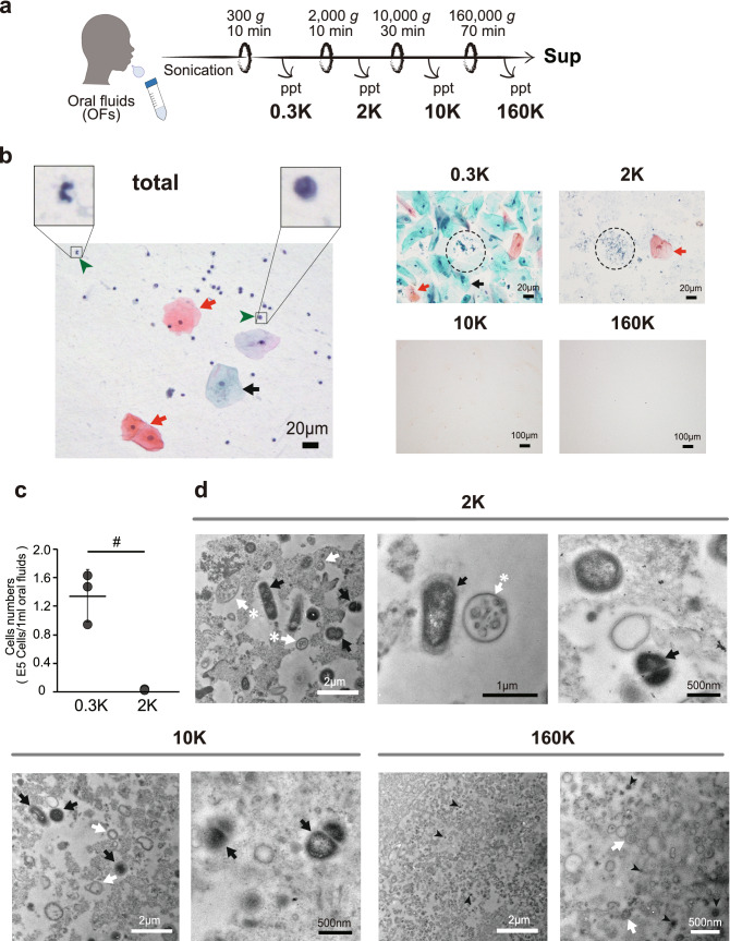
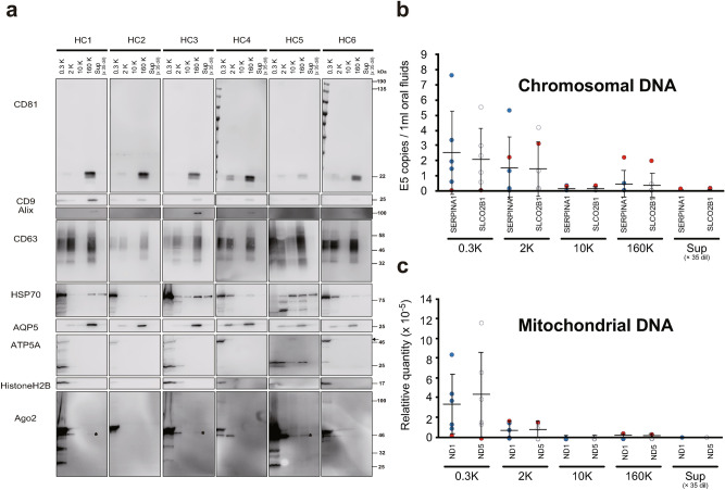
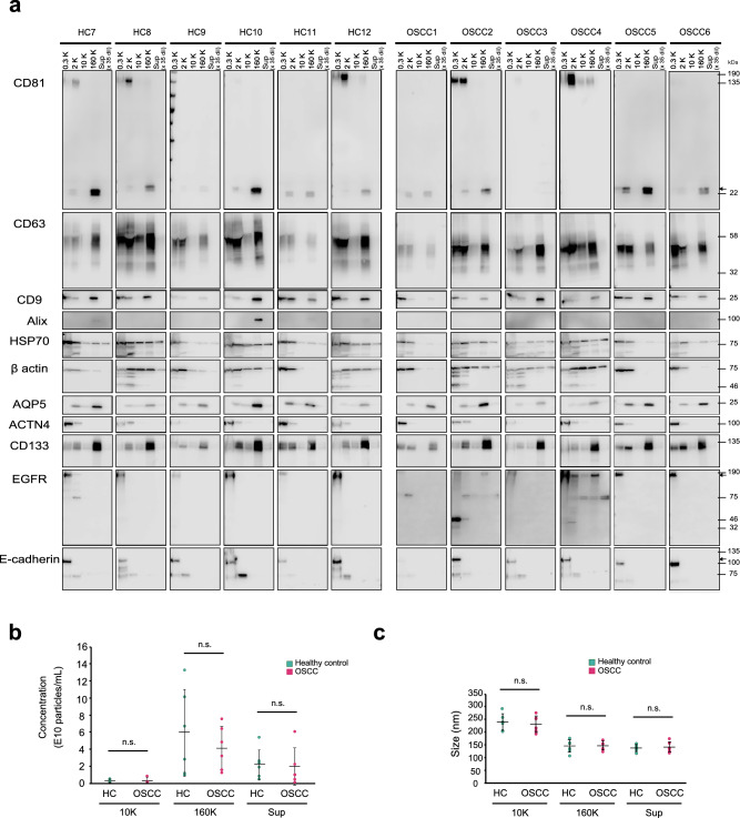
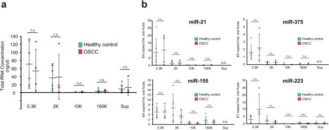
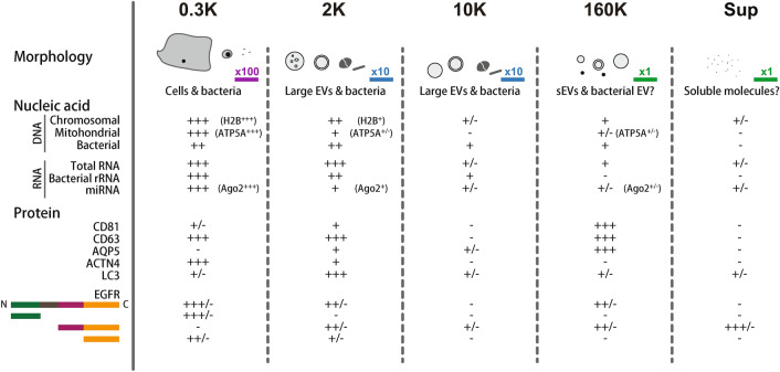
Oral fluids (OFs) contain small extracellular vesicles (sEVs or exosomes) that carry disease-associated diagnostic molecules. However, cells generate extracellular vesicles (EVs) other than sEVs, so the EV population is quite heterogeneous. Furthermore, molecules not packaged in EVs can also serve as diagnostic markers. For these reasons, developing a complete picture of particulate matter in the oral cavity is important before focusing on specific subtypes of EVs. Here, we used differential centrifugation to fractionate human OFs from healthy volunteers and patients with oral squamous cell carcinoma into 5 fractions, and we characterized the particles, nucleic acids, and proteins in each fraction. Canonical exosome markers, including CD63, CD9, CD133, and HSP70, were found in all fractions, whereas CD81 and AQP5 were enriched in the 160K fraction, with non-negligible amounts in the 2K fraction. The 2K fraction also contained its characteristic markers that included short derivatives of EGFR and E-cadherin, as well as an autophagosome marker, LC3, and large multi-layered vesicles were observed by electronic microscopy. Most of the DNA and RNA was recovered from the 0.3K and 2K fractions, with some in the 160K fraction. These results can provide guideline information for development of purpose-designed OF-based diagnostic systems.


Conflict of interest statement

The authors declare no competing interests.

Figures

Figure 1
Figure 1
Characterizations of the particulate matter contained in oral fluids (OFs). (a) Scheme of the pentapartite fractionation of OFs by differential centrifugation. Total OFs were sequentially centrifuged with increasing g force to prepare the 0.3K, 2K, 10K, and 160K pellets and the last supernatant (Sup) fraction. (b) Microscope images of Papanicolaou (Pap)-stained samples from the unfractionated OFs (total) and the 0.3K, 2K, 10K, and 160K fractions. Red and black arrows show superficial and intermediate epithelial squamous cells, respectively. Green arrowheads represent examples of leucocyte (Leu). Bacterial-like particles are circled by dash lines. Bars represent 20 µm and 100 µm for the total, 0.3K and 2K images and the 10K and 160K images, respectively. (c) Quantification of the cells contained in the 0.3K and 2K fractions by trypan blue staining. OFs were obtained from 3 healthy donors. Dots indicate the mean value of the three measurements for each subject. The mean values for the 3 healthy donors are represented as solid horizontal lines. The two-tailed unpaired t-test was used to evaluate statistical significance. # represents p < 0.05. (d) Transmission electron microscopy (TEM) images after positive staining of thin sections of the 2K, 10K, and 160K pellets (see “Methods”). Large particles with multiple bilayers (white arrows) are abundant in the 0.3K and 2K fractions; some of these contained intraluminal vesicle structures (labeled with asterisks). The 2K and 10K fractions also contained high-density (high contrast in TEM) particles, which likely represent oral bacteria cells (black arrows). The 160K fraction consisted of tiny particles, many of which has a vesicle structure with a single bilayer. As reported in other body fluids, some of these sEV had multiple bilayers (white arrows). The 160K fraction also contained tiny particles with high densities (black arrowheads), which may represent EVs from bacteria or tiny bacteria. Additional images are shown in Figure S2.
Figure 2
Figure 2
Distributions of protein markers, chromosomal DNA, and mitochondrial DNA in the pentapartite fractions of oral fluids (OFs) from 6 healthy volunteers. (a) Western blots of proteins markers for canonical exosomes (CD81, CD9, Alix, CD63 and HSP70), salivary gland (AQP5), mitochondria (ATP5A), chromatin (Histone H2B), and miRNA-associated protein (Ago2). Numbers on the right indicate molecular weight × 10–3. Black arrow shows the expected size of ATP5A. Asterisks in the 160K fractions of Ago2 are discussed in the text. Note that Sup was diluted 35-fold. The copy numbers of chromosomal DNA (b), and mitochondrial DNA (c), were determined by quantitative PCR. Target genes were SLCO2B1 and SERPINA1 for chromosomal DNA and ND1 and ND3 for mitochondrial DNA. The OFs were obtained from n = 6 healthy donors. Data are expressed as the mean ± standard deviation (SD). Dots indicate the mean value of the three measurements for each subject, while the mean values obtained from six subjects are represented as solid horizontal lines. Note that Sup was diluted 35-fold. The data obtained from HC45 are shown by red filled circles (see “Results” section).
Figure 3
Figure 3
Comparative analyses of pentapartite oral fluid (OF) fractions between healthy volunteers and patients with OSCC. (a) Western blot analyses of pentapartite fractions from six healthy subjects (HC7 to HC12) and six patients with OSCC (OSCC1 to OSCC6). Membranes were probed with antibodies against CD81, CD63, CD9, Alix, Hsp70, β-actin, AQP5, ACTN4, CD133, EGFR, and E-cadherin. Numbers on the right indicate molecular weight (MW) × 10–3 of for MW markers. Black arrows show the reported sizes of the proteins. Nanoparticle tracking analyses (NTA) of particles contained in the 10K, 160K, and Sup fractions derived from healthy controls and patients with OSCC. Particle concentrations (b), and mode diameters (c), are indicated by green and red dots, respectively. The mean value of each group is represented as a solid horizontal line. The two-tailed unpaired t-test was used to evaluate statistical significance. Statistical significance was defined as p < 0.05. n.s. not significant.
Figure 4
Figure 4
Distribution of RNA molecules among pentapartite fractions from oral fluids (OFs). (a) Total RNA concentration in each fraction from 6 healthy volunteers (green dots) and 4 patients with OSCC (red dots) estimated by Bioanalyzer. Each dot represents the mean value obtained from three measurements. (b) Copy numbers of miR-21, miR-375, miR-155, and miR-223 in each fraction were determined using chip-based Quant Studio 3D digital PCR. Green and pink dots indicate the mean values of the three measurements in 6 healthy volunteers and 4 patients with OSCC, respectively. The mean value of groups is represented as solid horizontal lines. The two-tailed unpaired t-test was used to evaluate statistical significance. n.s. not significance, p > 0.05.
Figure 5
Figure 5
Summary of pentapartite fractionation of human oral fluids. At the top, schematic illustrations of particles observed are shown. The scale bars labeled with × 100, × 10, and × 1 correspond to 20 μm, 2 μm, and 200 nm, respectively. Representative protein markers are shown. “+/−” represents some specimens gave positive signals. Details are described in the “Results” section.

Similar articles

Cited by

References

    1. Castagnola M, et al. Potential applications of human saliva as diagnostic fluid. Acta Otorhinolaryngol. Ital. 2011;31:347–357. - PMC - PubMed
    1. Pfaffe T, Cooper-White J, Beyerlein P, Kostner K, Punyadeera C. Diagnostic potential of saliva: Current state and future applications. Clin. Chem. 2011;57:675–687. doi: 10.1373/clinchem.2010.153767. - DOI - PubMed
    1. Kaczor-Urbanowicz KE, et al. Saliva diagnostics—Current views and directions. Exp. Biol. Med. (Maywood) 2017;242:459–472. doi: 10.1177/1535370216681550. - DOI - PMC - PubMed
    1. Chiabotto G, Gai C, Deregibus MC, Camussi G. Salivary extracellular vesicle-associated exRNA as cancer biomarker. Cancers (Basel) 2019 doi: 10.3390/cancers11070891. - DOI - PMC - PubMed
    1. Du M, Nair R, Jamieson L, Liu Z, Bi P. Incidence trends of lip, oral cavity, and pharyngeal cancers: Global burden of disease 1990–2017. J. Dent. Res. 2020;99:143–151. doi: 10.1177/0022034519894963. - DOI - PubMed