. 2021 May;5(5):455-466.
doi: 10.1038/s41551-020-00675-9. Epub 2021 Feb 8.

Prolonged activation of innate immune pathways by a polyvalent STING agonist

Affiliations

Prolonged activation of innate immune pathways by a polyvalent STING agonist

Suxin Li et al. Nat Biomed Eng. 2021 May.

Erratum in

Abstract

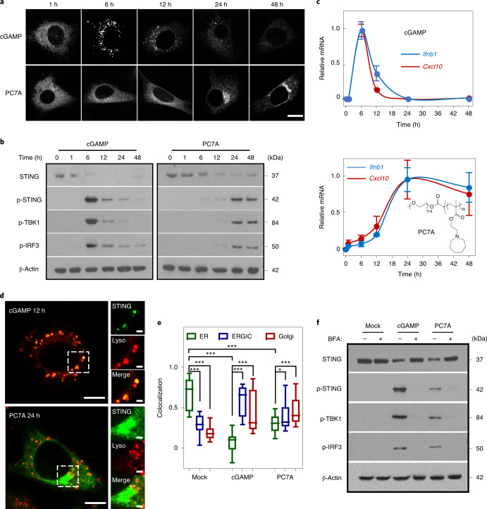
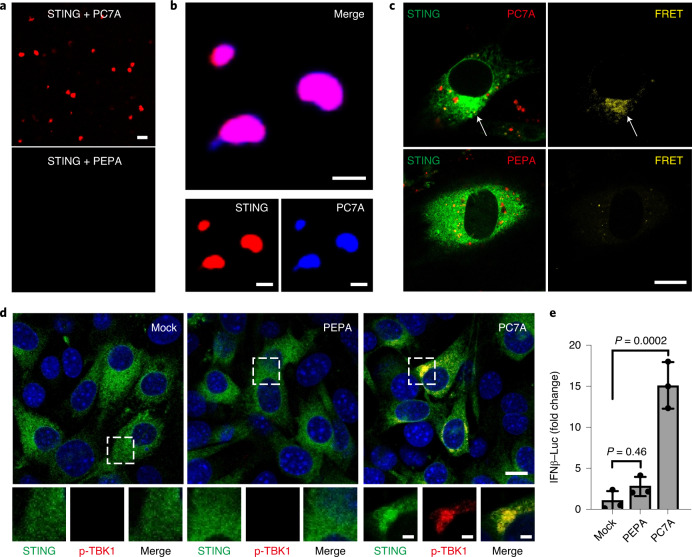
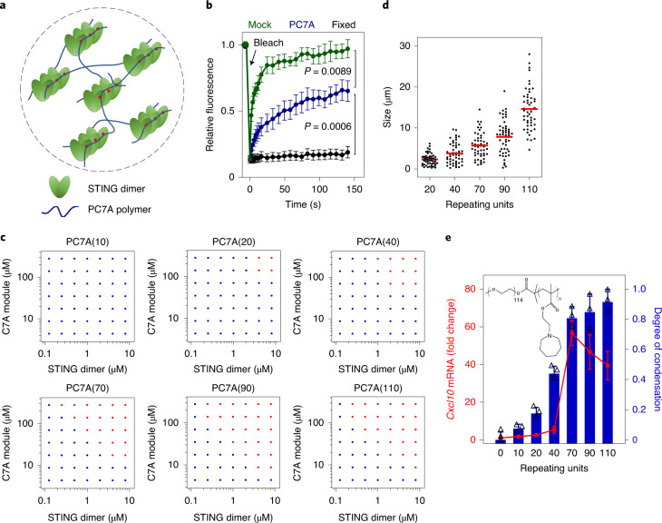
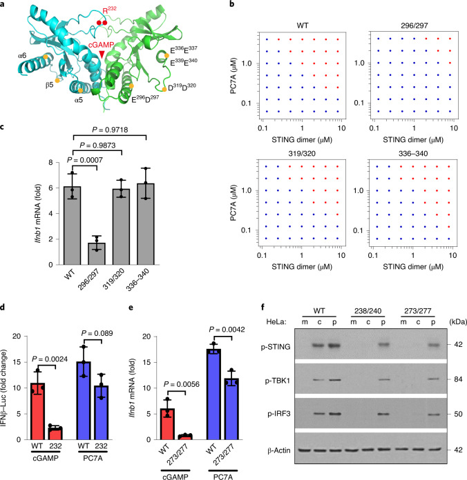
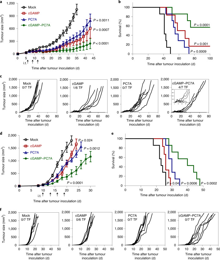
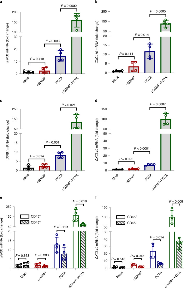
The stimulator of interferon genes (STING) is an endoplasmic reticulum transmembrane protein that is a target of therapeutics for infectious diseases and cancer. However, early-phase clinical trials of small-molecule STING agonists have shown limited antitumour efficacy and dose-limiting toxicity. Here, we show that a polyvalent STING agonist-a pH-sensitive polymer bearing a seven-membered ring with a tertiary amine (PC7A)-activates innate-immunity pathways through the polymer-induced formation of STING-PC7A condensates. In contrast to the natural STING ligand 2',3'-cyclic-GMP-AMP (cGAMP), PC7A stimulates the prolonged production of pro-inflammatory cytokines by binding to a non-competitive STING surface site that is distinct from the cGAMP binding pocket. PC7A induces antitumour responses that are dependent on STING expression and CD8+ T-cell activity, and the combination of PC7A and cGAMP led to synergistic therapeutic outcomes (including the activation of cGAMP-resistant STING variants) in mice bearing subcutaneous tumours and in resected human tumours and lymph nodes. The activation of the STING pathway through polymer-induced STING condensation may offer new therapeutic opportunities.


Conflict of interest statement

B.D.S. and J.G. are scientific co-founders and advisors of OncoNano Medicine, Inc.

Figures

Fig. 1
Fig. 1. PC7A polymer activates STING with a spatiotemporal profile that is distinct from cGAMP.
a, MEFs primed by cGAMP or PC7A exhibit different geometric and temporal patterns of GFP–STING-punctate formation and depletion. Cells were first incubated with cGAMP (10 μM, PEI was used for cytosolic delivery; Supplementary Fig. 1a,b) or PC7A micelles (10 μM) for 1 h, and the medium was then exchanged and cells were incubated for the indicated periods before imaging. Scale bar, 10 μm. b, THP1 cells that were treated with cGAMP display a burst effect of TBK1/IRF3 phosphorylation followed by rapid STING degradation, whereas treatment with PC7A led to sustained TBK1/IRF3 phosphorylation and slower STING degradation. c, Relative Ifnb1 and Cxcl10 mRNA levels show slower but prolonged STING activation in THP1 cells by PC7A compared with cGAMP. Data are mean ± s.d. n = 3 biologically independent experiments. d, GFP–STING colocalizes with lysosomes in MEFs 12 h after cGAMP treatment, supporting rapid degradation. By contrast, PC7A inhibits lysosomal degradation of GFP–STING, as indicated by the lack of colocalization and persistent GFP fluorescence. Scale bars, 5 μm (left images); 1 μm (right images). e, cGAMP and PC7A induce similar STING translocation from the ER to the ERGIC and Golgi apparatus. Colocalization was quantified using the Pearson’s correlation coefficient. For the box plot, the centre line is the mean, the box limits show the 25th to 75th percentile, and the whiskers show the minimum and maximum values. n = 20 cells examined over three independent experiments. Statistical analysis was performed using two-tailed Student’s t-tests (PC7A treatment group: ER versus ERGIC, *P = 0.029; ER versus Golgi, ***P = 0.0005; for all other comparisons, ***P < 0.0001). f, STING translocation is necessary for downstream signalling as BFA, which is an inhibitor of protein transport from ER to Golgi, prevents the phosphorylation of TBK1/IRF3 by cGAMP or PC7A. The confocal images in a and d are representative of at least three biologically independent experiments.
Fig. 2
Fig. 2. PC7A polymer induces STING condensation and immune activation.
a, PC7A, but not PEPA, induces STING (Cy5-labelled) phase condensation after 4 h incubation. Scale bar, 10 μm. b, STING (4 μM, Cy5-labelled) and PC7A polymer (2 μM, aminomethylcoumarin acetate-labelled) are colocalized within the condensates. Scale bars, 5 μm. c, Hetero-FRET from GFP–STING to TMR–PC7A shows the colocalization of STING and PC7A in MEFs. Energy transfer was not observed from GFP–STING to TMR–PEPA. Cell culture conditions were identical to those described in Fig. 1. GFP (λex/λem = 488 nm/515 nm) and TMR (λex/λem = 555 nm/580 nm) signals are shown on the left in green and red, respectively. FRET signals (λex/λem = 488 nm/580 nm) are shown in yellow on the right. Scale bar, 10 μm. d, p-TBK1 is recruited into the STING–PC7A condensates. Scale bar, 10 μm. Insets: magnified images of the areas indicated by the white boxes. Scale bars, 2 μm. e, PC7A, but not PEPA, induces the expression of IFNβ–luciferase in ISG-THP1 cells. Data are mean ± s.d. n = 3 biologically independent experiments. Statistical analysis was performed using one-way analysis of variance (ANOVA). The confocal images in ad are representative of at least three biologically independent experiments.
Fig. 3
Fig. 3. PC7A polymer induces STING condensation and immune activation through polyvalent interactions.
a, Schematic of STING oligomerization and condensation driven by PC7A through polyvalent interactions. b, PC7A decreases the molecular mobility of GFP–STING in the condensates compared with free GFP–STING in MEFs. Bleaching was performed 24 h after PC7A treatment, and recovery was monitored over 150 s. Untreated (mock) and fixed cells were used as mobile and stationary STING controls, respectively. Data are mean ± s.d. n = 5 cells examined over two independent experiments. Statistical analysis was performed using one-way ANOVA. c, Biomolecular condensation of STING and PC7A is dependent on PC7A valency. The red dots indicate phase separation, and the blue dots indicate no phase separation. d, Size distributions of STING condensates induced by an increase in PC7A with higher PC7A valency. Condensate size was calculated as the average of longest and shortest axes. n = 50 condensates examined over two independent experiments. The red lines represent the average. e, STING activation in THP1 cells is correlated with the PC7A valency, with optimal Cxcl10 expression induced by PC7A(70). Data are mean ± s.d. n = 3 biologically independent experiments. For the experiments shown in ce, polymers with different repeating units were used at the same C7A modular concentrations.
Fig. 4
Fig. 4. STING condensation and activation by PC7A polymer occurs through a distinct binding site from cGAMP.
a, Schematic of site-directed mutagenesis on the STING structure. Mutation sites are distinct from the cGAMP-binding pocket. b,c, STING mutation of E296A/D297A abolishes STING condensation (b) and immune activation (c) in response to PC7A. Other mutations in STING do not affect PC7A-induced STING activation. Data are mean ± s.d. n = 3 biologically independent experiments. Statistical analysis was performed using one-way ANOVA. df, PC7A retains immune activity in several cGAMP-resistant STING variants. R232H in THP1 cells (d) or R238A/Y240A in HeLa cells (f) abrogates cGAMP binding. Q273A/A277Q (e,f), which disrupts the tetramer interface and cGAMP-mediated STING oligomerization, abolishes STING activation by cGAMP but not by PC7A. Data are mean ± s.d. n = 3 biologically independent experiments. Statistical analysis was performed using two-tailed Student’s t-tests. c, cGAMP; m, mock; p, PC7A polymer.
Fig. 5
Fig. 5. PC7A and cGAMP show synergistic antitumour efficacy in tumour-bearing mice.
af, MC38 (ac) and TC-1 (df) tumour-bearing mice were injected intratumourally (i.t.; arrows) with 5% glucose (mock), cGAMP (2.5 μg), PC7A NPs (50 μg) or cGAMP-loaded PC7A NPs at the indicated time points. Mean tumour volume (a,d), Kaplan–Meier survival curves (b,e) and spider plots of individual tumour growth curves (c,f) are shown. PC7A NPs or cGAMP alone offer some degree of immune protection. cGAMP-loaded PC7A NPs confer a synergistic antitumour immune response, with significantly improved survival; 4 out of 7 mice in the MC38 model were tumour-free (TF). In the tumour growth studies (a and d), data are mean ± s.e.m. n = 7 (mock), n = 6 (cGAMP), n = 7 (PC7A) and n = 7 (cGAMP–PC7A) biologically independent mice in each tumour model. For a and d, statistical analysis was performed using two-tailed Student’s t-tests (versus mock). For b and e, statistical analysis was performed using Mantel–Cox tests.
Fig. 6
Fig. 6. PC7A and cGAMP show synergistic STING activation in fresh human tissues.
af, Free cGAMP alone is unable to activate STING, whereas PC7A NPs and cGAMP-loaded PC7A NPs demonstrate effective STING activation. Fresh surgically resected sentinel lymph nodes (SLN) (a,b) or squamous cell carcinoma samples from the base of tongue (SCC-BOT) (cf) were divided into multiple sections (1–5 mm3) and injected with 5% glucose, free cGAMP, PC7A NPs or cGAMP-loaded PC7A NPs in 5% glucose solutions. IFNB1 (a,c,e) and CXCL10 (b,d,f) gene expression was measured after 24 h incubation. e,f, The CD45+ cell population exhibited an enhanced level of STING activation compared with CD45 cells. Data are mean ± s.d. n = 4 SLN sections from the same patient in a and b. n = 4 SCC-BOT tumour sections from the same patient in cf. Statistical analysis was performed using two-tailed Student’s t-tests.

References

    1. Barber GN. STING: infection, inflammation and cancer. Nat. Rev. Immunol. 2015;15:760–770. doi: 10.1038/nri3921. - DOI - PMC - PubMed
    1. Chen Q, Sun L, Chen ZJ. Regulation and function of the cGAS–STING pathway of cytosolic DNA sensing. Nat. Immunol. 2016;17:1142–1149. doi: 10.1038/ni.3558. - DOI - PubMed
    1. Yum S, Li M, Frankel AE, Chen ZJ. Roles of the cGAS-STING pathway in cancer immunosurveillance and immunotherapy. Annu. Rev. Cancer Biol. 2019;3:323–344. doi: 10.1146/annurev-cancerbio-030518-055636. - DOI
    1. Ishikawa H, Ma Z, Barber GN. STING regulates intracellular DNA-mediated, type I interferon-dependent innate immunity. Nature. 2009;461:788–792. doi: 10.1038/nature08476. - DOI - PMC - PubMed
    1. Sun L, Wu J, Du F, Chen X, Chen ZJ. Cyclic GMP-AMP synthase is a cytosolic DNA sensor that activates the type I interferon pathway. Science. 2013;339:786–791. doi: 10.1126/science.1232458. - DOI - PMC - PubMed

Publication types