. 2021 Apr;46(4):1006-1018.
doi: 10.1007/s11064-021-03231-2. Epub 2021 Feb 8.

Cocaine-Specific Effects on Exosome Biogenesis in Microglial Cells

Affiliations

Cocaine-Specific Effects on Exosome Biogenesis in Microglial Cells

Sanjay Kumar et al. Neurochem Res. 2021 Apr.

Abstract

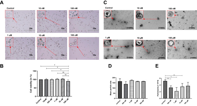
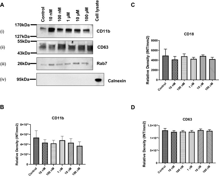
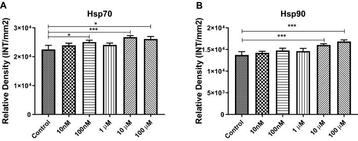
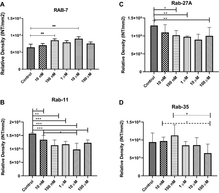
Cocaine is a highly addictive stimulant and a well-known drug, with multiple effects on physiology. Cocaine can have direct effects on all cell types in the brain, including microglia. Microglia can be activated by other conditions, such as infection, inflammation, or injury. However, how cocaine regulates microglia and the influence of cocaine on microglial-derived exosomes remains unknown. Exosomes are nanovesicles that are responsible for intercellular communications, signaling, and trafficking necessary cargo for cell homeostasis. In this study, we hypothesized that cocaine affects exosome biogenesis and composition in BV2 microglial cells. BV2 microglial cells were cultured in exosome-depleted RPMI-1640 media and were treated according to the experimental designs. We observed that cell viability decreased by 11% at 100 µM cocaine treatment but was unaffected at other concentrations. After treatments, the exosomes were isolated from the condition media. Purified exosomes were characterized and quantified using transmission electron microscope (TEM) and nanoparticle tracking analysis (NTA). By NTA, there was a significant decrease in particles/mL after cocaine treatment. There was a 39.5%, 58.1%, 32.3% and 28.1% decrease in particles/mL at 100 nM, 1 μM, 10 μM and 100 μM cocaine, respectively. The characterization of exosomes and exosomal protein was performed by western/dot blot analyses. Tetraspanins CD11b, CD18 and CD63 were relatively unchanged after cocaine treatment. The heat shock proteins (Hsps), Hsp70 and Hsp90, were both significantly increased at 10 μM and 100 μM, but only hsp70 was significantly increased at 10 nM. The Rab proteins were assessed to investigate their role in cocaine-mediated exosomal decrease. Rab11 was significantly decreased at 10 nM, 100 nM, 1 μM, 10 μM and 100 μM by 15%, 28%, 25%, 38% and 22%, respectively. Rab27 was decreased at all concentrations but only significantly decreased at 100 nM, 1 μM and 100 μM cocaine by 21%, 24% and 23%, respectively. Rab35 had no significant changes noted when compared to control. Rab7 increased at all cocaine concentrations but only a significant increase in expression at 100 nM and 10 μM by 1.32-fold and 1.4-fold increase. Cocaine was found to alter exosome biogenesis and composition in BV2 microglial cells. Western and dot blot analyses verified the identities of purified exosomes, and the specific protein compositions of exosomes were found to change in the presence of cocaine. Furthermore, cocaine exposure modulated the expression of exosomal proteins, such as Hsps and Rab GTPases, suggesting the protein composition and formation of microglial-derived exosomes were regulated by cocaine.

Keywords: BV2 microglia; Cocaine; Exosomes; Heat shock proteins; Lipids; rab GTPases.


Figures

Fig. 1
Fig. 1
Cocaine-specific effects on BV2 microglial cell viability and the mean size and number of particles. BV2 microglial cells were treated with 10 nM, 100 nM, 1 µM, 10 µM, and 100 µM cocaine. Cells were grown in exosome-free medium and the cocaine was added for a maximum of 24 h. a Microscopy, b cell viability, c TEM, d mean particle size and e particle/mL. Mean size is shown in nanometers, and particle numbers are shown as 108 per mL. Statistical significance is taken from 3 to 5 independent experiment in triplicates and indicated the mean of SD as follows: *p < 0.05; **p < 0.001; and ***p < 0.0001
Fig. 2
Fig. 2
Cocaine-specific effects on cell membrane molecules. BV2 microglial cells were treated with cocaine (10 nM, 100 nM, 1 µM, 10 µM, and 100 µM) for 24 h, and the expression of cell membrane molecules in exosomes was determined using western and dot blot analysis. a representative western blots; (i) CD11b, (ii) CD63, (iii) Rab7 and (iv) Calnexin, b CD11b expression, c CD18 expression and d CD63 expression. Statistical significance is taken from 5 independent experiment in triplicates and indicated the mean of SD as follows: *p < 0.05; and **p < 0.001
Fig. 3
Fig. 3
Cocaine-specific effects on Hsps in BV2 cells and exosomes. BV2 microglial cells were treated with cocaine (10 nM, 100 nM, 1 µM, 10 µM, and 100 µM) for 24 h, and the expression levels of was evaluated in in exosomes. a Hsp70 and b Hsp90 densities derived from dot blot. Statistical significance derived from 5 independent experiment in triplicates is indicated the mean of SD as follows: *p < 0.05; **p < 0.001; and ***p < 0.0001
Fig. 4
Fig. 4
Cocaine-specific effects on Rab GTPases. To examine the expression of Rab GTPases in microglial cells, cells were incubated with 10 nM, 100 nM, 1 µM, 10 µM, and 100 µM cocaine for 24 h, and Rab protein expression levels were evaluated in BV2 cells and exosomes using dot blot analysis. a Rab7, b Rab11, c Rab27A and d Rab35 expression in BV2 cell-derived exosomes. Statistical significance derived from 5 independent experiment in triplicates is indicated the mean of SD as follows: *p < 0.05; **p < 0.001; and ***p < 0.0001
Fig. 5
Fig. 5
Effects of cocaine on exosomal lipids. To examine the expression of various important lipids in exosomes, cells were exposed to various concentrations of cocaine for 24 h, and lipid components were tested in exosomes. a total lipids, b total cholesterol, c phospholipids, d phosphatidylserine, e phosphatidylcholine and f sphingomyelin were determined in exosomes by ELISA-based fluorometric assays. Graph showed the mean of SD derived from 5 independent experiment in triplicates

References

    1. Kreutzberg GW. Microglia: a sensor for pathological events in the CNS. Trends Neurosci. 1996;19:312–318. doi: 10.1016/0166-2236(96)10049-7. - DOI - PubMed
    1. Dickson DW, Lee SC. Microglia in HIV-related CNS neuropathology: an update. J NeuroAIDS. 1996;1:57–83. - PubMed
    1. Gehrmann J, Banati RB. Microglial turnover in the injured CNS: activated microglia undergo delayed DNA fragmentation following peripheral nerve injury. J Neuropathol Exp Neurol. 1995;54:680–688. doi: 10.1097/00005072-199509000-00010. - DOI - PubMed
    1. Thompson KK, Tsirka SE. The diverse roles of microglia in the neurodegenerative aspects of central nervous system (CNS) autoimmunity. Int J Mol Sci. 2017;18:504. doi: 10.3390/ijms18030504. - DOI - PMC - PubMed
    1. Tsuda M. Microglia in the CNS and neuropathic pain. Adv Exp Med Biol. 2018;1099:77–91. doi: 10.1007/978-981-13-1756-9_7. - DOI - PubMed

LinkOut - more resources