

Anatomic Repair vs Closed Reduction of the Syndesmosis
- PMID: 33559488
- PMCID: PMC8293725
- DOI: 10.1177/1071100721990008
Anatomic Repair vs Closed Reduction of the Syndesmosis
Abstract
Background: The goal of the study was to compare radiographic and functional outcomes between conventional closed syndesmotic reduction and screw fixation with open reduction, direct repair of the anterior inferior tibiofibular ligament (AiTFL) and screw fixation. We hypothesized that open reduction with restoration of the AiTFL would provide an improved reduction with better radiographic and functional outcomes.
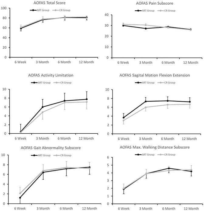
Methods: Fifty consecutive patients with OTA 44-C ankle fractures were enrolled. Treatment was nonrandomized and based on surgeon preference. Patients were treated with either open reduction, suture-anchor AiTFL repair, and screw fixation (ART group), or conventional closed reduction of the syndesmosis followed by screw fixation (CR group). The primary outcome measure was anteroposterior (AP) displacement of the fibula on CT scan at 3 months postoperatively. Secondary outcome measures included the Maryland Foot Score, the American Orthopaedic Foot & Ankle Society (AOFAS) Ankle Hindfoot Score, and the Foot and Ankle Outcome Score (FAOS).
Results: Mean AP difference between injured and noninjured ankles was decreased in the ART group compared with the CR group (0.7 ± 0.3 mm vs 1.5 ± 0.3 mm, P = .008). No differences were observed between groups in overall scores for secondary outcome measures. The ART group displayed a significant difference in Maryland Foot Shoe subscore at 12 months (ART = 9.5 vs CR = 8.3, P = .03) and FAOS Quality of Life subscore at 12 months (64.1 compared to 38.3, P = .04).
Conclusion: Open anatomic syndesmotic repair resulted in improved radiographic outcomes compared with closed reduction. Cosmesis was worse at 6 weeks compared to the CR group; however, quality of life and shoewear were improved in the ART group at 1 year postoperatively.
Level of evidence: Level II, prospective comparative study.
Keywords: arthritis; outcome studies; syndesmosis; trauma.
Conflict of interest statement
Figures


References
-
- Anand A, Wei R, Patel A, Vedi V, Allardice G, Anand BS. Tightrope fixation of syndesmotic injuries in Weber C ankle fractures: a multicentre case series. Eur J Orthop Surg Traumatol. 2017;27(4):461-467. - PubMed
-
- Burdett RG. Forces predicted at the ankle during running. Med Sci Sports Exerc. 1982;14(4):308-316. - PubMed
-
- Clanton TO, Williams BT, Backus JD, et al. Biomechanical analysis of the individual ligament contributions to syndesmotic stability. Foot Ankle Int. 2017;38(1):66-75. - PubMed
-
- Cottom JM, Hyer CF, Philbin TM, Berlet GC. Transosseous fixation of the distal tibiofibular syndesmosis: comparison of an interosseous suture and endobutton to traditional screw fixation in 50 cases. J Foot Ankle Surg. 2009;48(6):620-630. - PubMed
-
- Gardner MJ, Demetrakopoulos D, Briggs SM, Helfet DL, Lorich DG. Malreduction of the tibiofibular syndesmosis in ankle fractures. Foot Ankle Int. 2006;27(10):788-792. - PubMed
MeSH terms
LinkOut - more resources
Full Text Sources
Other Literature Sources
Medical
Research Materials
