Review
. 2021 Feb;103(2):e59-e64.
doi: 10.1308/rcsann.2020.7008.

Aggressive angiomyxoma of the ischioanal fossa in a post-menopausal woman

Affiliations
Review

Aggressive angiomyxoma of the ischioanal fossa in a post-menopausal woman

E Peterknecht et al. Ann R Coll Surg Engl. 2021 Feb.

Abstract

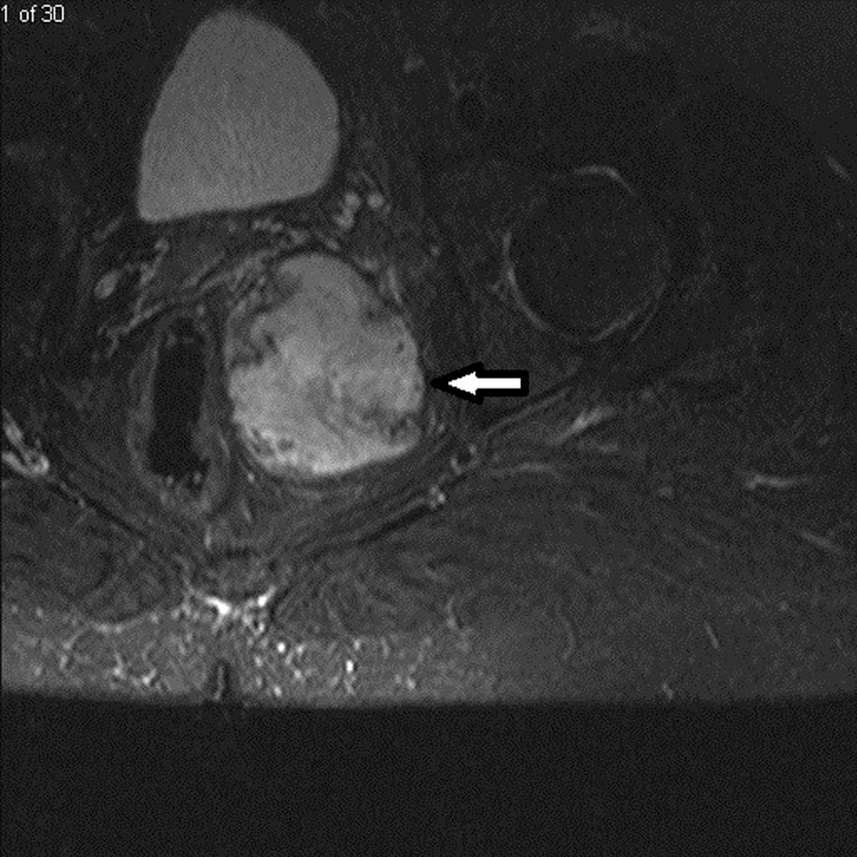
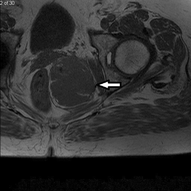
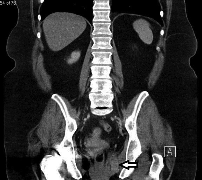
Aggressive angiomyxoma is a rare mesenchymal tumour, primarily arising in the soft tissue of the pelvis and perineum in women of reproductive age. There is a paucity of evidence on optimal management because of the rarity of these tumours, but the consensus has been for surgical excision. We present the case of a 65-year-old woman who was admitted with left-sided buttock pain and initially diagnosed with a perianal abscess. She underwent examination under anaesthesia rectum with surgical excision of the lesion, subsequent histopathological and immunochemical analysis was suggestive of aggressive angiomyxoma. To complement our case report, we also present a literature review focusing on aggressive angiomyxoma in the ischioanal fossa (also known as the ischiorectal fossa) with only eight cases of primary aggressive angiomyxoma involving the ischioanal fossa documented to date. The primary aims of this case report and literature review are to familiarise clinicians with the clinical, histopathological and immunochemical features of these tumours, and to increase appreciation that despite the rarity of aggressive angiomyxoma, it might be considered in the differential diagnosis of ischioanal lesions.

Keywords: Angiomyxoma; Ischioanal fossa; Mesenchymal tumour.


Figures

Figure 1
Figure 1
Magnetic resonance image of the pelvis; the axial short tau inversion recovery image showing a well-defined soft-tissue signal intensity mass (white arrow). The mass is predominantly located in the left inferior aspect of the pelvic cavity with extension into the ischioanal fossa, medial to the left obturator internus muscle and lateral to the distal rectum.
Figure 2
Figure 2
Magnetic resonance image of the pelvis (axial T1 view) showing a well-defined soft-tissue signal intensity mass (white arrow). The mass is predominantly located in the left inferior aspect of the pelvic cavity with extension into the ischioanal fossa, medial to the left obturator internus muscle and lateral to the distal rectum.
Figure 3
Figure 3
Intravenous-enhanced computed tomography of the abdomen and pelvis in portal venous phase. The coronal view shows a fairly well defined residual soft-tissue density mass (white arrow), following surgery.

References

    1. Steeper TA, Rosai J.. Aggressive angiomyxoma of the female pelvis and perineum: report of nine cases of a distinctive type of gynecologic soft-tissue neoplasm. Am J Surg Pathol 1983; 7: 463–476. 10.1097/00000478-198307000-00009 - DOI - PubMed
    1. Geng J, Cao B, Wang L.. Aggressive angiomyxoma: an unusual presentation. Korean J Radiol 2012; 13: 90. 10.3348/kjr.2012.13.1.90 - DOI - PMC - PubMed
    1. Sozutek A, Irkorucu O, Reyhan Eet al. . A giant aggressive angiomyxoma of the pelvis misdiagnosed as incarcerated femoral hernia: a case report and review of the literature. Case Rep Surg 2016; 2016: 1–6. 10.1155/2016/9256749 - DOI - PMC - PubMed
    1. Nakamura T, Miura K, Maruo Yet al. . Aggressive angiomyxoma of the perineum originating from the rectal wall. J Gastroenterol 2002; 37: 303–308. 10.1007/s005350200040 - DOI - PubMed
    1. Kelley SR. Robotic extralevator excision of a retrorectal giant aggressive angiomyxoma. Obstet Gynecol Sci 2018; 61: 693. 10.5468/ogs.2018.61.6.693 - DOI - PMC - PubMed

LinkOut - more resources