. 2021 Feb 9:10:e58534.
doi: 10.7554/eLife.58534.

Heterogeneity of murine periosteum progenitors involved in fracture healing

Affiliations

Heterogeneity of murine periosteum progenitors involved in fracture healing

Brya G Matthews et al. Elife. .

Abstract

The periosteum is the major source of cells involved in fracture healing. We sought to characterize progenitor cells and their contribution to bone fracture healing. The periosteum is highly enriched with progenitor cells, including Sca1+ cells, fibroblast colony-forming units, and label-retaining cells compared to the endosteum and bone marrow. Using lineage tracing, we demonstrate that alpha smooth muscle actin (αSMA) identifies long-term, slow-cycling, self-renewing osteochondroprogenitors in the adult periosteum that are functionally important for bone formation during fracture healing. In addition, Col2.3CreER-labeled osteoblast cells contribute around 10% of osteoblasts but no chondrocytes in fracture calluses. Most periosteal osteochondroprogenitors following fracture can be targeted by αSMACreER. Previously identified skeletal stem cell populations were common in periosteum but contained high proportions of mature osteoblasts. We have demonstrated that the periosteum is highly enriched with skeletal progenitor cells, and there is heterogeneity in the populations of cells that contribute to mature lineages during periosteal fracture healing.

Keywords: alpha smooth muscle actin; fracture; mouse; osteoblast; pdgfra; periosteum; regenerative medicine; skeletal stem cell; stem cells.


Conflict of interest statement

BM, SN, FS, JF, YC, EB, DG, IK No competing interests declared

Figures

Figure 1.
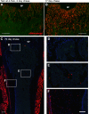
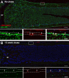
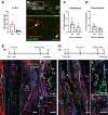
Figure 1.. Alpha smooth muscle actin (αSMA) identifies long-term progenitor cells in the periosteum.
Representative images of tibia mid-shaft periosteum: (A) 14 days or (B) 56 days after tamoxifen with the periosteal surface near the top and endosteal surface near the bottom of the image. Percentage of αSMA-labeled (Tom+) cells within the CD45- population at different time points in (C) periosteum and (D) endosteum as measured by flow cytometry (n = 2–5). Fracture histology of representative day 7 tibial fracture calluses in different groups. Diagrams indicate the experimental design. (E) αSMA-labeled with tamoxifen (Tam) at day −1, 0; (F) αSMA-labeled with tamoxifen at day −91, –90. Quantification of Col2.3GFP+ osteoblasts derived from αSMA-labeled cells in the fracture callus based on image analysis (Tom+GFP+ cells/GFP+ cells) in (G) day 7 fracture callus and (H) day 14 fracture callus at different times after tamoxifen administration. (I) Quantification of Sox9+ chondrocytes derived from αSMA-labeled cells in day 7 fracture callus. All images show mineralized bone by dark field (gray) and DAPI+ nuclei in blue. Scale bars are 100 µm (A, B) or 250 µm (E–F). In F, the boxed area is magnified and αSMA-labeled osteoblasts as quantified in G-H are indicated with arrowheads. Approximate callus area used for analysis is indicated by the dashed lines in E-F, cortical bone surface defines the inner boundary. *p<0.05 compared to no tamoxifen control with ANOVA followed by Dunnett’s posttest. #p<0.05 compared to tamoxifen day 0 group.
Figure 1—figure supplement 1.
Figure 1—figure supplement 1.. Alpha smooth muscle actin (αSMA) lineage tracing in the bone marrow compartment of juvenile mice.
Lineage tracing in the distal femur of αSMACreER/Ai9 mice at different time points after tamoxifen treatment at 4–5 weeks of age. (A) and (B) are replicates of data shown in Grcevic et al., 2012. Both show background signal in bone marrow in green and (C–F) have DAPI staining in blue. After 17 days, many osteoblasts and osteocytes in trabeculae are labeled, but at day 70 magnified areas show that few labeled cells are present in the primary spongiosa (D), but labeled osteocytes are retained in more proximal trabeculae (E) and in some cortical regions (F). Muscle is labeled due to targeting of some satellite cells by αSMACreER (Matthews et al., 2016). Scale bars are 250 µm (A–C) and 100 µm (D–F). gp, growth plate.
Figure 1—figure supplement 2.
Figure 1—figure supplement 2.. Alpha smooth muscle actin (αSMA) lineage tracing in the bone marrow compartment of adult mice.
Proximal tibia trabecular histology in αSMACreER/Ai9/Col2.3GFP mice 14 (A) and 100 days (B) after tamoxifen treatment during adulthood (at least 8 weeks of age). Note that there are few red cells and almost no contribution to osteoblasts or osteocytes. Scale bars are 250 µm. gp, growth plate.
Figure 1—figure supplement 3.
Figure 1—figure supplement 3.. Minimal αSMACreER leakiness in fracture callus and contribution to endosteal bone formation.
(A) Fracture histology of representative day 7 callus in an αSMACreER/Ai9/Col2.3GFP mouse never exposed to tamoxifen showing the red channel only and merged image. The magnified images show the boxed area. A small number of red cells are visible; however, very few colocalize with Col2.3GFP. Images show mineralized bone by dark field (gray) and DAPI+ nuclei in blue. Callus area is outlined by white dashed lines. Scale bar is 250 µm. (B) Contribution of αSMA-labeled cells to new Col2.3GFP+ osteoblasts in the bone marrow compartment (as indicated by pink box in A) in day 7 fractures and (C) in day 14 fractures. *p<0.05 compared to no tamoxifen control with ANOVA followed by Dunnett’s posttest. #p<0.05 compared to tamoxifen day 0 group. αSMA, alpha smooth muscle actin.
Figure 2.
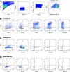
Figure 2.. Mesenchymal stem cell markers are enriched in periosteum.
Flow cytometry and cell sorting was used to evaluate marker expression in tissue from adult long bones. (A) Expression of cell surface markers in periosteum, endosteum, and bone marrow samples as a percentage of the CD45/Ter119/CD31- fraction, n = 4–50. Expression in periosteum is significantly different from both bone marrow and endosteum for all markers (two-way ANOVA with posttest comparing cell types). (B) Colony-forming unit fibroblast (CFU-F) formation in the total CD45/Ter119/CD31- fraction. (C) Representative dot plot showing gating of alpha smooth muscle actin (αSMA)-labeled cells. (D) Proportion of αSMA-labeled cells within the CD45/Ter119/CD31- gate 2–3 days after tamoxifen in different tissue compartments (n = 17). Cell surface marker expression in the Tom+ population 2–3 days after tamoxifen in (E) periosteum and (F) endosteum (n = 4–17). (G) Cell surface profile of αSMA-labeled periosteum cells at different time points (n = 2–7). *p<0.05 compared to CD45- population.
Figure 2—figure supplement 1.
Figure 2—figure supplement 1.. Flow cytometry plots of mesenchymal progenitor marker expression.
Representative plots showing (A) the initial gating scheme to enable analysis of CD45/CD31/Ter119- mesenchymal cell populations in periosteum and expression of indicated markers in (B) periosteum, (C) endosteum, and (D) bone marrow of representative samples in relation to Sca1. Quadrant percentages are specific to the sample that is shown. The periosteum, endosteum, and bone marrow samples in each column come from the same sample (obtained from two animals). FSC, forward scatter; SSC, side scatter.
Figure 2—figure supplement 2.
Figure 2—figure supplement 2.. Sca1+CD51+ and CD90+ periosteal mesenchymal cells are enriched for multipotent progenitor cells.
(A) Colony-forming unit fibroblast (CFU-F) formation of different CD45- periosteal populations (n = 3–4). *p<0.05 compared to total CD45- based on one-way ANOVA with Dunnett’s posttest. (B) Differentiation potential of cell populations grown in mixed osteogenic adipogenic conditions. Pink is ALP stain, red is Oil Red O, colonies indicated show osteogenic only (blue arrowhead), adipogenic only (black arrowhead), and mixed (*) colonies. (C) Quantification of colony differentiation from different populations. (D) Dot plots indicating different populations sorted and analyzed and percentage of αSMA+ cells (Tom+ cells 2–3 days after tamoxifen injection) within each of the CD45- cell populations indicated (n = 10–16).
Figure 3.
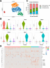
Figure 3.. Single cell RNAseq analysis of αSMA-Tom+ cells.
Plate-based RNAseq was performed on alpha smooth muscle actin (αSMA)-Tom+ cells at up to 13 weeks after tamoxifen delivery. (A) UMAP cluster plot indicating four clusters. (B) Violin plots of top markers for clusters 1–3. (C) Distribution of cells in clusters 1–3 at different time points following tamoxifen. (D) Heat map of expression of putative skeletal stem and periosteal progenitor markers.
Figure 3—figure supplement 1.
Figure 3—figure supplement 1.. Quality control data for single-cell RNAseq dataset.
(A) Gating for cell sorting of the 6-week chase cells. (B) Violin plots showing summary data for unique molecular identifier (UMI) counts, number of genes detected, and proportion of mitochondrial transcripts within the dataset.
Figure 3—figure supplement 2.
Figure 3—figure supplement 2.. Heat maps of cluster markers.
Heat map indicating expression of cluster markers with area under the receiver operating characteristics curve (AUROC) values >0.7 for (A) clusters 1–3 which appear to be periosteal populations and (B) cluster 4 which shows expression of skeletal muscle lineage genes.
Figure 4.
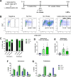
Figure 4.. Label-retaining cells are enriched with most progenitor markers.
(A) Experimental design for label-retaining cell experiments. (B) Representative gating of the CD45- population in different periosteum samples to define three populations based on GFP expression. (C) Quantification of different GFP populations in a representative experiment for periosteum and endosteum (n = 1, no doxycycline [dox]; n = 4, no chase; n = 6, 13-week chase). (D) Colony-forming unit fibroblast (CFU-F) frequency in different CD45- GFP populations in periosteal no chase samples (n = 3) and (E) following chase (n = 6). Cell surface marker expression is shown in different CD45- GFP subsets following chase (populations equivalent to right-hand label-retaining cell plot in B) in (F) periosteum cells and (G) endosteal cells. Data is pooled from three separate experiments involving 13–22 weeks chase (n = 19). *p<0.05, repeated measures ANOVA with Dunnett’s posttest compared to GFP- population.
Figure 4—figure supplement 1.
Figure 4—figure supplement 1.. Histology of H2B-GFP/αSMACreER/Ai9 femurs.
(A) H2B-GFP expression in a distal femur following 5-week doxycycline (dox) and 2 days after tamoxifen. (B) H2B-GFP label-retaining cells after 13-week chase; tamoxifen was given at the time of dox withdrawal. Boxed areas are magnified. Dashed lines indicate the periosteum and arrowheads indicate GFP+ αSMA-Tom+ cells in the cambium layer of the periosteum. gp, growth plate. Scale bars are 250 or 100 µm (high magnification images).
Figure 4—figure supplement 2.
Figure 4—figure supplement 2.. Alpha smooth muscle actin (αSMA)-labeled periosteal label-retaining cells are enriched with colony-forming unit fibroblast (CFU-F).
(A) Representative flow plots showing identification of αSMA-labeled label-retaining cells. Values represent an average of n = 3–10. (B) CFU-F formation and (C) differentiation of colonies under mixed osteogenic/adipogenic conditions of αSMA-labeled label-retaining cells and other subpopulations (n = 5 for CFU-F, n = 3 for differentiation). (D) Cell surface marker expression in αSMA-labeled subpopulations. *p<0.05 compared to negative population, #p<0.05 compared to Tom+GFP- population.
Figure 5.
Figure 5.. Bone, cartilage, and stromal progenitor (BCSP) and skeletal stem cell (SSC) populations in adult bone contain mature osteoblasts.
(A) Marker expression on Col2.3GFP+ osteoblasts, n = 4–32. (B) Frequency of BCSP and SSC populations in different tissue compartments and populations (n = 8–12). (C–D) Gating to identify BCSPs, pre-BCSPs, SSCs, and presence of Col2.3GFP+ osteoblasts as a major component of these populations in periosteum (C) and endosteum (D). Mean ± standard deviation for % GFP+ cells are shown.
Figure 6.
Figure 6.. Alpha smooth muscle actin (αSMA)-labeled cells are transplantable self-renewing osteochondroprogenitors.
(A) Colony-forming unit fibroblast (CFU-F) frequency of periosteal αSMA-labeled cells sorted 2 days after tamoxifen. (B) Engraftment of transplanted αSMA-labeled cells in a calvarial defect 5 weeks after transplantation. Arrowheads indicate cells that are Col2.3GFP+. Quantification of αSMA-labeled cell contribution to (C) osteoblasts and (D) chondrocytes in day 7 secondary fracture calluses is shown. (E) Experimental design and fracture callus histology following a second tibia fracture in αSMACreER/Ai9/Col2.3GFP mice showing contribution of Tom+ cells to callus tissue, including osteoblasts (F) and Sox9+ chondrocytes (white) (G). (H–J) Histology of a similar secondary fracture where tamoxifen was given 14 days prior to the primary fracture. White arrowheads indicate αSMA-labeled Col2.3GFP+ osteoblasts, orange arrowheads indicate αSMA-labeled Sox9+ chondrocytes. DAPI stain is shown in blue. Approximate callus area used for analysis is indicated by the dashed lines in E and H. Scale bars indicate 250 µm (low magnification) and 100 µm (high magnification).
Figure 7.
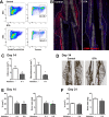
Figure 7.. Ablation of alpha smooth muscle actin (αSMA)+ cells impairs fracture healing.
(A) Flow cytometry of day 4 fracture callus from control αSMACreER/Ai9 mice and DTA animals (αSMACreER/Rosa-DTA/Ai9) indicating significantly fewer (p<0.01) αSMA-labeled cells in the ablation model (mean ± SD shown, n = 4). (B) Representative histological sections from day 4 fracture callus in controls or with DTA-mediated ablation. The outline of the callus is indicated. (C) Histological analysis of callus area and mineralized area at day 14, and (D) representative images of von-Kossa staining (n = 4–7). (E) Micro-computed tomography (MicroCT) analysis of day 14 fracture callus in DTA- and DTA+ mice that had undergone two different tamoxifen regimens to ablate αSMA+ cells, day 0, 1 of fracture or day 0, 2, 4, 6, 8. Callus tissue volume and bone mass were measured. Cre-: n = 14, 8 male (M), 6 female (F); DTA D0-1: n = 10, 4M 6F; DTA D0-8: n = 8, 4M 4F. (F) MicroCT analysis of day 21 fracture callus in mice treated with tamoxifen on day 0, 2, and 4 (n = 14–15).
Figure 8.
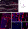
Figure 8.. Contribution of mature osteoblasts to fracture healing and PDGFRα distribution.
(A) Fracture histology of a representative day 7 tibial fracture callus from a Col2.3CreER/Ai9 mouse treated with tamoxifen at days −4 and −3. Mineralized bone is shown by dark field (gray) and DAPI+ nuclei in blue. Scale bar is 250 µm. (B) Quantification of Col2.3-labeled cell contribution to Osx+ osteoblasts. *p<0.05 based on ANOVA with Dunnett’s posttest compared to no tamoxifen group. (C) Tibia histology of PdgfraCreER/Ai9 mice given three daily doses of tamoxifen and euthanized 1 day later. Scale bar is 200 µm. Magnified areas of (D) trabecular and (E) cortical bone are shown. (F) Tibial histology of a PdgfraCreER/Ai9 mouse that had not been exposed to tamoxifen showing minimal leakiness of the CreER. Representative images of n = 3 aged 5–8 weeks of age are shown. (G) PDGFRα immunostaining of bone. Scale bar is 50 µm. bm, bone marrow; gp, growth plate; p, periosteum.
Author response image 1.
Author response image 1.. Dot plot of periosteum sample used for sorting αSMA-labeled cells for calvarial defect transplantation.
Note that almost all Tom+ cells are GFP-.

References

    1. Abou-Khalil R, Yang F, Lieu S, Julien A, Perry J, Pereira C, Relaix F, Miclau T, Marcucio R, Colnot C. Role of muscle stem cells during skeletal regeneration. Stem Cells. 2015;33:1501–1511. doi: 10.1002/stem.1945. - DOI - PubMed
    1. Ambrosi TH, Scialdone A, Graja A, Gohlke S, Jank AM, Bocian C, Woelk L, Fan H, Logan DW, Schürmann A, Saraiva LR, Schulz TJ. Adipocyte accumulation in the bone marrow during obesity and aging impairs stem Cell-Based hematopoietic and bone regeneration. Cell Stem Cell. 2017;20:771–784. doi: 10.1016/j.stem.2017.02.009. - DOI - PMC - PubMed
    1. Arthur A, Nguyen TM, Paton S, Zannettino ACW, Gronthos S. Loss of EfnB1 in the osteogenic lineage compromises their capacity to support hematopoietic stem/progenitor cell maintenance. Experimental Hematology. 2019;69:43–53. doi: 10.1016/j.exphem.2018.10.004. - DOI - PubMed
    1. Becht E, McInnes L, Healy J, Dutertre C-A, Kwok IWH, Ng LG, Ginhoux F, Newell EW. Dimensionality reduction for visualizing single-cell data using UMAP. Nature Biotechnology. 2019;37:38–44. doi: 10.1038/nbt.4314. - DOI - PubMed
    1. Bolisetty MT, Stitzel ML, Robson P. CellView: interactive exploration of high dimensional single cell RNA-seq data. bioRxiv. 2017 doi: 10.1101/123810. - DOI

Publication types

Associated data