. 2021 Jul 1;23(7):1056-1071.
doi: 10.1093/neuonc/noab020.

Consensus recommendations for MRI and PET imaging of primary central nervous system lymphoma: guideline statement from the International Primary CNS Lymphoma Collaborative Group (IPCG)

Affiliations

Consensus recommendations for MRI and PET imaging of primary central nervous system lymphoma: guideline statement from the International Primary CNS Lymphoma Collaborative Group (IPCG)

Ramon F Barajas et al. Neuro Oncol. .

Abstract

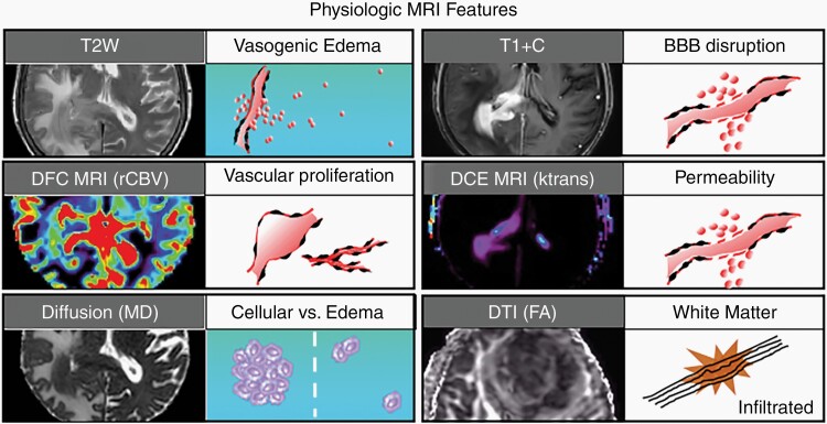
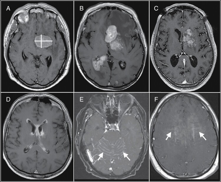
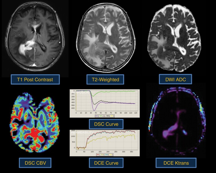
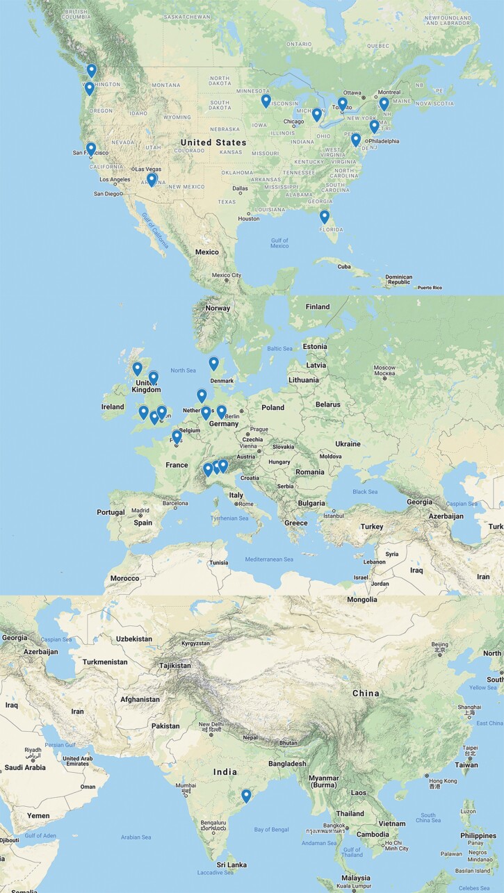
Advanced molecular and pathophysiologic characterization of primary central nervous system lymphoma (PCNSL) has revealed insights into promising targeted therapeutic approaches. Medical imaging plays a fundamental role in PCNSL diagnosis, staging, and response assessment. Institutional imaging variation and inconsistent clinical trial reporting diminishes the reliability and reproducibility of clinical response assessment. In this context, we aimed to: (1) critically review the use of advanced positron emission tomography (PET) and magnetic resonance imaging (MRI) in the setting of PCNSL; (2) provide results from an international survey of clinical sites describing the current practices for routine and advanced imaging, and (3) provide biologically based recommendations from the International PCNSL Collaborative Group (IPCG) on adaptation of standardized imaging practices. The IPCG provides PET and MRI consensus recommendations built upon previous recommendations for standardized brain tumor imaging protocols (BTIP) in primary and metastatic disease. A biologically integrated approach is provided to addresses the unique challenges associated with the imaging assessment of PCNSL. Detailed imaging parameters facilitate the adoption of these recommendations by researchers and clinicians. To enhance clinical feasibility, we have developed both "ideal" and "minimum standard" protocols at 3T and 1.5T MR systems that will facilitate widespread adoption.

Keywords: MRI; PCNSL; PET; imaging; primary central nervous system lymphoma.


Figures

Fig. 1
Fig. 1
Biophysical features characterized by conventional and advanced physiologic MRI techniques. Shown are 6 MRI techniques that are commonly employed in neuro-oncologic imaging, along with their respective corresponding tumor phenotypes. T2-weighted (T2W) signal is typically used to define vasogenic edema. T1-weighted post-contrast enhancement (T1 + C) shows areas of disrupted blood-brain barrier (BBB). Dynamic susceptibility contrast (DSC) MRI measures of relative cerebral blood volume (rCBV) define microvascular volume as an indicator of tumor-related angiogenesis. Dynamic contrast-enhanced (DCE) MRI measures of vascular permeability (Ktrans). Diffusion-weighted imaging apparent diffusion coefficient (ADC) correlates with cellular density and proliferative indices and can aid in distinguishing tumor from vasogenic edema. Diffusion tensor imaging (DTI) fractional anisotropy (FA) measures the integrity of white matter tracts, which can be used to identify regions of tumor infiltration.
Fig. 2
Fig. 2
Challenges of bidirectional measurements of primary central nervous system lymphoma (PCNSL) tumor burden. (A) A single-rounded PCNSL mass lends itself to straightforward bidirectional measurement (yellow lines). (B) These bidirectional measurements can also be applied to multiple discrete masses. However, bidirectional measurements become more challenging with varied imaging patterns, such as with (C) heterogeneous enhancement, (D) linear ependymal enhancement, (E) leptomeningeal enhancement (arrows), and (F) linear perivascular enhancement (arrows). These varied imaging patterns would be more amenable to measurement using volumetric-based approaches to define tumor burden. Abbreviations: ADC, apparent diffusion coefficient; CBV, cerebral blood volume; DCE, dynamic contrast-enhanced; DSC, dynamic susceptibility contrast; PCNSL, primary central nervous system lymphoma.
Fig. 3
Fig. 3
Typical morphologic and physiologic MRI appearance of PCNSL. PCNSL classically appears as a diffuse often periventricular enhancing mass (top left). T2-weighted imaging is often heterogenous but frequently demonstrates a mass like hypointense component (top middle) within enhancing regions. Increasing tumor cellularity is associated with decreasing T2 and ADC hypointensity (top right). Likewise, the degree of angiogenesis is reflected by DSC and DCE perfusion MRI sequences. CBV (bottom left) and Ktrans (bottom right) are quite heterogenous in PCNSL and may be reflective of tumor aggressiveness.
Fig. 4
Fig. 4
Demographic distribution of IPCG survey response. The IPCG imaging subcommittee surveyed members to determine clinical imaging practices for the evaluation of PCNSL at institutions across the world. About 147 institutions were invited to participate in this electronic survey. A 20% response rate was received from institutions delineated on the map from North America (N = 11), Europe (N = 17), and India (N = 1). Abbreviations: IPCG, International PCNSL Collaborative Group; PCNSL, primary central nervous system lymphoma.

References

    1. Ostrom QT, Gittleman H, Liao P, et al. . CBTRUS Statistical Report: primary brain and other central nervous system tumors diagnosed in the United States in 2010–2014. Neuro Oncol. 2017;19(suppl_5):v1–v88. - PMC - PubMed
    1. Grommes C, DeAngelis LM. Primary CNS lymphoma. J Clin Oncol. 2017;35(21):2410–2418. - PMC - PubMed
    1. Abrey LE, Batchelor TT, Ferreri AJ, et al. ; International Primary CNS Lymphoma Collaborative Group . Report of an international workshop to standardize baseline evaluation and response criteria for primary CNS lymphoma. J Clin Oncol. 2005;23(22):5034–5043. - PubMed
    1. Ellingson BM, Bendszus M, Boxerman J, et al. ; Jumpstarting Brain Tumor Drug Development Coalition Imaging Standardization Steering Committee . Consensus recommendations for a standardized brain tumor imaging protocol in clinical trials. Neuro Oncol. 2015;17(9):1188–1198. - PMC - PubMed
    1. Kaufmann TJ, Smits M, Boxerman J, et al. . Consensus recommendations for a standardized brain tumor imaging protocol for clinical trials in brain metastases. Neuro Oncol. 2020;22(6):757–772. - PMC - PubMed

Publication types