. 2021 Apr 1;132(4):e50-e55.
doi: 10.1213/ANE.0000000000005387.

Activation of GABAergic Neurons in the Rostromedial Tegmental Nucleus and Other Brainstem Regions Promotes Sedation and Facilitates Sevoflurane Anesthesia in Mice

Affiliations

Activation of GABAergic Neurons in the Rostromedial Tegmental Nucleus and Other Brainstem Regions Promotes Sedation and Facilitates Sevoflurane Anesthesia in Mice

Ksenia Vlasov et al. Anesth Analg. .

Abstract

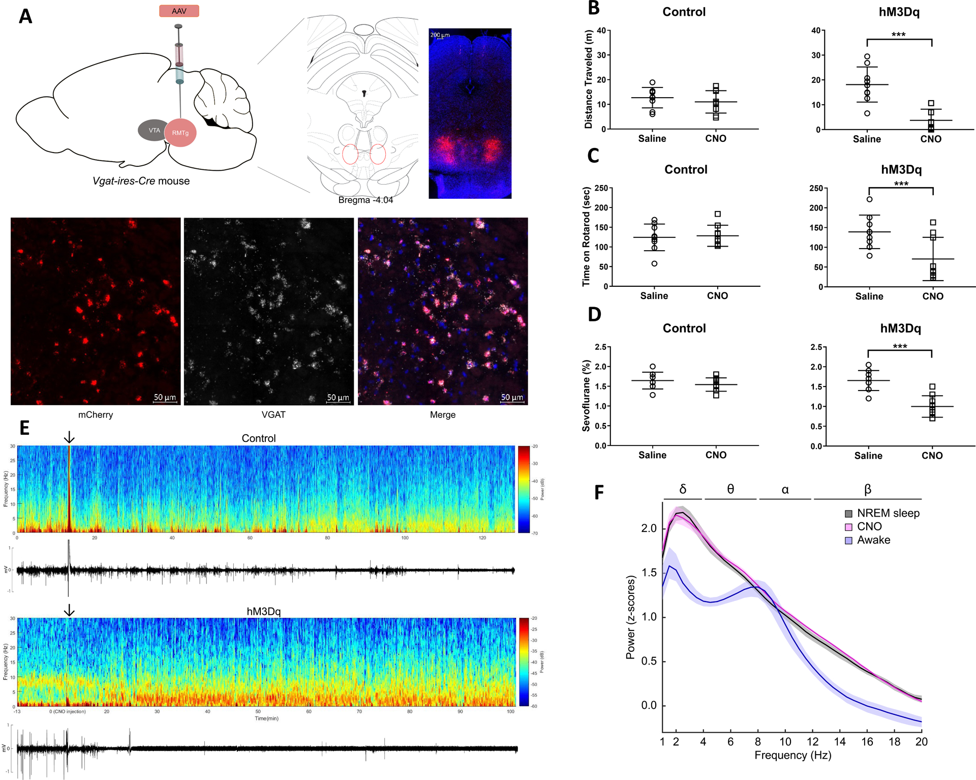
Many general anesthetics potentiate gamma-aminobutyric acid (GABA) A receptors but their neuroanatomic sites of action are less clear. GABAergic neurons in the rostromedial tegmental nucleus (RMTg) send inhibitory projections to multiple arousal-promoting nuclei, but the role of these neurons in modulating consciousness is unknown. In this study, designer receptors exclusively activated by designer drugs (DREADDs) were targeted to RMTg GABAergic neurons of Vgat-ires-Cre mice. DREADDs expression was found in the RMTg and other brainstem regions. Activation of these neurons decreased movement and exploratory behavior, impaired motor coordination, induced electroencephalogram (EEG) oscillations resembling nonrapid eye movement (NREM) sleep without loss of righting and reduced the dose requirement for sevoflurane-induced unconsciousness. These results suggest that GABAergic neurons in the RMTg and other brainstem regions promote sedation and facilitate sevoflurane-induced unconsciousness.


Conflict of interest statement

Conflicts of Interest: See Disclosures at the end of the article.

Figures

Fig.
Fig.
A. Bilateral expression of hM3Dq-mCherry in RMTg GABAergic neurons of Vgat-ires-Cre mice. Representative expression pattern of hM3Dq-mCherry in the RMTg (top). Co-localization of mCherry and VGAT (visualized with fluorescent in situ hybridization) demonstrates selective expression of hM3Dq in GABAergic neurons (bottom). B. Activation of GABAergic neurons decreases the total distance traveled in the open field test. Scatter plots showing total distance traveled (meters) in the 5-minute open field test after i.p. administration of saline (vehicle) or CNO (1 mg/kg) for control and hM3Dq mice (lines represent mean and SD). Although there was no significant change in control mice after CNO, total distance traveled was significantly decreased in hM3Dq mice. C. Activation of GABAergic neurons decreases the total time on an accelerating rotarod. Scatter plots showing total time (seconds) on the accelerating rotarod (lines represent mean and SD). CNO (1 mg/kg) significantly decreased total rotarod time in hM3Dq mice compared to saline, but there was no significant difference in control mice. D. Activation of GABAergic neurons decreases the dose requirement for sevoflurane-induced unconsciousness. Scatter plots show that in control mice, CNO (1 mg/kg) had no significant effect on sensitivity to sevoflurane-induced loss of righting reflex (LORR) compared to saline (lines represent mean and SD). In hM3Dq mice, however, CNO significantly reduced the dose of sevoflurane required for LORR. E. Activation of GABAergic neurons increases EEG spectral power at low frequencies and decreases EMG activity. Spectrogram of a continuous EEG recording from a representative control mouse before and after CNO injection (top). The arrow indicates the time of CNO administration. The animal remained awake and active, as demonstrated by low spectral power in the delta range (1–4 Hz), and high EMG activity. However, in a representative hM3Dq mouse, approximately 20 minutes after CNO, spectral power increased at low frequencies (1–4 Hz) and persisted for the duration of the recording. The corresponding EMG activity below the spectrogram shows that the increase in EEG delta power coincided with decreased movement. F. Activation of GABAergic neurons produces an EEG power spectrum similar to NREM sleep. Group power spectral density estimates comparing EEG recordings taken during wakefulness, NREM sleep, and after CNO. The 95% bootstrapped confidence intervals are shown in the shaded areas. CNO (pink) induced a power spectrum very similar to NREM sleep (black) and distinct from the awake state (blue).

References

    1. Hemmings HC Jr, Riegelhaupt PM, Kelz MB, et al. Towards a Comprehensive Understanding of Anesthetic Mechanisms of Action: A Decade of Discovery. Trends Pharmacol Sci. 2019;40(7):464–481. - PMC - PubMed
    1. Franks NP. General anaesthesia: from molecular targets to neuronal pathways of sleep and arousal. Nat Rev Neurosci. 2008;9(5):370–386. - PubMed
    1. Pal D, Dean JG, Liu T, et al. Differential Role of Prefrontal and Parietal Cortices in Controlling Level of Consciousness. Curr Biol. 2018;28(13):2145–2152 e2145. - PMC - PubMed
    1. Kelz MB, Garcia PS, Mashour GA, Solt K. Escape From Oblivion: Neural Mechanisms of Emergence From General Anesthesia. Anesth Analg. 2019;128(4):726–736. - PMC - PubMed
    1. Eban-Rothschild A, Rothschild G, Giardino WJ, Jones JR, de Lecea L. VTA dopaminergic neurons regulate ethologically relevant sleep-wake behaviors. Nat Neurosci. 2016;19(10):1356–1366. - PMC - PubMed

Publication types