. 2021 Feb 9;16(2):e0246872.
doi: 10.1371/journal.pone.0246872. eCollection 2021.

Internal defect scanning of sweetpotatoes using interactance spectroscopy

Affiliations

Internal defect scanning of sweetpotatoes using interactance spectroscopy

Michael W Kudenov et al. PLoS One. .

Abstract

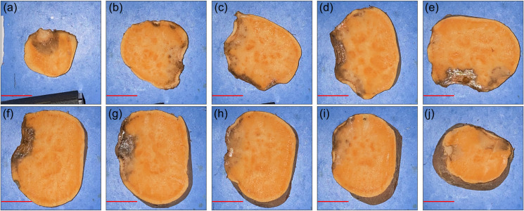
While standard visible-light imaging offers a fast and inexpensive means of quality analysis of horticultural products, it is generally limited to measuring superficial (surface) defects. Using light at longer (near-infrared) or shorter (X-ray) wavelengths enables the detection of superficial tissue bruising and density defects, respectively; however, it does not enable the optical absorption and scattering properties of sub-dermal tissue to be quantified. This paper applies visible and near-infrared interactance spectroscopy to detect internal necrosis in sweetpotatoes and develops a Zemax scattering simulation that models the measured optical signatures for both healthy and necrotic tissue. This study demonstrates that interactance spectroscopy can detect the unique near-infrared optical signatures of necrotic tissues in sweetpotatoes down to a depth of approximately 5±0.5 mm. We anticipate that light scattering measurement methods will represent a significant improvement over the current destructive analysis methods used to assay for internal defects in sweetpotatoes.


Conflict of interest statement

The authors have declared that no competing interests exist.

Figures

Fig 1
Fig 1. Schematic overview of the interactance spectroscopy setup.
Optical fibers were placed directly onto the sweetpotato target that was positioned a distance a apart.
Fig 2
Fig 2. Experimental setup.
A cut tissue section from one sweetpotato was measured with the optical fibers, which were placed into direct contact with the (a) healthy or (b) necrotic tissues.
Fig 3
Fig 3. Interactance spectrum sampling of tissue slices.
(a) Sweetpotatoes were sampled towards their proximal ends in approximately 10 mm thick sections. (b) The probe was positioned adjacent to the necrotic tissue, and the depth to the defect was measured.
Fig 4
Fig 4. Sampling of full sweetpotatoes with skin removed.
A patch of skin was removed from each of the three sections (proximal, middle, and distal). Four measurements were taken with the probes positioned at locations denoted 1a, 1b, 2a, and 2b corresponding to green, magenta, blue, and cyan, respectively. Note that 1a and 2b were measured with the skin in place and 1b and 2a were measured with the skin removed.
Fig 5
Fig 5. Pure tissue spectra.
Normalized interactance spectra from the two tissue types. Error bars are based on the signal to noise ratio.
Fig 6
Fig 6. Pictures of healthy and necrotic tissues at different depths.
(a) necrotic tissue observed at the proximal end of a sweetpotato at a depth of 3.5±0.5 mm; (b) necrotic tissue at a depth of 5.5±0.5 mm towards the middle of the SP, (c) mid-section of a healthy sweetpotato; and (d) mid-section of a necrotic sweetpotato with necrosis at a depth of 3.0±0.5 mm.
Fig 7
Fig 7. Measured interactance spectra.
Data were acquired from healthy and necrotic tissues at the different depths as reported in Fig 6. Error bars are based on the signal to noise ratio.
Fig 8
Fig 8. RGB imagery of the two batches of SPs that were measured.
(a) top and (b) bottom side of the first batch and (c) top and (d) bottom side of the second batch.
Fig 9
Fig 9. Interactance spectra from the 22 samples.
(a) First batch containing 10 samples; and (b) second batch containing 12 samples.
Fig 10
Fig 10. Healthy tissue RGB cross-sectional images of SP 3 from batch 2.
Slices are depicted from the top to bottom (with reference to the orientation in Fig 8) for panels (a-j). The red scale bar in each panel represents a distance of 25 mm.
Fig 11
Fig 11. Necrotic tissue RGB cross-sectional images of SP 11 from batch 2.
Slices are depicted from the top to bottom (with reference to the orientation in Fig 8) for panels (a-j). The red scale bar in each panel represents a distance of 25 mm.
Fig 12
Fig 12. Interactance spectra of the skin and flesh.
Recall that 1a and 2a were taken through the skin while 1b and 2b were taken with the skin removed.
Fig 13
Fig 13. Zemax scattering simulation.
Healthy and necrotic tissues are configured with thicknesses of t1 and t2, respectively. An annular detector with an active area defined by inner and outer radii r1 and r2, respectively.
Fig 14
Fig 14. Data used in the model for fitting.
(a) Graphical solution for τ versus the normalized detected power; (b) Mean skin transmission spectrum (double-pass) from Fig 12(c).
Fig 15
Fig 15. Bulk scattering transmission coefficients for pure healthy and necrotic tissue.
Fig 16
Fig 16. Simulation results for necrotic tissues at depths t1 spanning 1 to 8 mm in 1 mm increments (red through violet sold lines, respectively).
Error bars from the measured data are included on the simulated healthy spectrum (black solid line) out to one standard deviation.
Fig 17
Fig 17. Simulation and measured results after subtracting the healthy reference spectra.
Simulation results are provided for necrotic tissues at depths t1 spanning 1 to 8 mm in 1 mm increments (red through violet sold lines, respectively). Healthy and necrotic tissues.

References

    1. Kinnunen M, Karmenyan A. Overview of single-cell elastic light scattering techniques. JBO. 2015;20: 051040 10.1117/1.JBO.20.5.051040 - DOI - PubMed
    1. Qiu L, Perelman LT. Confocal Light Absorption and Scattering Spectroscopic Microscopy Tuchin VV, editor. Handbook of Photonics for Biomedical Science. Boca Raton: Crc Press-Taylor & Francis Group; 2010. pp. 465–480.
    1. Qiu L, Turzhitsky V, Chuttani R, Pleskow DK, Goldsmith JD, Guo L, et al. Spectral Imaging With Scattered Light: From Early Cancer Detection to Cell Biology. IEEE J Sel Top Quantum Electron. 2012;18: 1073–1083. 10.1109/JSTQE.2011.2161575 - DOI - PMC - PubMed
    1. Lu R, Van Beers R, Saeys W, Li C, Cen H. Measurement of optical properties of fruits and vegetables: A review. Postharvest Biology and Technology. 2020;159: UNSP 111003 10.1016/j.postharvbio.2019.111003 - DOI
    1. Noordam JC, Otten GW, Timmermans TJ, van Zwol BH. High-speed potato grading and quality inspection based on a color vision system Machine Vision Applications in Industrial Inspection VIII. International Society for Optics and Photonics; 2000. pp. 206–217.

Publication types

LinkOut - more resources