. 2021 Feb 27;27(3):gaab009.
doi: 10.1093/molehr/gaab009.

Motility of efferent duct cilia aids passage of sperm cells through the male reproductive system

Affiliations

Motility of efferent duct cilia aids passage of sperm cells through the male reproductive system

Isabella Aprea et al. Mol Hum Reprod. .

Abstract

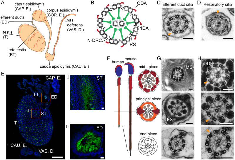
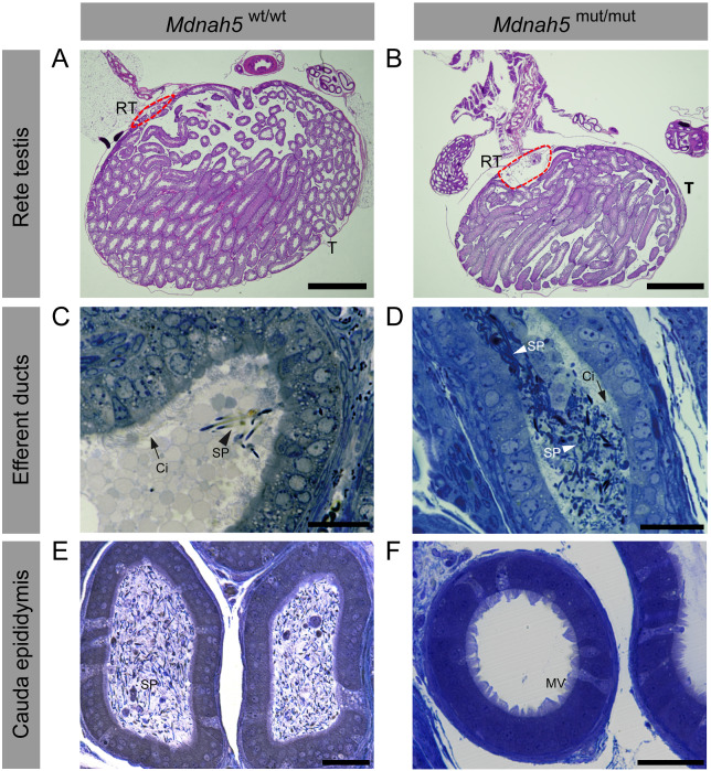
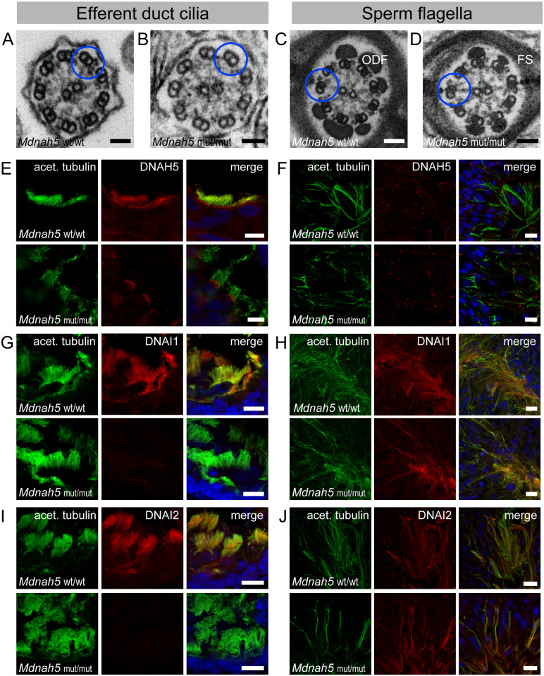
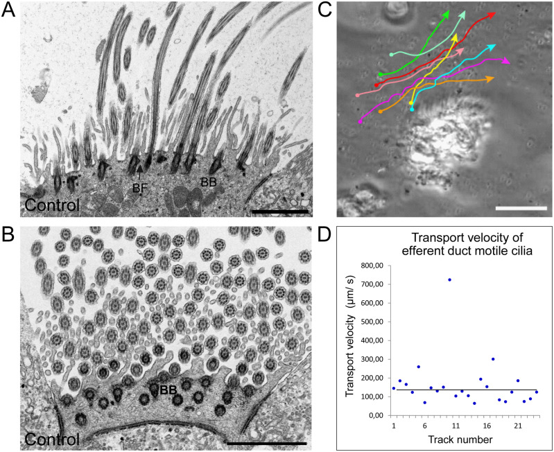
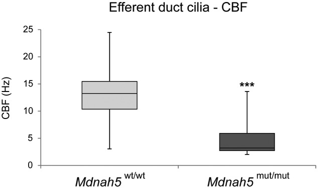
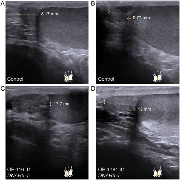
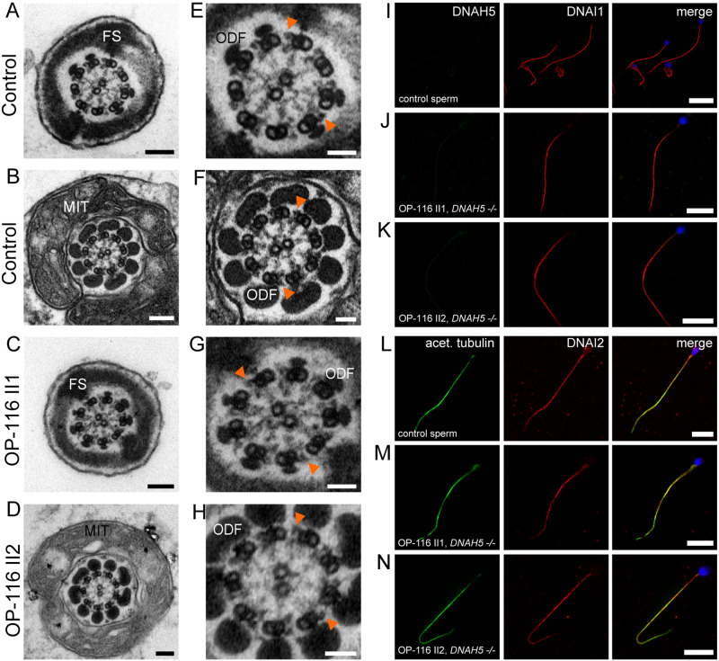
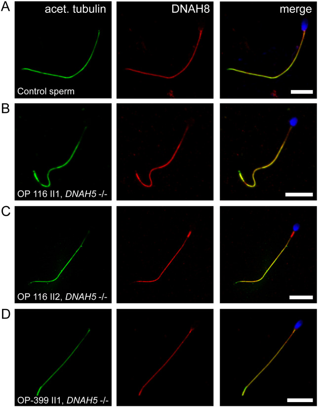
Motile cilia line the efferent ducts of the mammalian male reproductive tract. Several recent mouse studies have demonstrated that a reduced generation of multiple motile cilia in efferent ducts is associated with obstructive oligozoospermia and fertility issues. However, the sole impact of efferent duct cilia dysmotility on male infertility has not been studied so far either in mice or human. Using video microscopy, histological- and ultrastructural analyses, we examined male reproductive tracts of mice deficient for the axonemal motor protein DNAH5: this defect exclusively disrupts the outer dynein arm (ODA) composition of motile cilia but not the ODA composition and motility of sperm flagella. These mice have immotile efferent duct cilia that lack ODAs, which are essential for ciliary beat generation. Furthermore, they show accumulation of sperm in the efferent duct. Notably, the ultrastructure and motility of sperm from these males are unaffected. Likewise, human individuals with loss-of-function DNAH5 mutations present with reduced sperm count in the ejaculate (oligozoospermia) and dilatations of the epididymal head but normal sperm motility, similar to DNAH5 deficient mice. The findings of this translational study demonstrate, in both mice and men, that efferent duct ciliary motility is important for male reproductive fitness and uncovers a novel pathomechanism distinct from primary defects of sperm motility (asthenozoospermia). If future work can identify environmental factors or defects in genes other than DNAH5 that cause efferent duct cilia dysmotility, this will help unravel other causes of oligozoospermia and may influence future practices in genetic and fertility counseling as well as ART.

Keywords: efferent ducts / cilia / sperm / motor proteins / outer dynein arm-defects / DNAH5 / 45 male infertility / oligozoospermia / motile ciliopathy / primary ciliary dyskinesia.


Figures

Figure 1.
Figure 1.
Cilia and flagella in the mouse male reproductive tract.  A, overview of the mouse male reproductive tract. B, ciliary/flagellar axonemes consist of microtubules and functional complexes. ODA: outer dynein arm; IDA: inner dynein arm; RS: radial spoke; N-DRC: nexin-dynein regulatory complex. C, axoneme ultrastructure of control mouse efferent duct and D, respiratory cilia. E, IF labeling for acetylated alpha-tubulin (green; DAPI-labelled nuclei: blue) in the mouse male reproductive tract. (I) spermatozoa in seminiferous tubules (ST). (II) efferent duct cilia. Nuclei (blue) were stained with DAPI. F, schematic and G,TEM cross sections of sperm flagella with axonemes surrounded by accessory structures. H, detail of G. Biological replicate: n=3; Scale bars represent 100 nm (C, D, G and H), 1000 µm (e), 10 µm (I and II). C, D, H, orange arrowheads indicate ODAs.
Figure 2.
Figure 2.
Adult Mdnah5 homozygous mutant male mice display enlargement of the rete testes and sperm accumulation in the efferent duct tract.  A and B, hematoxylin and eosin staining of (A) control and (B) Mdnah5mut/mut testes (T) show that compared to control littermates, Mdnah5mut/mut males present a dilatation of the rete testes (RT, red line). CF, toluidine blue staining of efferent duct and cauda epididymis semi thin sections.C and D,cilia (Ci) line efferent duct lumen of control (Mdnah5wt/wt) and Mdnah5mut/mut mice.Controls present few isolated spermatozoa (SP). Sperm accumulation is clearly visualized in the Mdnah5mut/mut mouse. E and F, opposite distribution of spermatozoa is observed within the cauda epididymis:a large amount of spermatozoa is present in the control but only few are visible in the Mdnah5mut/mut male. One example is representatively shown for a biological replicate of three adult mice (n=3). Scale bars represent 1000 µm (A, B), 50 µm (C–F). MV: microvilli.
Figure 3.
Figure 3.
Mdnah5 homozygous mutant male mice show loss of ODAs in efferent duct cilia but not sperm flagella.  A, TEM of control (Mdnah5wt/wt) efferent duct cilia. B, absence of ODAs in mutant (Mdnah5mut/mut) efferent duct cilia; ODA presence in (C) control and (D) mutant sperm flagella. E, IF co-staining confirms DNAH5 (red) presence in control efferent duct cilia along the entire ciliary length marked with an antibody against acetylated alpha-tubulin (green) and DNAH5 loss in Mdnah5-mutant tissue. F, IF staining on testicular sections show that DNAH5 (red) is not present in control and Mdnah5-mutant mouse sperm flagella. G, DNAI1 (red) localizes to control efferent duct cilia, but not in Mdnah5mut/mut mice. H, IF staining on testicular sections show that DNAI1 (red) localizes to sperm flagella in control and Mdnah5mut/mut male. I, as for the other ODA components, also DNAI2 (red) localizes to control efferent duct cilia, but not in Mdnah5mut/mut mice. J, IF staining on testicular sections show that control sperm cells (upper panel) and those of the Mdnah5-mutant male (lower panel), display both panaxonemal localization for DNAI2 (depicted in red). Biological replicate for efferent duct cilia: n= 9 control mice; n=11 Mdnah5 homozygous mutant mice. Biological replicate for sperm flagella: n=3/group. Nuclei (blue) were stained with DAPI. Scale bars represent 50 nm (A–D), 10 µm (E–J).
Figure 4.
Figure 4.
Mouse efferent duct cilia display a regular orientation and directed beating pattern.  A and B, TEM analysis of ciliary cross sections in control mouse efferent ducts.A, longitudinal sections of efferent duct cilia displaying regular disposed basal bodies and basal foot processes. B, central pairs of efferent duct cilia (Ci) are oriented in a regular fashion as indicated by the red bars. C, manual tracking of 0.5 µm plastic beads applied on efferent duct cilia confirms that this cilia type displays a directed beating pattern. D, evaluation of the transport velocity of control efferent duct cilia shows that applied beads are mobilized with a median velocity of 130 µm/s. Biological replicate: n=3; Scale bars represent 2 µm (A and B), 50 µm (C).
Figure 5.
Figure 5.
CBF of efferent duct cilia is significantly reduced in Mdnah5 homozygous mutant male mice. Compared to control mice (Mdnah5wt/wt), statistical analysis by unpaired two-tailed student’s t-test with Welch’s correction reveals a significant reduction of CBF (in Hz: Hertz) of motile efferent duct cilia in Mdnah5mut/mut mice. Biological replicate: n=5 control group; n=7 Mdnah5 homozygous mutant mice, P<0.001: ***.
Figure 6.
Figure 6.
DNAH5-mutant male individuals present enlargement of the epididymal head.  A and B, sonographs of respectively right (A) and left (B) testis and epididymal head, in a healthy control individual. Epididymal head height measures 8.17 mm (A) and 9.77 mm (B). C, individual OP-116 II1 and D, individual OP-1781 II1, carrying disease causing mutations in DNAH5, present an epididymal head height of 17.7 mm (OP-116 II1) and 13 mm (OP-1781 II1), respectively. This indicates an enlargement of the epididymal head size, compared to control (A, B) and published maximum reference values of 11.6 mm (right epididymal head) and 11.3 mm (left epididymal head) (Pilatz et al., 2013).
Figure 7.
Figure 7.
ODA protein DNAH5 is not present in human sperm flagella.  A and B, TEM of control sperm cells showing the 9 + 2 axoneme with clearly visible ODAs and accessory structures (FS: fibrous sheath, MIT: mitochondria, and ODF: outer dense fibers). C and D, DNAH5 deficiency does not cause ODA defects of sperm flagellar axonemes. E–H, magnifications of A–D, orange arrowheads exemplarily indicate ODAs. I, J, K, IF co-staining assessing DNAH5 absence and panaxonemal localization of DNAI1 in human control and DNAH5-deficient sperm cells. L, control sperm cells display a panaxonemal flagellar localization of DNAI2 (red) that co-localizes (yellow in the merged image) with the flagellar marker acetylated alpha-tubulin (depicted in green). M and N, sperm flagella of individuals with PCD causing compound heterozygous mutations in DNAH5 display an unaltered localization pattern of DNAI2, indicating that DNAH5 is not a component of the ODA in sperm flagella. Nuclei (blue) were stained with DAPI. Scale bars represent 100 nm (A–D), 50 nm (E–H), 10 µm (I–N).
Figure 8.
Figure 8.
DNAH8 is present in sperm flagella of DNAH5-deficient individuals.  A, IF analysis showing DNAH8 panaxonemal localization in human control sperm cells. B–D, same staining procedure shows that sperm flagella of DNAH5-mutant individuals present an unaltered DNAH8 localization along the entire flagellar length. This is consistent with the observed unaltered sperm ultrastructure of DNAH5-deficient individuals and the hypothesis that DNAH8, as a paralogue of DNAH5, developed a specific function in sperm flagella. Flagellar marker: acet. tubulin (acetylated alpha-tubulin). Nuclei (blue) were stained with DAPI. Scale bars represent 10 µm.

Similar articles

Cited by

References

    1. Adzhubei IA, Schmidt S, Peshkin L, Ramensky VE, Gerasimova A, Bork P, Kondrashov AS, Sunyaev SR. A method and server for predicting damaging missense mutations. Nat Methods 2010;7:248–249. - PMC - PubMed
    1. Becker O. Ueber Flimmerepithelium im Nebenhoden des Menschen. Wiener Medizinische Wochenschrift 1856;6:184.
    1. Bellve AR, Cavicchia JC, Millette CF, O'Brien DA, Bhatnagar YM, Dym M. Spermatogenic cells of the prepuberal mouse. Isolation and morphological characterization. J Cell Biol 1977;74:68–85. - PMC - PubMed
    1. Björndahl L, Barratt CLR, Mortimer D, Jouannet P. ‘How to count sperm properly’: checklist for acceptability of studies based on human semen analysis. Hum Reprod 2016;31:227–232. - PubMed
    1. Bracke A, Peeters K, Punjabi U, Hoogewijs D, Dewilde S. A search for molecular mechanisms underlying male idiopathic infertility. Reprod Biomed Online 2018;36:327–339. - PubMed

Publication types

MeSH terms

LinkOut - more resources