. 2021 Mar 22;31(6):1192-1205.e6.
doi: 10.1016/j.cub.2021.01.001. Epub 2021 Feb 8.

The actin networks of chytrid fungi reveal evolutionary loss of cytoskeletal complexity in the fungal kingdom

Affiliations

The actin networks of chytrid fungi reveal evolutionary loss of cytoskeletal complexity in the fungal kingdom

Sarah M Prostak et al. Curr Biol. .

Abstract

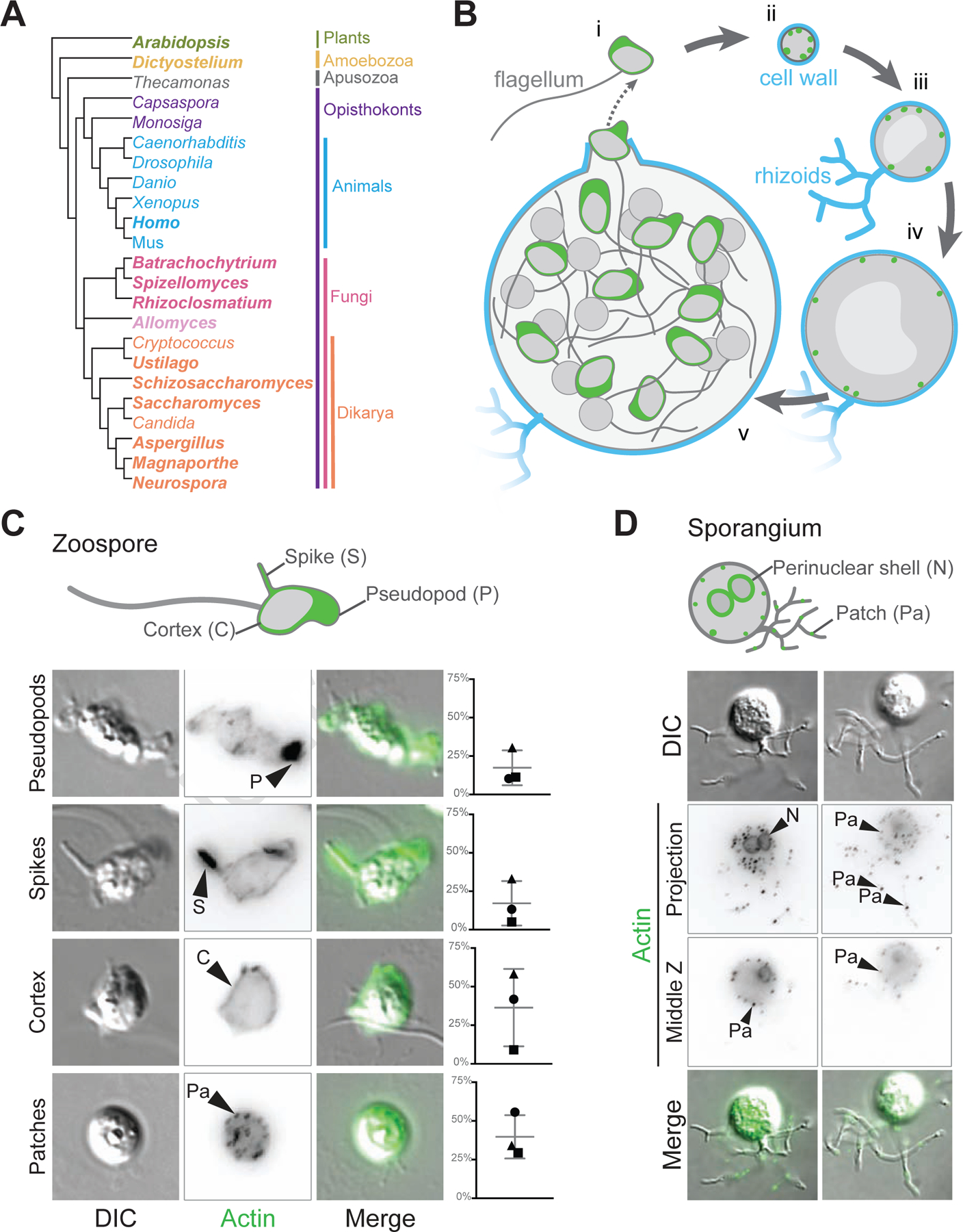
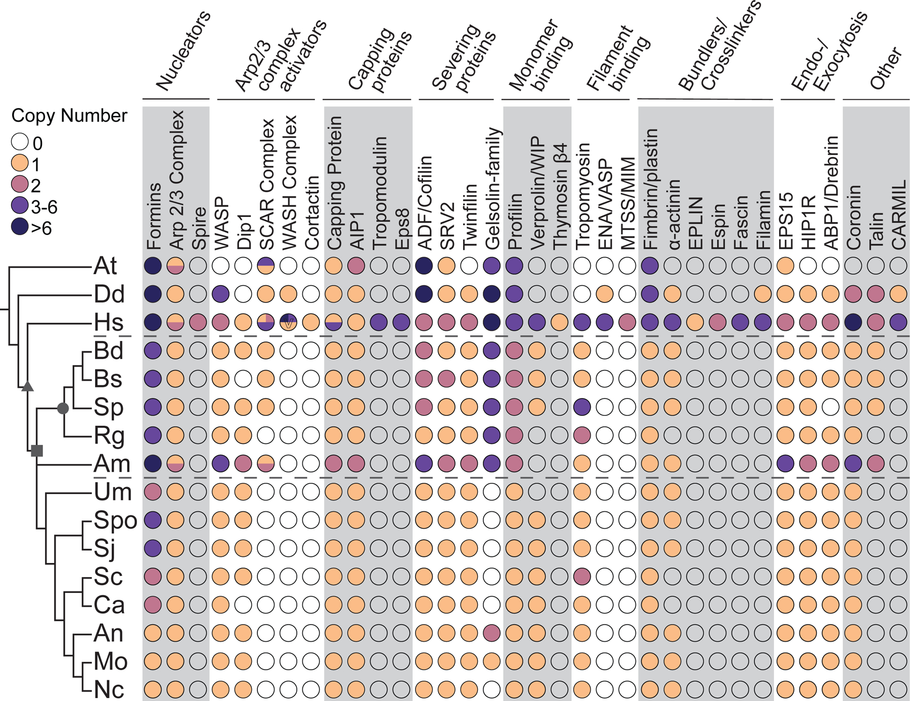
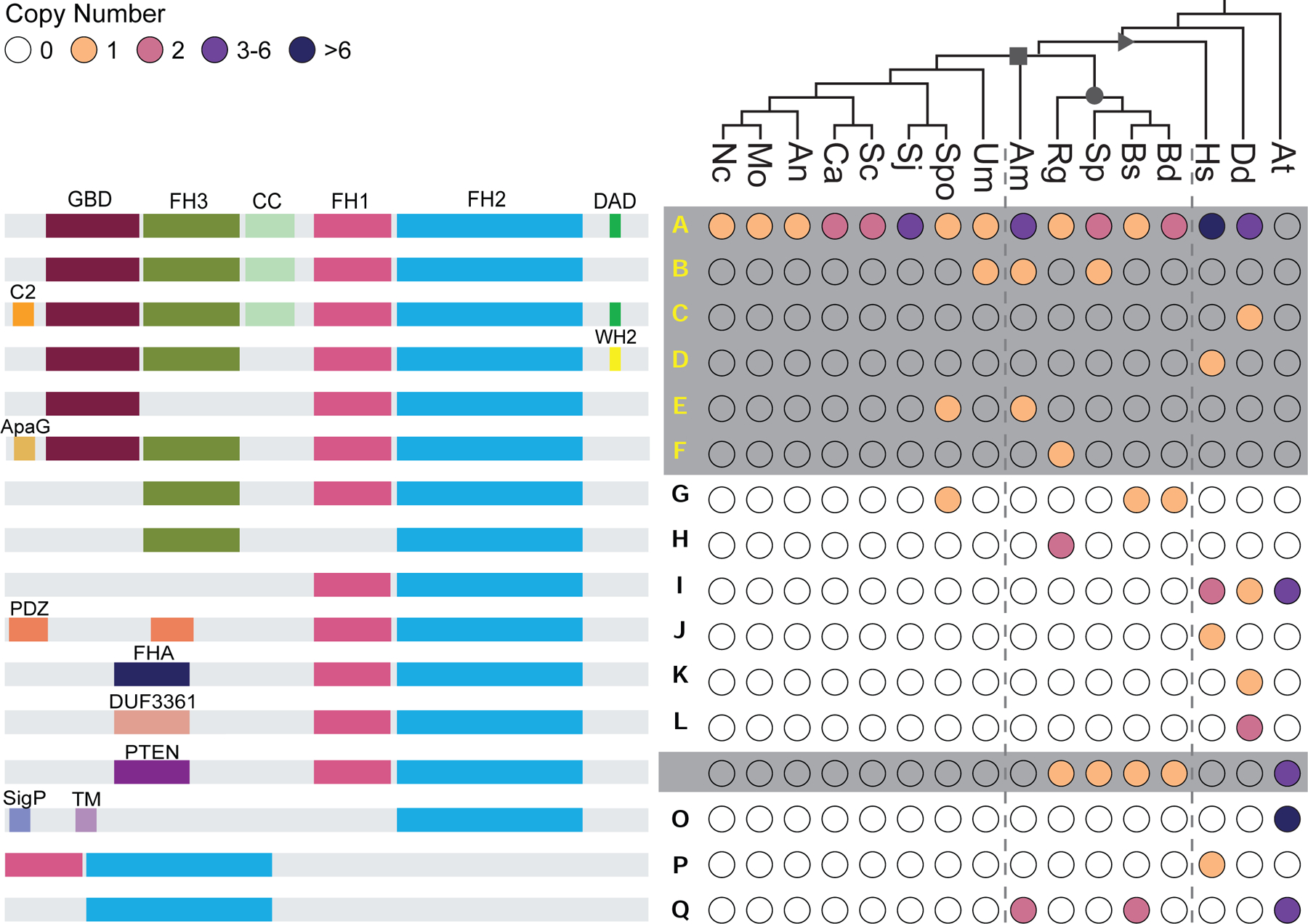
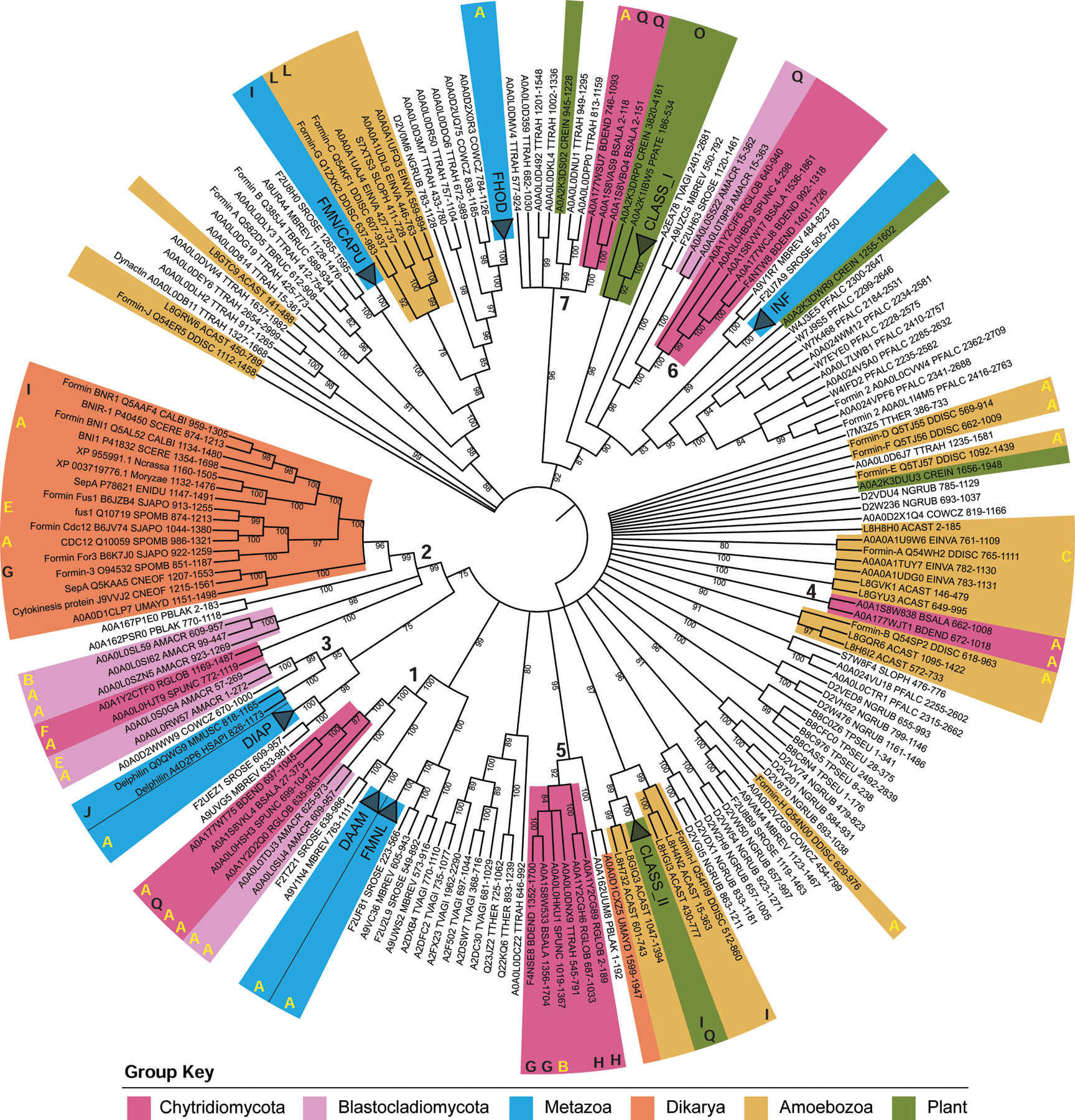
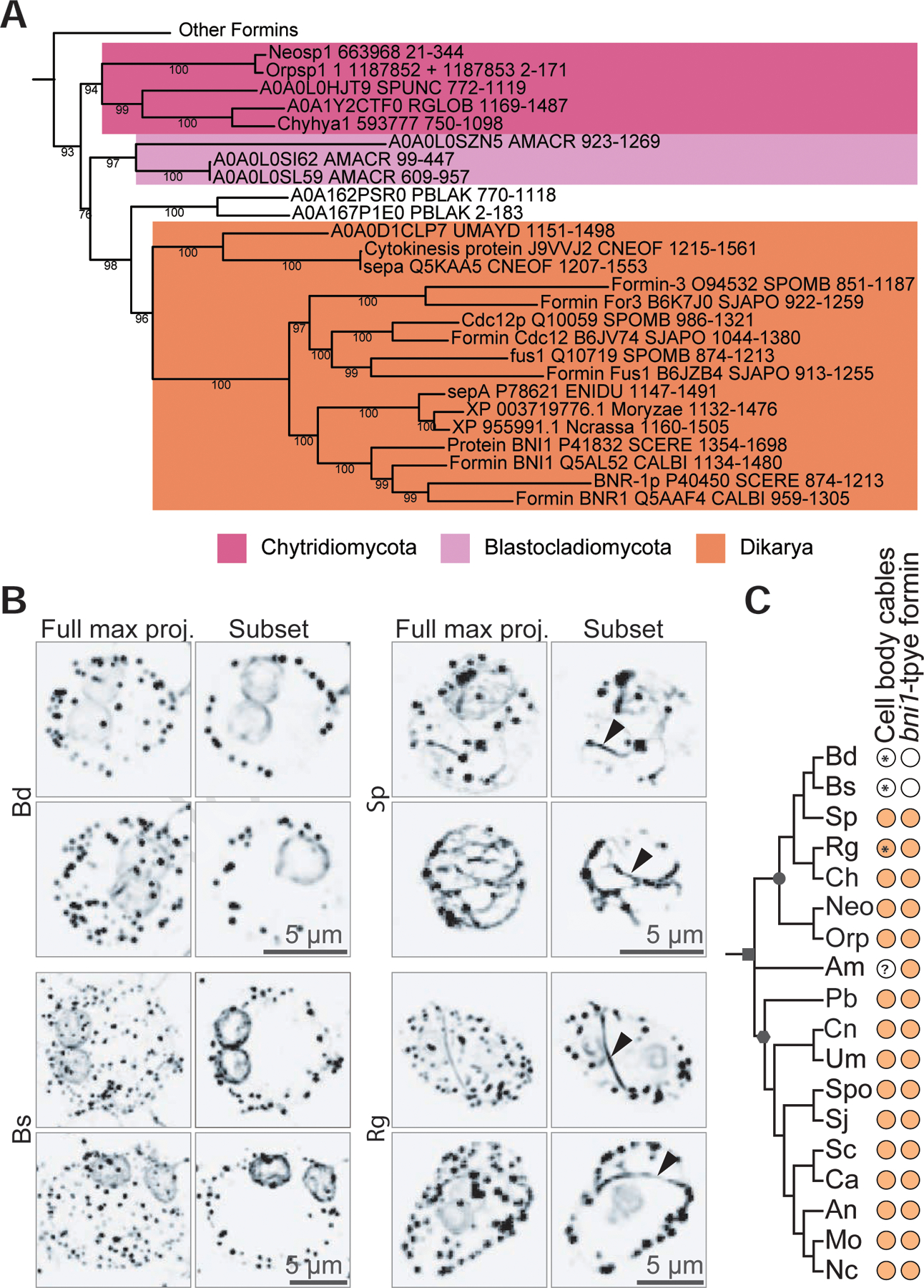
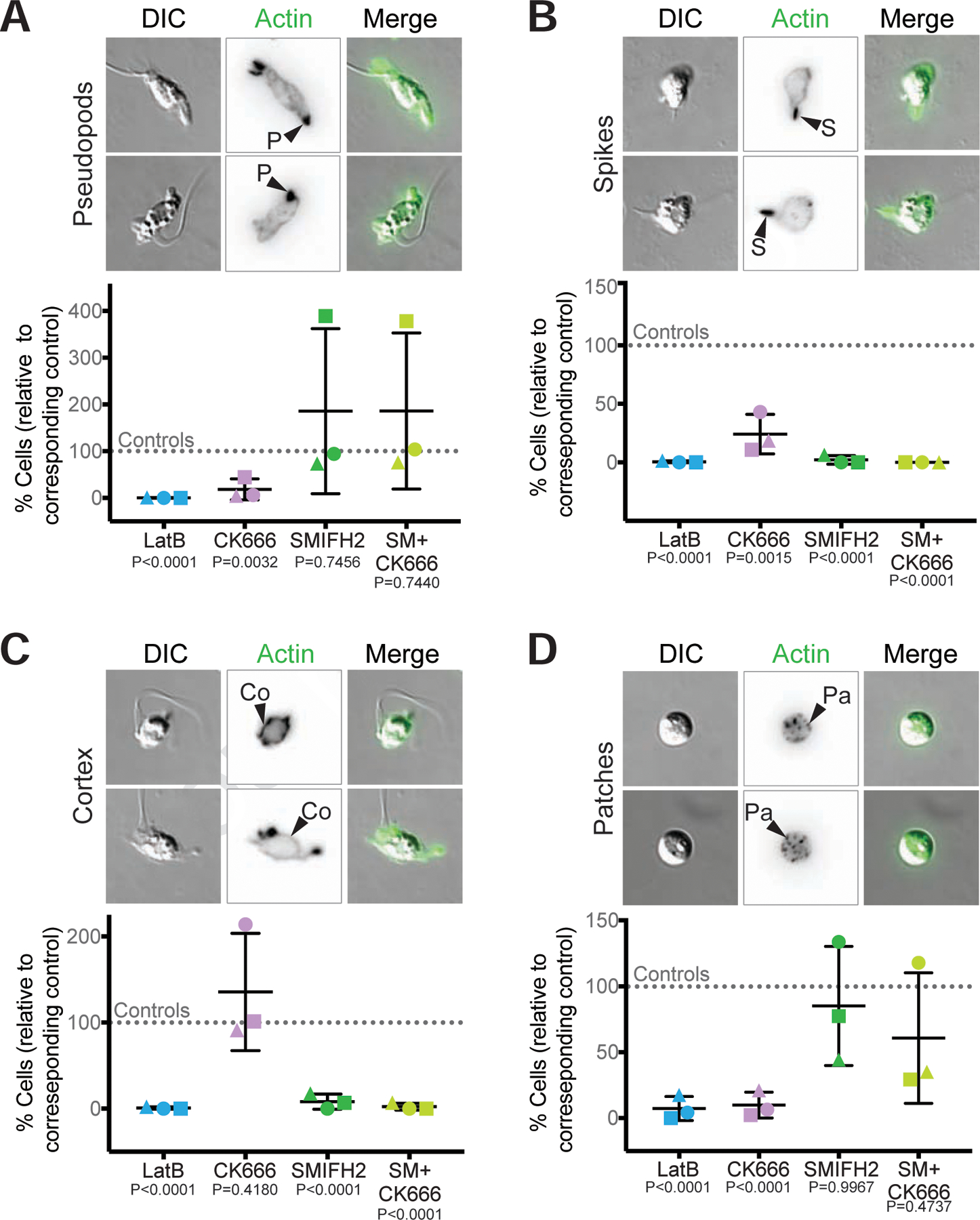
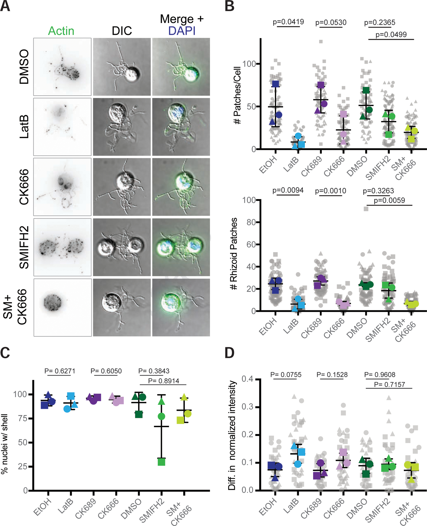
Cells from across the eukaryotic tree use actin polymer networks for a wide variety of functions, including endocytosis, cytokinesis, and cell migration. Despite this functional conservation, the actin cytoskeleton has undergone significant diversification, highlighted by the differences in the actin networks of mammalian cells and yeast. Chytrid fungi diverged before the emergence of the Dikarya (multicellular fungi and yeast) and therefore provide a unique opportunity to study actin cytoskeletal evolution. Chytrids have two life stages: zoospore cells that can swim with a flagellum and sessile sporangial cells that, like multicellular fungi, are encased in a chitinous cell wall. Here, we show that zoospores of the amphibian-killing chytrid Batrachochytrium dendrobatidis (Bd) build dynamic actin structures resembling those of animal cells, including an actin cortex, pseudopods, and filopodia-like spikes. In contrast, Bd sporangia assemble perinuclear actin shells and actin patches similar to those of yeast. The use of specific small-molecule inhibitors indicate that nearly all of Bd's actin structures are dynamic and use distinct nucleators: although pseudopods and actin patches are Arp2/3 dependent, the actin cortex appears formin dependent and actin spikes require both nucleators. Our analysis of multiple chytrid genomes reveals actin regulators and myosin motors found in animals, but not dikaryotic fungi, as well as fungal-specific components. The presence of animal- and yeast-like actin cytoskeletal components in the genome combined with the intermediate actin phenotypes in Bd suggests that the simplicity of the yeast cytoskeleton may be due to evolutionary loss.

Keywords: Batrachochytrium dendrobatidis; actin; chytrid; cytoskeleton; development; evolution; formin; fungi; motility; myosin.


Conflict of interest statement

Declaration of interests The authors declare no competing interests.

Figures

Figure 1.
Figure 1.. The chytrid fungus Batrachochytrium dendrobatidis is an early branching fungus with an archetypal chytrid life cycle and animal-like and fungal-like actin structures.
(A) This cladogram shows the relationships between representative genera of major eukaryotic groups. Chytrids are represented by Batrachochytrium, Spizellomyces, Rhizoclosmatium, and Allomyces (magenta, lavender), diverging before the diversification of Dikarya (orange), and are in a sister clade to animals (cyan). Bold type indicates genera used for the majority of the homologous sequence analyses in this paper. (B) In vitro life cycle of Batrachochytrium dendrobatidis (Bd). Bd has a motile stage known as a zoospore (i) with a flagellum made of microtubules, no cell wall, and can crawl using actin (green) based protrusions. Zoospores encyst and build a cell wall (cyan), this stage is referred to as a germling (ii). The germling grows in size, becoming a sporangium. Sporangia develop hyphal-like structures called rhizoids used for nutrient uptake and undergo synchronous rounds of mitosis (iii-iv) before cellularization and release of the next generation of zoos ores (v). This life cycle takes approximately three days in laboratory culture conditions. (C) Representative examples of zoospores (DIC: grey) and the phalloidin stained actin Z-projections of cells at this stage (inverted, black), with an overlay of the two (actin, green). Actin structures in Bd zoospores are: actin-filled pseudopods (P), actin-filled spikes (S), cortical actin (Co), and actin patches (Pa). Graphs on the right indicate the raw percent of cells with the phenotype in 3 independent experiments, shapes here match the shapes for the replicates in Figure 6. (D) Representative examples of Bd sporangia (DIC: grey) and the phalloidin stained actin structures at this stage (inverted, black; both a max intensity z projection and a single slice), with an overlay of the DIC and fluorescence (actin, green). Actin structures in sporangia are: actin patches (Pa), and perinuclear actin shells (N). See also Figure S1.
Figure 2.
Figure 2.. Chytrid actin regulatory protein networks are intermediate to those of animals and Dikarya.
The distribution of actin regulatory proteins across taxa. Color-filled circles indicate the presence of clear homologs found, with the number of homologs for each protein in each species shown in the colors specified in the key. Unfilled circles indicate that no homolog was detected in that species. Circles with multiple colors indicate complexes with different copy numbers for multiple complex members. Circles for Capping Protein represent the copy number for both α and β subunits. Dashed lines mark the chytrids. Symbols on the tree represent: opisthokonts (triangle); fungi (square); chytridiomycota (circle). Symbols in the circles represent: V, copy number of WASH varies individually, as many WASH genes are subtelomeric. O, Arabidopsis has 5 villin-like genes and an additional gelsolin-domain containing protein, none of which are phylogenetically related to metazoan gelsolin/villin family members. +, See Data S2 for details and additional potential homologs with caveates. At, Arabidopsis thaliana; Dd, Dictyostelium discoideum; Hs, Homo sapiens; Bd, Batrachochytrium dendrobatidis; Bs, Batrachochytrium salamandrivorans; Sp, Spizellomyces punctatus; Rg, Rhizoclosmatium globosum; Am, Allomyces macrogynus; Sc, Saccharomyces cerevisiae; Spo, Schizosaccharomyces pombe; Sj, Schizosaccharomyces japonicus; Ca, Candida albicans; An, Aspergillus nidulans; Mo, Magnaporthe oryzae; Nc, Neurospora crassa; Um, Ustilago maydis. See also Data S1, Data S2, Figure S2, and Figure S3.
Figure 3.
Figure 3.. Chytrid formins share similar domain architectures to those of animals, Dikarya, and plants.
The distribution of given formin domain architectures (left, not to scale), across taxa (right). Each domain architecture is assigned a letter (middle) which is mapped onto Figure 4; yellow letters (A-F) indicate diaphanous-like architectures, white letters (M,N) indicate PTEN-domain-containing architectures (both plant and non-plant formins), black letters indicate architectures which did not fall into either of these classes. Color-filled circles (right) indicate the presence of at least one formin with the given domain architecture in that species, with color indicating the number of formins according to the key. Unfilled circles indicate that no formin with the indicated domain architecture was found in the given species. Symbols on the tree represent: opisthokonts (triangle); fungi (square); chytridiomycota (circle). Symbols in the circles represent: *, for at least one formin sequence in the indicated species, the DAD domain does not perfectly fit the consensus motif, but could potentially function as an autoregulatory domain. -, for at least one formin sequence, little to no sequence is present after the FH2 domain. #, although no region of this protein met the formal definition of an FH1 domain, a proline rich region (containing 4 polyproline stretches: 8 prolines/14 amino acids; 4/5; 4/6; and 7/11; total of 22 prolines over 197 amino acids) is found N-terminal to the FH2 domain in this protein (Genbank: ORY46833.1). ^, Dictyostelium formin ForC has no polyproline stretches and therefore no FH1 domain. At, Arabidopsis thaliana; Dd, Dictyostelium discoideum; Hs, Homo sapiens; Bd, Batrachochytrium dendrobatidis; Bs, Batrachochytrium salamandrivorans; Sp, Spizellomyces punctatus; Rg, Rhizoclosmatium globosum; Am, Allomyces macrogynus; Sc, Saccharomyces cerevisiae; Spo, Schizosaccharomyces pombe; Sj, Schizosaccharomyces japonicus; Ca, Candida albicans; An, Aspergillus nidulans; Mo, Magnaporthe oryzae; Nc, Neurospora crassa; Um, Ustilago maydis. See also Data S3.
Figure 4.
Figure 4.. Chytrids have animal-like formins related to DAAM a well as other diaphanous-related formins.
A maximum likelihood consensus tree was inferred using the FH2 domains of 266 formin proteins, rooted at the midpoint, with bootstrap values as shown, and nodes with <75% bootstrap support collapsed to polytomies. Metazoan clades and Arabidopsis thaliana clades were collapsed and named according to their formin group, except for Delphilin. Taxa of interest are colored according to the key, bold numbers indicate chytrid-containing clades. The bold letters around the outside of the tree correspond to the domain architectures in Figure 3; yellow letters indicate diaphanous-like architectures, white letters indicate PTEN-domain-containing architectures, black letters indicate architectures which did not fall into either of these classes. Protein names, Uniprot accession numbers, Full species name, Uniprot 5-letter species codes, position of the FH2 domain, and additional details can be found in Data S6. See also Data S4, Data S5, and Data S6.
Figure 5.
Figure 5.. Bni1-type formins are associated with actin cables in the cell body.
(A) The evolutionary history of 291 FH2 domain sequences from formin homologs was inferred by the maximum likelihood method for 349 amino acid positions in ≥80% of the sequences. This tree is the same as the tree in Figure 4, but includes the FH2 domains from the formins of three additional chytrid species (or their relatives) that have been observed to assemble actin cables in the cell body [Neocallimastix patriciarum, Orpinomyces joyonii, and Chytriomyces hyalinus]. N. particiarum and O. joyonii did not have available genomes, so we used the genomes of species in the same genus [Neocallimastix californiae G165; Orpinomyces sp. strain C1A v1.066, assuming that the presence of cables is consistent across a genus. Consensus tree shown, pruned to highlight clade 2 (the main fungal clade) from Figure 4. Nodes with <75% bootstrap support were collapsed to polytomies. All bootstrap values are shown. (B) Representative examples of Batrachochytrium dendrobatidis (Bd), Batrachochytrium salamandrivorans (Bs), Spizellomyces punctatus (Sp), and Rhizoclosmatium globosum (Rg) sporangia stained for polymerized actin (inverted, black). Images are shown as a full maximum intensity projection as well as a subset of the z-stacks to highlight the middle sections of the cell body. Rg and Sp sporangia have actin patches, perinuclear actin shells, and actin cables within the cell body (arrowhead). Bd and Bs sporangia have actin patches and perinuclear actin shells, but no actin cables in the cell body. Scale bars, 5 µm. (C) Distribution of actin cables and bni1-type formins across fungi. Color-filled dots indicate the presence of the given component in the given species. Symbols on the tree represent: fungi (square); Dikarya (hexagon); chytridiomycota (circle). ? indicates the presence of cables in this species is unknown in the literature. * indicates a finding from this paper. Neo/Neosp1, Neocallimastix spp.; Orp/Orpsp1, Orpinomyces spp.; Bd/BDEND, Batrachochytrium dendrobatidis; Bs/BSALA, Batrachochytrium salamandrivorans; Sp/SPUNC, Spizellomyces punctatus; Rg/RGLOB, Rhizoclosmatium globosum; Ch/Chyhya1, Chytriomyces hyalinus; Am/AMACR, Allomyces macrogynus; Pb/PBLAK, Phycomyces blakesleeanus; Um/UMAYD, Ustilago maydis; Cn/CNEOF, Cryptococcus neoformans; An/ENIDU, Aspergillus nidulans; Sc/SCERE, Saccharomyces cerevisiae; Spo/SPOMB, Schizosaccharomyces pombe; Sj/SJAPO, Schizosaccharomyces japonicus; Ca/CALBI, Candida albicans; Mo/Moryzae, Magnaporthe oryzae; Nc/Ncrassa, Neurospora crassa.
Figure 6.
Figure 6.. Actin networks in Bd zoospores are dynamic and use distinct nucleators.
Synchronized populations of Bd zoospores were treated with LatrunculinB (LatB) to identify dynamic actin structures, or with Arp2/3 and/or formin inhibitors for 30 minutes. Cells were then fixed and stained for polymerized actin with fluorescent phalloidin, imaged, and quantified for presence of actin-filled pseudopods (P, part A), actin spikes (S, part B), cortical actin (Co, part C), and actin patches (Pa, part D). Each panel shows examples of cells (DIC: grey) and phalloidin-stained actin structures (alone inverted, black; overlay, green) with relative percent of cells with each structure quantified below. Percent of cells with the indicated actin phenotype for each drug treatment is normalized to its respective control: 1 µM LatrunculinB (LatB) normalized to the ethanol carrier control (EtOH); 100 µM CK666 normalized to its inactive analog CK689; 25 µM SMIFH2 normalized to a DMSO carrier control; and the combination treatment of 25 µM SMIFH2 + 100 µM CK666 (SM+CK666) was also normalized to the DMSO control. For raw data, see Data S7. Three independent experiments were performed, each represented by a different shape, the means and standard deviations shown in black. P-values for each treatment, relative to its respective control, are shown (unpaired Student’s T-tests for EtOH vs. LatB and CK689 vs. CK666; one-way ANOVA with Tukey’s multiple comparisons test for DMSO vs. SMIFH2 and the double treatment). Fluorescent images for (A), (B), and (D) are maximum intensity projections, fluorescent images for (C) are single z-slices to highlight the cortex. Brightness and contrast are not the same across images. Scale bars, 5 µm. See also Figure S4, Figure S5, and Data S7.
Figure 7.
Figure 7.. Actin patches in Bd sporangia are dynamic and use the Arp2/3 complex.
Populations of Bd sporangia seeded 1 day prior were treated with drugs using the same concentrations as in Figure 6, then fixed and stained for polymerized actin with phalloidin and for DNA with DAPI. (A) Examples of sporangia (DIC: grey) and phalloidin stained actin patches (inverted, black; green in overlay), with an overlay including the nucleus (blue) after treatment with each drug. Though DMSO has an effect on patches (see Figure S4), all controls looked phenotypically similar (see Figure S7), so only a DMSO treated cell is shown. (B) quantification of the number of actin patches per sporangia (top) and the number of patches in the rhizoids alone (bottom). Larger, colored shapes indicate the average number of patches per cell in each treatment from three independent experiments, each represented by a different shape. Each gray shape represents the number of patches in a single cell, or in a cell’s rhizoids in that experiment. Means and standard deviations of these averages are shown in black. (C) Percent of nuclei encased within an actin shell per treatment for three independent experiments. (D) Difference in the normalized intensity of actin shells on each side of the nucleus. The intensity along lines drawn through each nucleus were normalized to the center of each line. The average normalized intensity for each side was calculated, with the difference between the brightest half and the other half plotted here. Each gray shape represents the difference in normalized actin intensity for a single nucleus. Statistical tests were performed using the averages of the three experiments (i.e., the three colored shapes). P-values for each treatment, relative to its respective control, are shown (unpaired Student’s T-tests for EtOH vs. LatB and CK689 vs. CK666; one-way ANOVA with Tukey’s multiple comparisons test for DMSO vs. SMIFH2 and the double treatment). Brightness and contrast are not the same across images. Scale bar, 5 µm. See also Figure S6 and Figure S7.

References

    1. Blanchoin L, Boujemaa-Paterski R, Sykes C, and Plastino J (2014). Actin dynamics, architecture, and mechanics in cell motility. Physiol. Rev 94, 235–263. - PubMed
    1. Medina EM, Robinson KA, Bellingham-Johnstun K, Ianiri G, Laplante C, Fritz-Laylin LK, and Buchler NE (2020). Genetic transformation of Spizellomyces punctatus, a resource for studying chytrid biology and evolutionary cell biology. elife 9. - PMC - PubMed
    1. Velle KB, and Fritz-Laylin LK (2019). Diversity and evolution of actin-dependent phenotypes. Curr. Opin. Genet. Dev 58–59, 40–48. - PubMed
    1. Breitsprecher D, and Goode BL (2013). Formins at a glance. J. Cell Sci 126, 1–7. - PMC - PubMed
    1. Gallop JL (2020). Filopodia and their links with membrane traffic and cell adhesion. Semin. Cell Dev. Biol 102, 81–89. - PubMed

Publication types