. 2021 Mar 17;109(6):1055-1066.e4.
doi: 10.1016/j.neuron.2021.01.016. Epub 2021 Feb 8.

Interhemispheric transfer of working memories

Affiliations

Interhemispheric transfer of working memories

Scott L Brincat et al. Neuron. .

Abstract

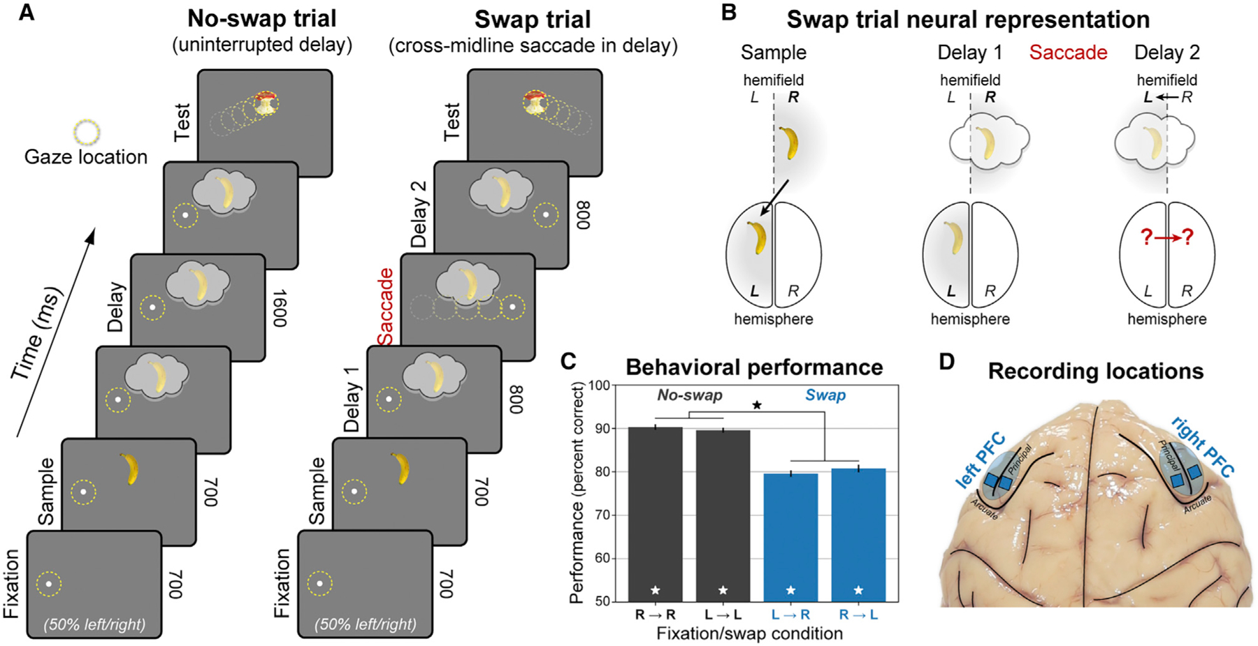
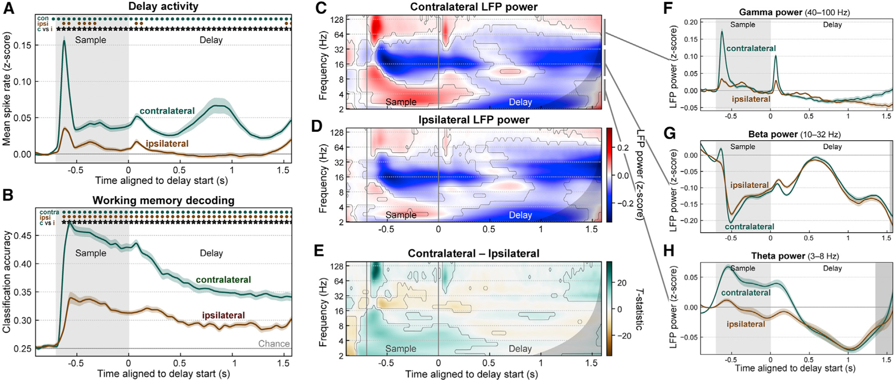
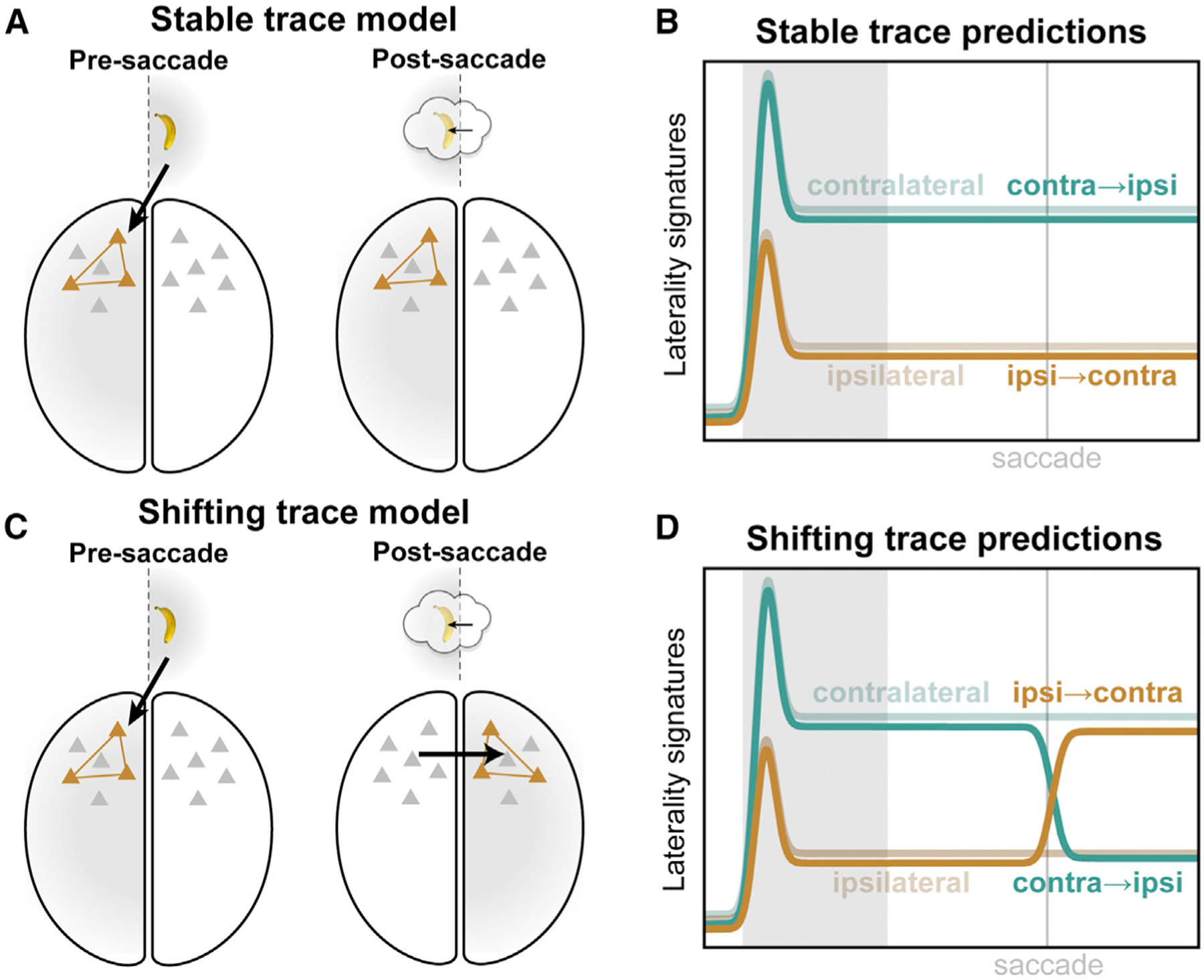
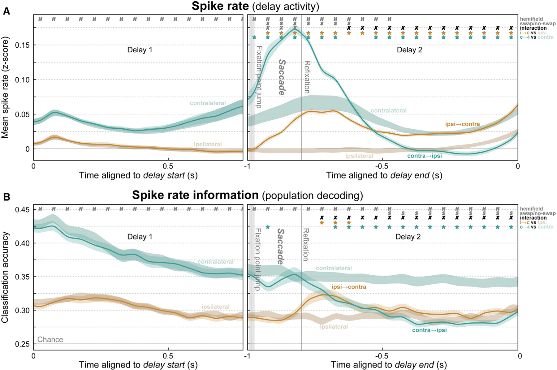
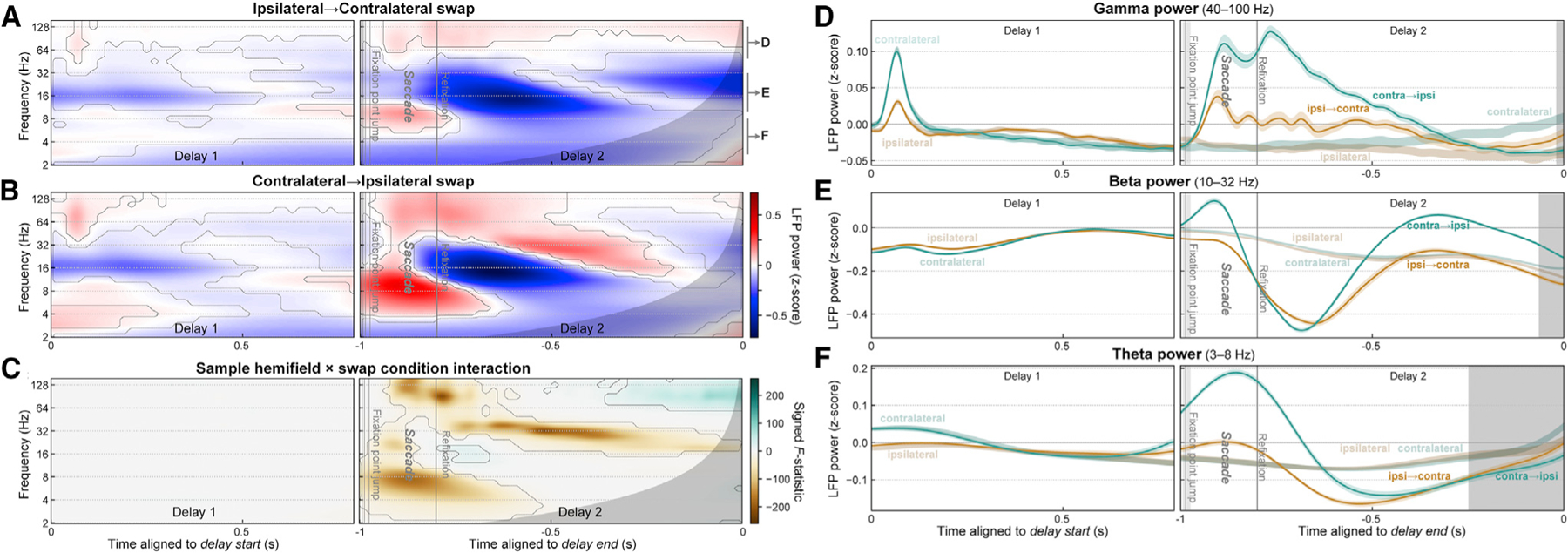
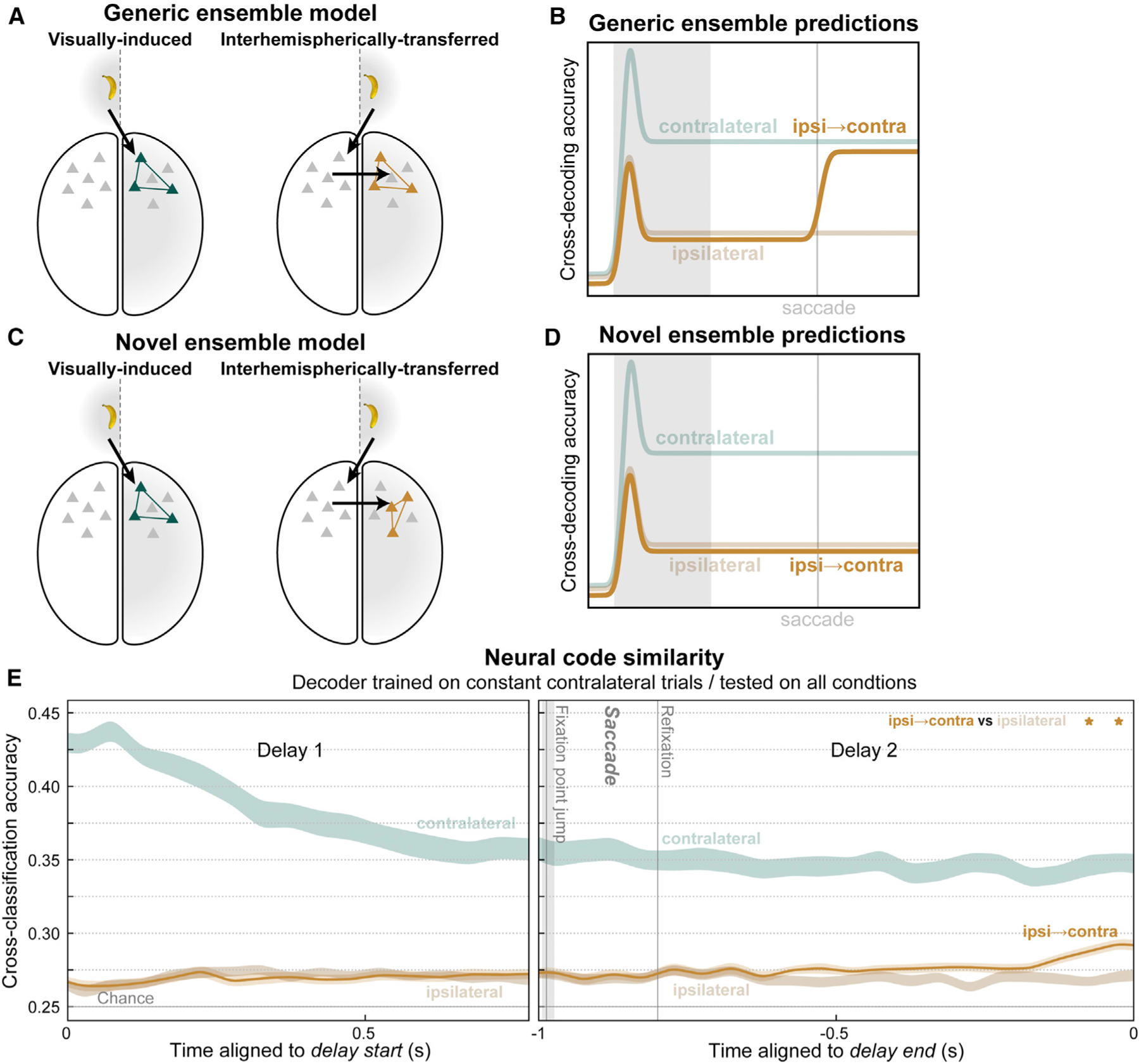
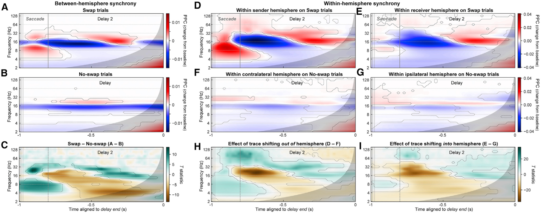
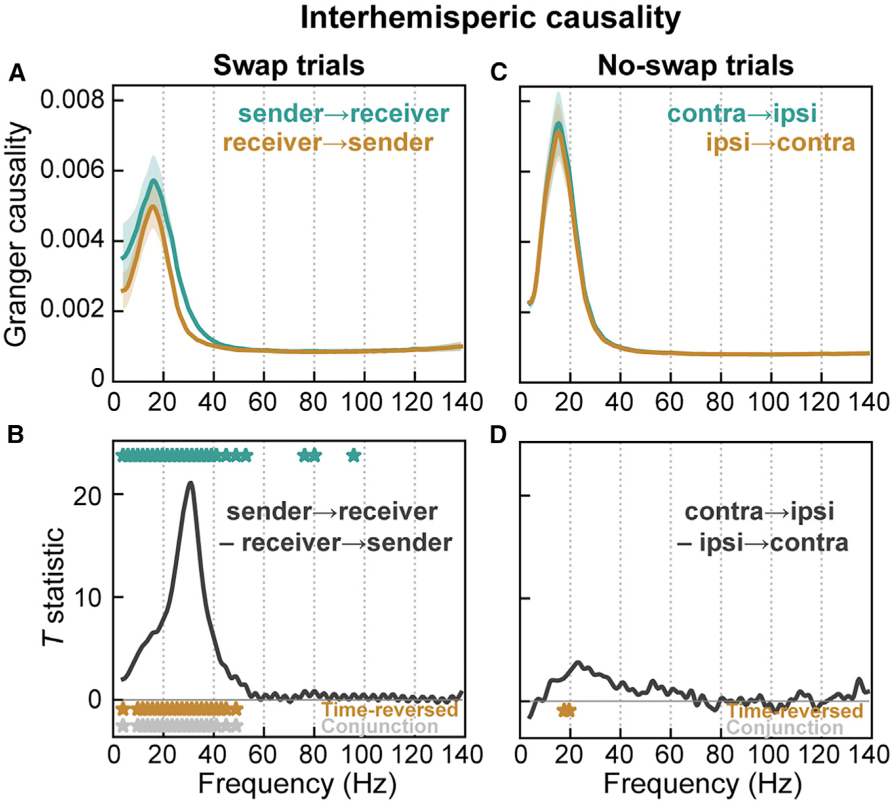
Visual working memory (WM) storage is largely independent between the left and right visual hemifields/cerebral hemispheres, yet somehow WM feels seamless. We studied how WM is integrated across hemifields by recording neural activity bilaterally from lateral prefrontal cortex. An instructed saccade during the WM delay shifted the remembered location from one hemifield to the other. Before the shift, spike rates and oscillatory power showed clear signatures of memory laterality. After the shift, the lateralization inverted, consistent with transfer of the memory trace from one hemisphere to the other. Transferred traces initially used different neural ensembles from feedforward-induced ones, but they converged at the end of the delay. Around the time of transfer, synchrony between the two prefrontal hemispheres peaked in theta and beta frequencies, with a directionality consistent with memory trace transfer. This illustrates how dynamics between the two cortical hemispheres can stitch together WM traces across visual hemifields.

Keywords: cognition; interhemispheric; neural synchrony; prefrontal cortex; working memory.


Conflict of interest statement

Declaration of interests The authors declare no competing interests.

Figures

Figure 1.
Figure 1.. Behavioral and electrophysiological methods
(A) Hemifield-swap working memory (WM) task. Subjects fixated to the left or right while a sample object was presented in the center, placing it in the right or left visual hemifield, respectively. Samples could be one of two objects, presented in one of two locations (above or below center). Both the object and its location needed to be remembered over a blank 1.6 s delay. After the delay, a series of two test objects was displayed, and subjects responded to the one that did not match the sample in object identity or upper/lower location (response to first object shown for brevity). In ‘‘no-swap’’ trials (left), the WM delay was uninterrupted. In ‘‘swap’’ trials (right), subjects were instructed to saccade to the opposite side mid-delay, switching the visual hemifield of the remembered location relative to gaze. (B) Neural representation of WM in swap trials. The WM trace is initially encoded in the prefrontal hemisphere contralateral to the sample. We tested whether the change in gaze on swap trials caused the WM traces to transfer to the other hemisphere. (C) Mean performance (± SEM across 56 sessions) for each swap condition and visual hemifield. Monkeys performed the task well (white stars, significant versus chance), with a small but significant decrease (black star) on swap trials. (D) Electrophysiological signals were recorded bilaterally from 256 electrodes in lateral prefrontal cortex (PFC).
Figure 2.
Figure 2.. Contralateral bias in prefrontal cortex
(A) Population mean spike rates (multi-unit activity Z-scored to baseline, ± SEM across 56 sessions) for sample objects contralateral (green) and ipsilateral (brown) to the recorded prefrontal hemisphere (pooled across left and right). Activity for contralateral samples was greater than baseline (green dots) and greater than activity for ipsilateral samples (stars). See also Figure S1. (B) Mean (± SEM) accuracy for decoding the item held in WM (object identity and upper/lower location) from prefrontal population spike rates, for samples in the contralateral (green) and ipsilateral (brown) visual hemifield. Contralateral decoding accuracy was greater than ipsilateral (stars). (C and D) Mean time-frequency LFP power (Z-scored to baseline) for contralateral (C) and ipsilateral (D) samples. Contours indicate significant change from baseline. Gray regions to right of (C)–(H) indicate time points with possible temporal smearing of test-period effects. Gamma (~40–100 Hz) and theta (~3–8 Hz) power increased from baseline (red), while beta (~10–32 Hz) power was suppressed from baseline (blue). (E) Contrast (paired-observation t-statistic map) between contralateral and ipsilateral power. Contours indicate significant difference. (F–H) Summary of LFP power for contralateral (green) and ipsilateral (brown) sample objects, pooled within frequency bands: gamma (F), beta (G), and theta (H). Because of off-axis structure in the time-frequency data, these one-dimensional (1D) plots cannot fully capture it and are thus included only to aid visualization. All modulations from baseline were stronger for contralateral samples, but only gamma showed a difference during the delay period. See also Figure S2.
Figure 3.
Figure 3.. Competing models of swap effects
(A) The stable trace model posits that once a working memory is encoded in a given cortical hemisphere (left), it will remain there (right), despite the remembered location shifting from one hemifield to the other (inset). (B) This model predicts that neural signatures of memory trace laterality will be unaltered by the middelay saccade in our task. (C) The shifting trace model assumes that when the hemifield of the remembered location is swapped, the memory trace will be transferred from one cortical hemisphere to the other. (D) This model predicts a post-saccadic inversion of the neural signatures of laterality: shifting the remembered location into the contralateral hemifield (orange) should come to approximate the constant contralateral location (desaturated green), while shifting it ipsilateral (green) should come to look like constant ipsilateral trials (desaturated brown).
Figure 4.
Figure 4.. Evidence for interhemispheric transfer of working memory traces in prefrontal spiking activity
(A) Mean (± SEM) multi-unit spike rates for all trials where the remembered location was constant in the contralateral (desaturated green) or ipsilateral (desaturated brown) hemifield or where it swapped from ipsilateral to contralateral (orange) or from contralateral to ipsilateral (green). Before the mid-delay saccade, there was only a significant effect of the sample hemifield (H symbols). Around the saccade, activity was greater overall for swap than no-swap trials (S symbols). Later, the swap trials inverted and approximated activity in the corresponding no-swap trials (X symbols, significant hemifield × swap condition interaction). Stars indicate significant difference of swap conditions from their respective no-swap baseline. (B) Mean (± SEM) accuracy for decoding the item held in WM from spike rates. As predicted, post-saccade accuracy decreased in contralateral-to-ipsilateral trials. Decoding on ipsilateral-to-contralateral trials also significantly improved above baseline, but only transiently, and it never reached the level of constant contralateral trials.
Figure 5.
Figure 5.. Evidence for interhemispheric transfer of working memory traces in prefrontal LFP power
(A and B) Mean time-frequency LFP power for trials where the remembered location shifted from the ipsilateral to contralateral (A) or from the contralateral to ipsilateral (B) hemifield. Contours indicate significant difference from pre-sample baseline. Gray regions at right of all panels indicate time points with possible temporal smearing of test-period effects. (C) Swap inversion effect. F-statistic map for sample hemifield × swap condition interaction, signed to indicate if power was greater when remembered location ends up in contralateral (green) or ipsilateral (brown) hemifield. Contours indicate significant interaction. (D–F) Summary of LFP power for ipsilateral-to-contralateral (green) and contralateral-to-ipsilateral (orange) trials, pooled within frequency bands labeled in (A): gamma (D), beta (E), and theta (F). Around the time of the saccade, LFP power in all bands showed strong effects of the swap condition (saccade versus no saccade). Later in the post-saccade delay, signatures in all bands inverted, as predicted by the shifting trace model. See also Figure S2.
Figure 6.
Figure 6.. Transferred working memory traces used novel ensembles but converged toward visually induced ensembles at delay end
(A) The generic ensemble model assumes that a given memory trace will activate the same neural ensemble (colored neurons) whether it arrives in prefrontal cortex via feedforward inputs from visual cortex (left) or via interhemispheric inputs from the opposite cortical hemisphere (right). (B) It predicts that a classifier trained to decode working memory traces on constant contralateral trials will also be able to decode contralateral-shifting swap trials (orange). (C) The novel ensemble model posits that interhemispheric inputs activate a distinct ensemble (right) from visual inputs (left), even for the same memory trace. (D) It predicts failure of contralateral-trained decoders to generalize to contralateral-shifting trials. (E) For most of the post-saccadic delay, cross-decoding accuracy for ipsilateral-to-contralateral swap trials (orange) did not significantly differ from constant ipsilateral trials, as predicted by the novel ensemble model. Near the end of the delay, a significant difference emerged (stars), indicating contralateral-shifting trials became more similar to constant contralateral trials, as predicted by the generic ensemble model. See also Figure S3.
Figure 7.
Figure 7.. Interhemispheric beta/theta synchrony may mediate working memory trace transfer
(A and B) Mean phase synchrony (pairwise phase consistency [PPC]) between all pairs of LFPs in the two prefrontal hemispheres, for swap (A) and no-swap (B) trials, expressed as the change in PPC from the pre-sample fixation-period baseline. Contours indicate significant differences from baseline. Gray regions indicate time points with possible influence of test-period effects. (C) Contrast (paired t-statistic map) between swap and no-swap conditions. Contours indicate significant between-condition difference. During the time period of putative interhemispheric memory trace transfer (–1 to –0.8 s), there was a significant increase (green) in interhemispheric synchrony in the theta (~4 to 10 Hz) and beta (~18 to 40 Hz) bands and a decrease (brown) in the alpha/low-beta band (~11 to 17 Hz). (D and E) PPC between LFP pairs within the sender (D) and receiver (E) hemisphere on swap trials. (F and G) PPC between LFP pairs within the contralateral (F) and ipsilateral (G) hemispheres on no-swap trials. (H and I) t-Statistic maps for contrasts between swap and no-swap results for each hemisphere. These contrasts represent the effect of a WM trace shifting out of and into (I) a hemisphere.
Figure 8.
Figure 8.. Granger causality indicates flow between prefrontal hemispheres in same direction as putative memory trace transfer
(A and B) Spectral Granger causality in swap trials during time period of putative memory trace transfer from prefrontal hemisphere contralateral to initial sample location (‘‘sender’’) to hemisphere contralateral to post-saccade location (‘‘receiver’’). (A) Mean (± SEM) causality in the sender-to-receiver direction (green) and in the receiver-to-sender direction (orange). (B) t-Statistic for contrast between causal directions. Causality was significantly greater in the sender-to-receiver direction across all frequencies ~10–40 Hz (green stars). This directional asymmetry reversed, as expected, for time-reversed data (orange stars; full results in Figure S4). Gray stars indicate frequencies significant for both forward and time-reversed data. (C and D) No asymmetry in interhemispheric causality was observed during analogous time points (relative to delay end) in no-swap trials between contralateral and ipsilateral hemispheres.

Comment in

  • Hemispheres in harmony.
    Fries P. Fries P. Neuron. 2021 Mar 17;109(6):916-917. doi: 10.1016/j.neuron.2021.02.026. Neuron. 2021. PMID: 33735614

References

    1. Alvarez GA, and Cavanagh P (2005). Independent resources for attentional tracking in the left and right visual hemifields. Psychol. Sci 16, 637–643. - PubMed
    1. Alvarez GA, Gill J, and Cavanagh P (2012). Anatomical constraints on attention: hemifield independence is a signature of multifocal spatial selection. J. Vis 12, 9. - PMC - PubMed
    1. Barbas H, and Pandya DN (1984). Topography of commissural fibers of the prefrontal cortex in the rhesus monkey. Exp. Brain Res 55, 187–191. - PubMed
    1. Bastos AM, and Schoffelen J-M (2016). A tutorial review of functional connectivity analysis methods and their interpretational pitfalls. Front. Syst. Neurosci 9, 175. - PMC - PubMed
    1. Bastos AM, Vezoli J, Bosman CA, Schoffelen J-M, Oostenveld R, Dowdall JR, De Weerd P, Kennedy H, and Fries P (2015). Visual areas exert feedforward and feedback influences through distinct frequency channels. Neuron 85, 390–401. - PubMed

Publication types

LinkOut - more resources