. 2021 Apr;29(4):547-557.
doi: 10.1016/j.joca.2021.01.005. Epub 2021 Feb 6.

ADAMTS5 is required for normal trabeculated bone development in the mandibular condyle

Affiliations

ADAMTS5 is required for normal trabeculated bone development in the mandibular condyle

A W Rogers-DeCotes et al. Osteoarthritis Cartilage. 2021 Apr.

Abstract

Objective: Determine the role of the extracellular matrix protease ADAMTS5 in development of the trabeculated bone of the mandibular condyle.

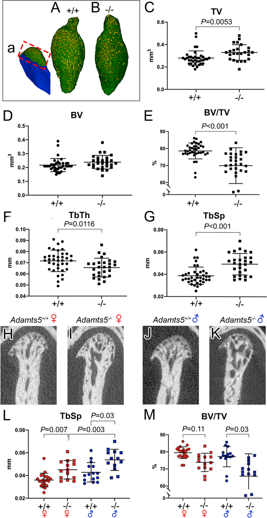
Methods: The mandibular condyles of wild type and mice deficient in the protease ADAMTS5 were examined for histopathology with Safranin O staining. Microcomputed tomography was performed to analyze the developing bone of the mandibular condyle. RNAscope and immunohistochemistry were utilized to investigate cell type and extracellular matrix expression.

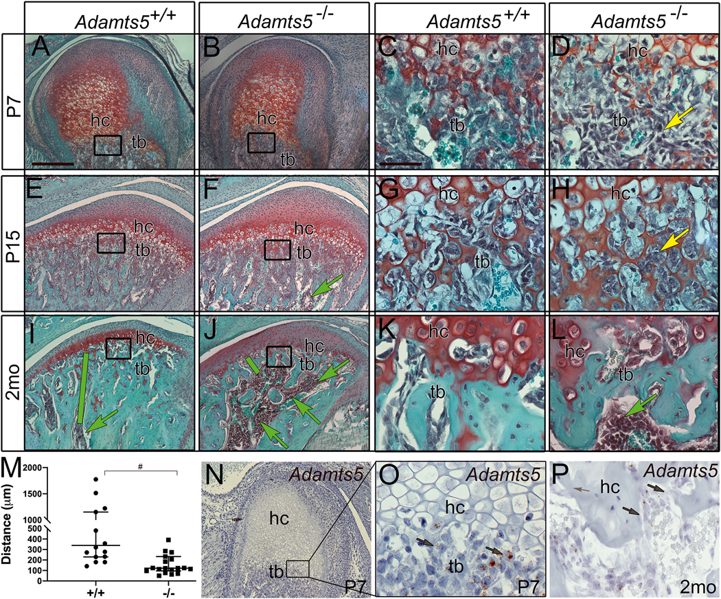
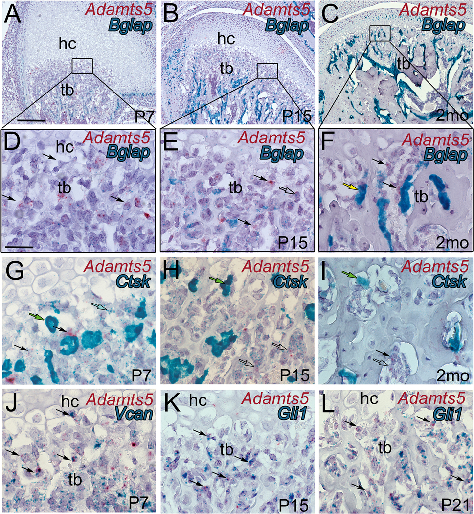
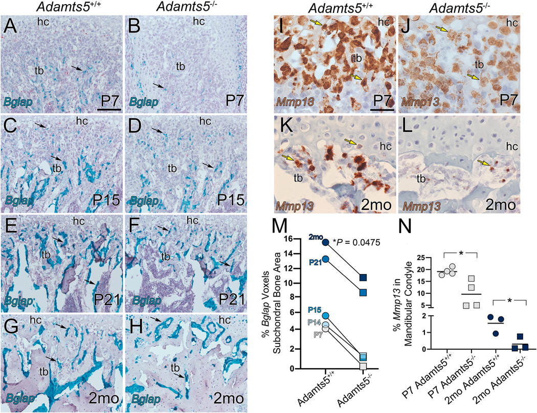
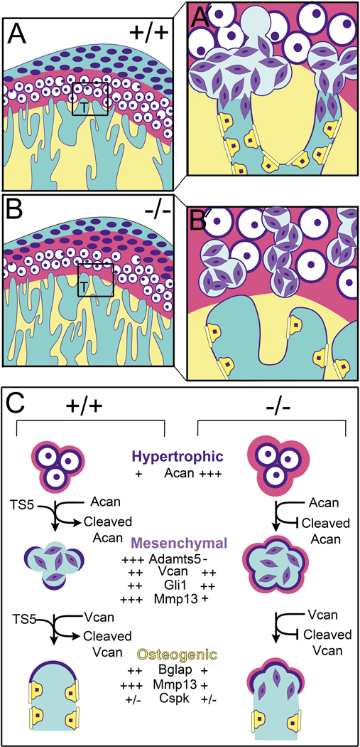
Results: Mice deficient in Adamts5, (Adamts5tm1Dgen/J) exhibit an increase in trabecular separation (n = 37 wild type; n = 27: P < 0.0001) and reduction of trabecular thickness P = 0.0116 and bone volume fraction P = 0.0869 in the mandibular condylar head compared to wild type littermates. The altered bone parameters were more pronounced in male Adamts5-/- mice compared to female Adamts5-/- mice (TbSp; P = 0.03). Adamts5 was co-expressed with versican and Gli1 in mesenchymal, stem-like cells in the transition zone where the trabeculated bone is adjacent to mature hypertrophic chondrocytes. Loss of Adamts5 caused a reduction of Bglap expressing osteoblasts throughout mandibular condylar development and in young adult mice. The protease Mmp13, that is involved in mineralization and is expressed by hypertrophic chondrocytes and osteoblasts, was reduced in the mandibular condyle of Adamts5 deficient mice.

Conclusion: This is the first report of a novel and critical role for Adamts5 in bone formation within the mandibular condyle of the temporomandibular joint. These data indicate Adamts5 may be required in the transdifferentiation of hypertrophic chondrocytes to osteoblasts during trabecular bone formation in development of the mandibular condyle.

Keywords: ADAMTS; Bone; Extracellular matrix; Mandibular condyle; Temporomandibular joint (TMJ).


Figures

Figure 1:
Figure 1:. The interface of hypertrophic chondrocytes and trabeculated bone in the mandibular condyle is altered during development in Adamts5−/− mice and exhibits an increase in bone marrow.
Safranin O stained sections of the developing condyle at postnatal day 7 (P7) (A-D), P15 (E-H), and adult 2 month (2mo) (I-L), in Adamts5+/+ (A, C, E, G, I, K, N-O) and Adamts5−/− (B, D, F, H, J, L). Cartilage bone interface region of hypertrophic chondrocytes (hc) and trabeculated bone (tb) (A, B, E, F, I, J black boxes magnified in C, D, G, H, K, L, respectively. Yellow arrow (D) indicates changes in mesenchymal cells in the transition zone of Adamts5−/− mandibular condyles (D, H). Green arrows (F, I, J, L) -bone marrow infiltration in the condylar head. Green bars (E, F) denote distance of bone marrow from the hc, graphed in I. Graph in M indicates the distance of the bone marrow from the transition zone containing the hypertrophic chondrocytes. Each point on the graph indicates data from one mouse. #- P<0.0001, Mann-Whitney U, 142; 95% CI −661.1 to −245.6, R= 0.2819. Expression of Adamts5 using RNAscope™ (N-P, brown, arrows) at postnatal P7 (N, box in N magnified and shown in O). Adamts5 expression in cells of the trabeculated bone in an adult 2 month (2mo) (P, brown arrows) mandibular condyle. Bar in A = 200μm applies to B, E, F, I, J, N; bar in C = 50 μm applies to D, G, H, K, L, O, P.
Figure 2:
Figure 2:. Young adult female and male Adamts5−/− mice exhibit changes in the subchondral bone of the mandibular condyle.
Three dimensional reconstructions of microtomography (μCT) of mandibular condyles in wild type (a, A) and Adamts5/− mice (B). Red box in ‘a’ refers to the region of μCT analysis in C-M. Each point on the graph represents measurements from one condyle. TV-trabecular volume; BV-bone volume; BV/TV-bone volume/trabecular volume = bone density; Tb. Th-trabecular thickness; Tb. Sp. –trabecular spacing. H-K are two dimensional μCT slices of female Adamts5+/+ (H), male Adamts5+/+ (I), female Adamts5−/− (J) and male Adamts5−/− (K). μCT data was analyzed by sorting the sexes of mice (L, M), there was a significant difference between male and female Adamts5−/− mice in the trabecular spacing of the mandibular condyle, and the bone density (BV/TV) was more significantly reduced in male mice compared to female mice. The longer bars on the graphs represent the average of the mean, with the shorter bars denoting standard deviation. Bar in C, P=0.0053; E, P<0.001; F, P=0.0116; G, P<0.001; L (left to right), P=0.007,P=0.003, P=0.03; M (left to right), P=0.11, 0.03.
Figure 3:
Figure 3:. Adamts5 expression overlaps with mesenchymal and stem cell markers in the trabeculated bone of the developing mandibular condyle.
Histological sections of the developing murine mandibular condyles Adamts5 expression depicted in red (A-L) from a dual RNAscope™ assay with bone gamma-carboxyglutamic acid-containing protein (Bglap) (A-F, blue-green), Cathepsin K (Ctsk) (G-I, blue-green), versican (Vcan) (J, blue-green) and glioma associated oncogene 1 (Gli1) (K, L, blue-green). Postnatal day 7 (P7); Postnatal day 15 (P15); postnatal day 21 (P21); hc-hypertrophic chondrocytes; tb-trabeculated bone; black arrows in A-L-Adamts5 (red) expressing cells; open arrow in E, colocalization of Adamts5 and Bglap; yellow arrow-differentiated osteoblast; green arrows-osteoclasts; blue arrow-low expressing Cstk cell co-expressed with Adamts5; open arrows in H, I, -low level expressing Cstk cells with co-expression of Adamts5; black arrows in J -co-expression of Vcan and Adamts5; black arrows in K, L -co-expression of Gli1 and Adamts5. Bar in A = 200 μm applies to B, C; bar in D = 50 μm applies to E-L.
Figure 4:
Figure 4:. There is a reduction of osteoblast marker Bglap and MMP13 expression in the transition zone of the Adamts5 deficient mandibular condyles.
Bglap (osteocalcin precursor) expression was evaluated in the developing mandibular condylar bone of wildtype and Adamts5 deficient mice at P7, P14, P15, P21, and 2mo (A-H, blue-green). At each developmental stage there was significantly more Bglap expression in the wildtype condyles than Adamts5 deficient shown quantified in the graph (M). The grey lines in M are drawn from the wildtype and Adamts5 deficient values to highlight the age-matched reduction of Bglap expression in the Adamts5 deficient condyle; 95%CI 0.07840 to 0.9773. Mmp13 expression was also evaluated at P7 and 2mo (I-L), at each stage there was a significant reduction in MMP13 expression in the Adamts5 deficient mandibular condyles compared to wildtype (N). In graphs circles depict wildtype and squares indicate Adamts5 deficient values. Each symbol on the graph represents measurements from one condyle. Black arrows in A-H-expression of Bglap; yellow arrows in I-L-expression of Mmp13. Bar in A = 100 μm applies to B-H; bar in I = 50 μm applies to J-L. Bglap 95%CI 0.07840 to 0.9773; Mmp13, P7, 95%CI −16.68 to −2.373; Mmp13, 2mo, 95%CI −2.314 to −0.1828.
Figure 5.
Figure 5.. Schematic model depicting a role for ADAMTS5 in the developing trabeculated bone of the mandibular condyle.
At the cartilage bone interface (transition zone) in the developing mandibular condyle hypertrophic cells undergo ‘transdifferentiation’ into bone-producing cells, a process that is poorly understood but that we reveal a role for ADAMTS5. In mice devoid of Adamts5 there is a reduction of trabeculated bone in the mandibular condyle and less bone-forming cells during postnatal development. Adamts5 is expressed by stem cell-like mesenchymal cell progenitors (purple cells) that are derived from hypertrophic chondrocytes at the cartilage-bone interface in the condylar head (boxes in A, B, in high magnification in A′ and B′). Mesenchymal cells in this transition zone also express Vcan, a proteoglycan substrate of ADAMTS5; VCAN cleavage in other contexts facilitates differentiation and reduces proliferation. ADAMTS5 generated by the mesenchymal cells degrades the ACAN-rich cartilage matrix and pericellular ECM of hypertrophic chondrocytes (magenta) at the transition zone. Once this cartilage ECM is degraded and the pericellular matrix shell of the hypertrophic chondrocytes is partially removed, fusion occurs with blood vessels at the interface with the trabeculated bone. Next the mesenchymal cells appear to migrate into and adhere to the vessel walls, differentiate into osteoblast-like cells (yellow cells) and deposit osteoid (yellow bars) on the endosteal surface of bone (yellow). Fully differentiated osteoblasts (Bglap expressing) do not express detectable levels of Adamts5 indicating a transient role for this protease in the stem-like mesenchymal cell stages. The reduction of Mmp13 in Adamts5 deficient condyles likely contributes to the bone phenotype since Mmp13 is a key factor in bone mineralization.

References

    1. Wang XD, Zhang JN, Gan YH, Zhou YH. Current understanding of pathogenesis and treatment of TMJ osteoarthritis. J Dent Res 2015; 94: 666–673. - PubMed
    1. Milam SB. Pathogenesis of degenerative temporomandibular joint arthritides. Odontology 2005; 93: 7–15. - PubMed
    1. Zhang J, Jiao K, Zhang M, Zhou T, Liu XD, Yu SB, et al. Occlusal effects on longitudinal bone alterations of the temporomandibular joint. J Dent Res 2013; 92: 253–259. - PMC - PubMed
    1. Mizoguchi I, Toriya N, Nakao Y. Growth of the manddible and biological characteristics of the mandibular condylar cartilage. Japanese Dental Science Review 2013; 49: 139–150.
    1. Hinton RJ, Jing Y, Jing J, Feng JQ. Roles of Chondrocytes in Endochondral Bone Formation and Fracture Repair. J Dent Res 2017; 96: 23–30. - PMC - PubMed

Publication types

MeSH terms

LinkOut - more resources