. 2021 Feb 7;13(2):234.
doi: 10.3390/pharmaceutics13020234.

Thermo-Responsive PLGA-PEG-PLGA Hydrogels as Novel Injectable Platforms for Neuroprotective Combined Therapies in the Treatment of Retinal Degenerative Diseases

Affiliations

Thermo-Responsive PLGA-PEG-PLGA Hydrogels as Novel Injectable Platforms for Neuroprotective Combined Therapies in the Treatment of Retinal Degenerative Diseases

José Javier López-Cano et al. Pharmaceutics. .

Abstract

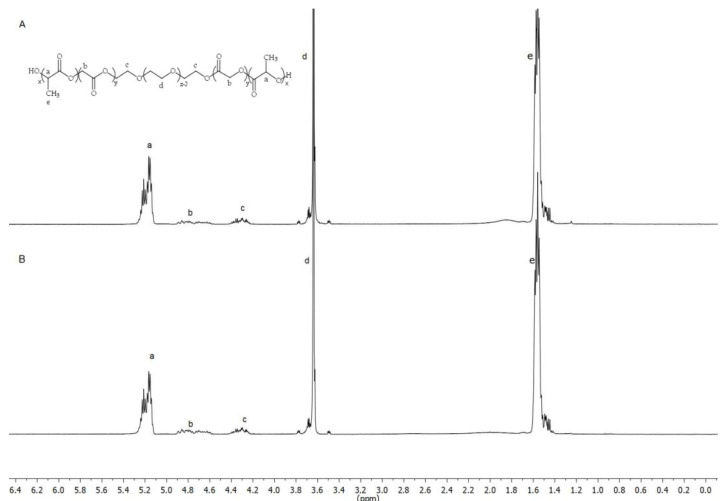
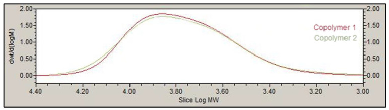
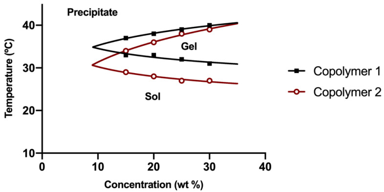
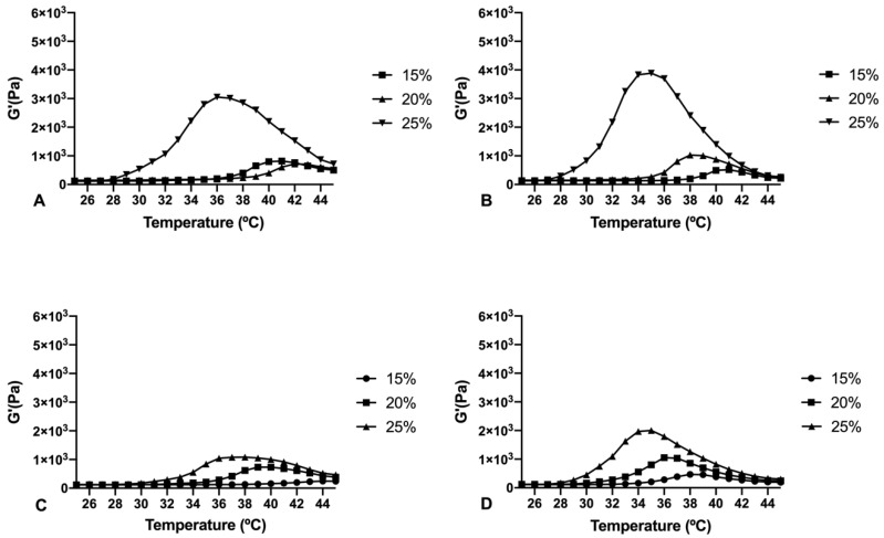
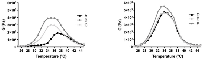
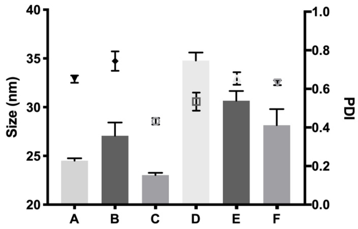
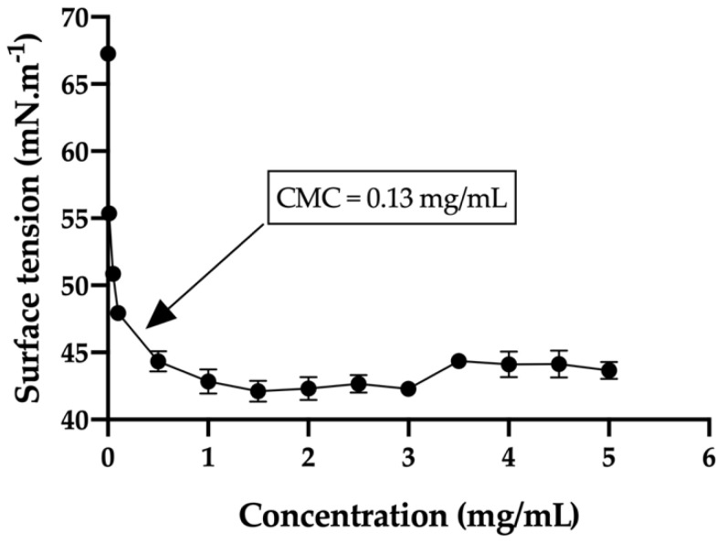
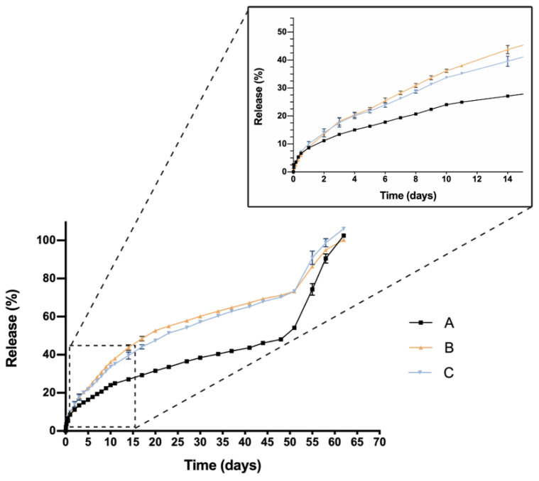
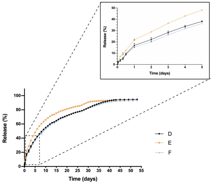
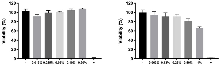
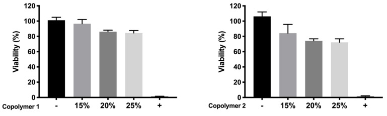
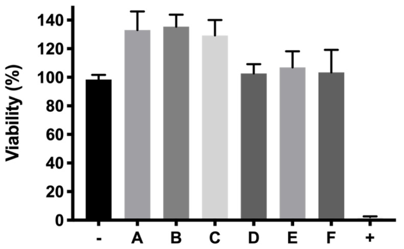
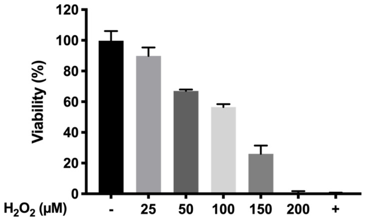
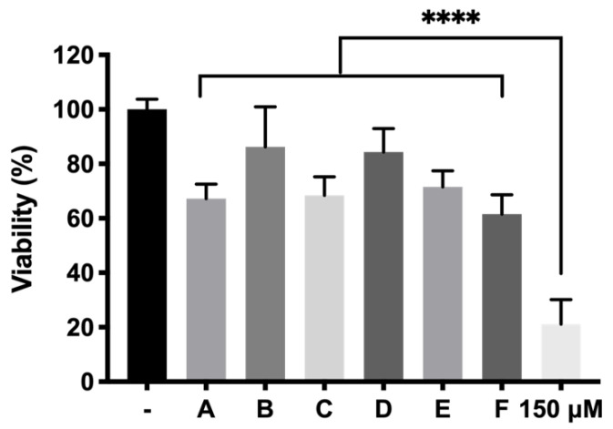
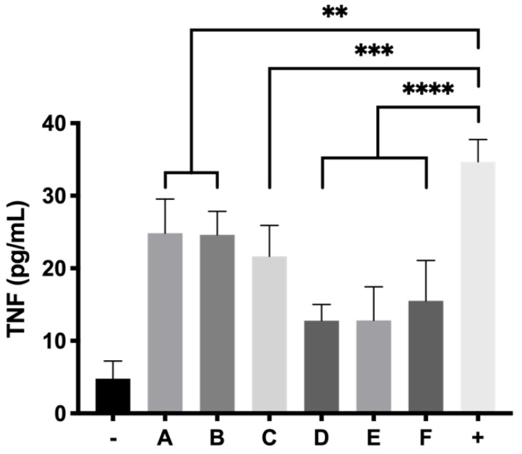
The present study aims to develop a thermo-responsive-injectable hydrogel (HyG) based on PLGA-PEG-PLGA (PLGA = poly-(DL-lactic acid co-glycolic acid); PEG = polyethylene glycol) to deliver neuroprotective agents to the retina over time. Two PLGA-PEG PLGA copolymers with different PEG:LA:GA ratios (1:1.54:23.1 and 1:2.25:22.5) for HyG-1 and HyG-2 development respectively were synthetized and characterized by different techniques (gel permeation chromatography (GPC), nuclear magnetic resonance (NMR), dynamic light scattering (DLS), critical micelle concentration (CMC), gelation and rheological behaviour). According to the physicochemical characterization, HyG-1 was selected for further studies and loaded with anti-inflammatory drugs: dexamethasone (0.2%), and ketorolac (0.5%), alone or in combination with the antioxidants idebenone (1 µM) and D-α-Tocopherol polyethylene glycol 1000 succinate (TPGS) (0.002%). In vitro drug release and cytotoxicity studies were performed for the active substances and hydrogels (loaded and drug-free). A cellular model based on oxidative stress was optimized for anti-inflammatory and antioxidant screening of the formulations by using retinal-pigmented epithelial cell line hTERT (RPE-1). The copolymer 1, used to prepare thermo-responsive HyG-1, showed low polydispersity (PDI = 1.22) and a strong gel behaviour at 25% (w/v) in an isotonic buffer solution close to the vitreous temperature (31-34 °C). Sustained release of dexamethasone and ketorolac was achieved between 47 and 62 days, depending on the composition. HyG-1 was well tolerated (84.5 ± 3.2%) in retinal cells, with values near 100% when the anti-inflammatory and antioxidant agents were included. The combination of idebenone and dexamethasone promoted high oxidative protection in the cells exposed to H2O2, with viability values of 86.2 ± 14.7%. Ketorolac and dexamethasone-based formulations ameliorated the production of TNF-α, showing significant results (p ≤ 0.0001). The hydrogels developed in the present study entail a novel biodegradable tool to treat neurodegenerative processes of the retina overtime.

Keywords: PLGA-PEG-PLGA; dexamethasone; inflammation; intravitreal drug delivery; ketorolac; micelles; neurodegenerative diseases; oxidative stress; thermo-responsive hydrogel.


Conflict of interest statement

The authors declare no conflict of interest.

Figures

Figure 1
Figure 1
Design of the in vitro release experiments for dexamethasone and ketorolac.
Figure 2
Figure 2
Nuclear magnetic resonance (NMR) spectrum of copolymer 1 and 2 (A,B) showing their chemical structure.
Figure 3
Figure 3
Gel permeation chromatography (GPC) dwt/d(logM) vs. log MW showing the molecular weight distribution for copolymers 1 and 2.
Figure 4
Figure 4
Phase diagram of copolymer 1 (HyG-1) and 2 (HyG-2) in the final bicarbonate buffer.
Figure 5
Figure 5
Rheological behaviour values (G’ modulus) of PLGA-PEG-PLGA HyG-1 at different concentrations dispersed in water (A) and the bicarbonate buffer (B) and HyG-2 hydrogels also dispersed in water (C) and the bicarbonate buffer (D).
Figure 6
Figure 6
Rheology of Dexamethasone (left) and Ketorolac (right) formulations with selected HyG-1 in bicarbonate buffer at 25% of PLGA-PEG-PLGA. DX 0.2% (A), DX 0.2% + Idebenone 1 µM (B), DX 0.2% + TPGS 0.02% (C), KT 0.5% (D), KT 0.5% + Idebenone 1 µM (E) and KT 0.5% + TPGS 0.02% (F).
Figure 7
Figure 7
Size of the micelles in nanometers in comparison with the single icons indicating the polydispersity index of sizes (PDI) of DX 0.2% (A), DX 0.2% + Idebenone 1 µM (B), DX 0.2% + TPGS 0.02% (C), KT 0.5% (D), KT 0.5% + Idebenone 1 µM (E) and KT 0.5% + TPGS 0.02% (F), respectively.
Figure 8
Figure 8
Surface tension diagram of the selected copolymer 1 showing the critical micelle concentration (CMC).
Figure 9
Figure 9
In vitro drug release profile of dexamethasone-based formulations with TPGS or Idebenone respectively. DX 0.2% (A), DX 0.2% + Idebenone 1 µM (B), DX 0.2% + TPGS 0.02% (C). Data are represented as mean ± standard deviation (SD) from 3 different batches (n = 3).
Figure 10
Figure 10
In vitro drug release profile of ketorolac-based formulations with TPGS or Idebenone respectively; KT 0.5% (D), KT 0.5% + Idebenone 1 µM (E) and KT 0.5% + TPGS 0.02% (F). Data are represented as mean ± standard deviation (SD) from three different batches (n = 3).
Figure 11
Figure 11
Cell viability of dexamethasone phosphate (left) and ketorolac tris salt (right) at different concentrations in RPE-1 cells. Data are expressed as mean ± standard deviations. Cell viability (%) is calculated in relation to the negative control (100% of viability).
Figure 12
Figure 12
Viability of copolymers 1 and 2 respectively in bicarbonate buffer without drugs.
Figure 13
Figure 13
Viability of final formulations (%) prepared with copolymer 1. DX 0.2% (A), DX 0.2% + Idebenone 1 µM (B), DX 0.2% + TPGS 0.02% (C), KT 0.5% (D), KT 0.5% + Idebenone 1 µM (E) and KT 0.5% + TPGS 0.02% (F), respectively.
Figure 14
Figure 14
Optimization of cell viability in the oxidative stress model at different H2O2 concentrations.
Figure 15
Figure 15
In vitro protection of the final formulations DX 0.2% (A), DX 0.2% + Idebenone 1 µM (B), DX 0.2% + TPGS 0.02% (C), KT 0.5% (D), KT 0.5% + Idebenone 1 µM (E) and KT 0.5% + TPGS 0.02% (F), respectively, in response to 150 µM of (positive control, +) oxidative stress in RPE-1 cells. High statistically significant values (p ≤ 0.0001; ****).
Figure 16
Figure 16
TNFα activity of RPE-1 cells exposed to final formulations DX 0.2% (A), DX 0.2% + Idebenone 1 µM (B), DX 0.2% + TPGS 0.02% (C), KT 0.5% (D), KT 0.5% + Idebenone 1 µM (E) and KT 0.5% + TPGS 0.02% (F), respectively, in response to LPS induced inflammation. p ≤ 0.01 (**), p ≤ 0.001 (***) and p ≤ 0.0001 (****).

References

    1. Bressler N.M. Age-related macular degeneration is the leading cause of blindness. JAMA. 2004;291:1900–1901. doi: 10.1001/jama.291.15.1900. - DOI - PubMed
    1. Flaxman S.R., Bourne R.R.A., Resnikoff S., Ackland P., Braithwaite T., Cicinelli M.V., Das A., Jonas J.B., Keeffe J., Kempen J., et al. Global causes of blindness and distance vision impairment 1990–2020: A systematic review and meta-analysis. Lancet Glob. Health. 2017;5:e1221–e1234. doi: 10.1016/S2214-109X(17)30393-5. - DOI - PubMed
    1. Bhutto I., Lutty G. Understanding age-related macular degeneration (AMD): Relationships between the photoreceptor/retinal pigment epithelium/Bruch’s membrane/choriocapillaris complex. Mol. Asp. Med. 2012;33:295–317. doi: 10.1016/j.mam.2012.04.005. - DOI - PMC - PubMed
    1. Baba T., Grebe R., Hasegawa T., Bhutto I., Merges C., McLeod D.S., Lutty G.A. Structural and functional maturation of the fetal human choriocapillaris. Investig. Ophthalmol. Vis Sci. 2009;50:3503–3511. doi: 10.1167/iovs.08-2614. - DOI - PMC - PubMed
    1. Garcia-Layana A., Cabrera-López F., García-Arumí J., Arias-Barquet L., Ruiz-Moreno J.M. Early and intermediate age-related macular degeneration: Update and clinical review. Clin. Interv. Aging. 2017;12:1579–1587. doi: 10.2147/CIA.S142685. - DOI - PMC - PubMed

LinkOut - more resources