

Deep Neural Network-Based Semantic Segmentation of Microvascular Decompression Images
- PMID: 33562275
- PMCID: PMC7915571
- DOI: 10.3390/s21041167
Deep Neural Network-Based Semantic Segmentation of Microvascular Decompression Images
Abstract
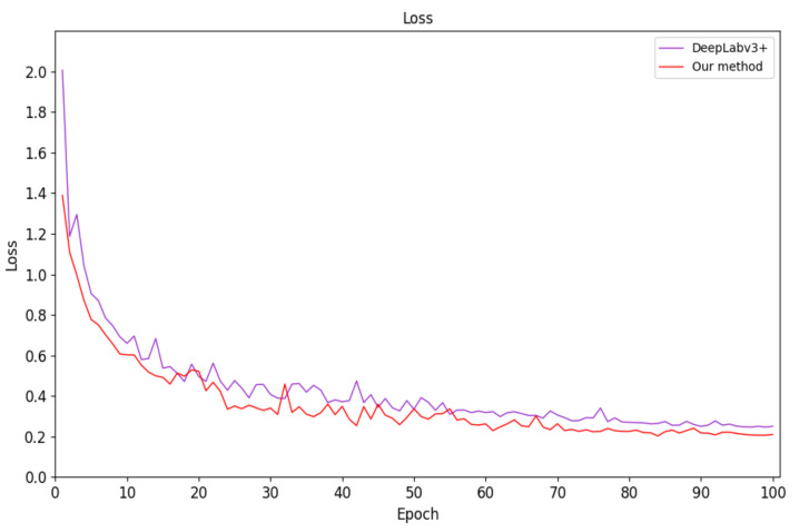
Image semantic segmentation has been applied more and more widely in the fields of satellite remote sensing, medical treatment, intelligent transportation, and virtual reality. However, in the medical field, the study of cerebral vessel and cranial nerve segmentation based on true-color medical images is in urgent need and has good research and development prospects. We have extended the current state-of-the-art semantic-segmentation network DeepLabv3+ and used it as the basic framework. First, the feature distillation block (FDB) was introduced into the encoder structure to refine the extracted features. In addition, the atrous spatial pyramid pooling (ASPP) module was added to the decoder structure to enhance the retention of feature and boundary information. The proposed model was trained by fine tuning and optimizing the relevant parameters. Experimental results show that the encoder structure has better performance in feature refinement processing, improving target boundary segmentation precision, and retaining more feature information. Our method has a segmentation accuracy of 75.73%, which is 3% better than DeepLabv3+.
Keywords: DeepLabv3+; decoder structure; encoder structure; microvascular decompression image; semantic segmentation.
Conflict of interest statement
The authors declare no conflict of interest.
Figures


References
-
- Li Q., Cai W., Wang X., Zhou Y., Feng D.D., Chen M. Medical image classification with convolutional neural network; Proceedings of the 2014 13th International Conference on Control Automation Robotics & Vision (ICARCV); Singapore. 10–12 December 2014; pp. 844–848.
MeSH terms
LinkOut - more resources
Full Text Sources
Other Literature Sources
