Review
. 2021 Feb 7;11(2):236.
doi: 10.3390/biom11020236.

The Ins and Outs of RAS Effector Complexes

Affiliations
Review

The Ins and Outs of RAS Effector Complexes

Christina Kiel et al. Biomolecules. .

Abstract

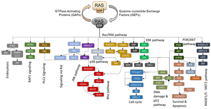
RAS oncogenes are among the most commonly mutated proteins in human cancers. They regulate a wide range of effector pathways that control cell proliferation, survival, differentiation, migration and metabolic status. Including aberrations in these pathways, RAS-dependent signaling is altered in more than half of human cancers. Targeting mutant RAS proteins and their downstream oncogenic signaling pathways has been elusive. However, recent results comprising detailed molecular studies, large scale omics studies and computational modeling have painted a new and more comprehensive portrait of RAS signaling that helps us to understand the intricacies of RAS, how its physiological and pathophysiological functions are regulated, and how we can target them. Here, we review these efforts particularly trying to relate the detailed mechanistic studies with global functional studies. We highlight the importance of computational modeling and data integration to derive an actionable understanding of RAS signaling that will allow us to design new mechanism-based therapies for RAS mutated cancers.

Keywords: RAS in human cancer; RAS oncogene; RAS signaling networks; computational modeling; personalized therapies; targeting RAS.


Conflict of interest statement

The authors declare no conflict of interest.

Figures

Figure 1
Figure 1
Known RAS effectors and signaling pathways. The RAS activation cycle comprises GTP binding aided by Guanine nucleotide Exchange Factors (GEFs), which puts RAS proteins into an active conformation where it can bind to effectors and activate downstream signaling pathways. The main and currently best-characterized RAS signaling pathways are shown. Effectors are released when GTP is hydrolyzed to GDP, and this intrinsic, but low, catalytic RAS activity is vastly accelerated by GTPase Activating Proteins (GAPs).
Figure 2
Figure 2
RAS and 56 classical effectors. (A) Classification of effectors based on the binding affinity of the RBD in complex with RAS∙GTP. High-affinity binders are defined by Kd values of ≤1 μM, and low-affinity binders by Kd values of >1 μM). (B) Schematic representation of the recruitment of effectors to RAS∙GTP via their RBD. Five effectors are predicted to be recruited efficiently to RAS∙GTP via their RBD only in all/most of the 29 human tissues, and four effectors are predicted to be recruited efficiently to RAS∙GTP via their RBD only in some of the 29 human tissues [23].
Figure 3
Figure 3
Mechanisms enhancing RAS effector recruitment. (A) The reduction of a three-dimensional space to the two-dimensions of a membrane can enhance the affinity between RAS and its effectors by >100 fold [29]. (B) Schematic representation of the recruitment of effectors to RAS∙GTP via their RBD and indication of the actin meshwork of the cell cortex. All effectors are predicted to increase their residence time in the PM once recruited via their RBD. Four effectors have been shown to bind to proteins that are located in the actin meshwork. (C) Representation of the recruitment of effectors to the membrane via their RBD and via membrane-binding domains. Twenty-six effectors contain domains that have the ability to bind to membranes (the names of the domains are indicated in green). (D) Recruitment of effectors via their RBD and via domains (or linear motifs) that bind to membrane-associated proteins.
Figure 4
Figure 4
Summary of PM recruitment of 56 effectors grouped into 12 effector pathways. Classification of effectors into 12 effector pathways [17] and summary of the proposed membrane recruitment mechanisms (based on Figure 2 and Figure 3).
Figure 5
Figure 5
RAS regulation by posttranslational modifications. (A) RAS regulation by mono-ubiquitination. KRAS mono-ubiquitination at lysine 147 renders KRAS refractory to GAPs, while HRAS mono-ubiquitination at lysine 117 stimulates GEF independent GTP binding. Both mechanisms lead to the activation of downstream pathways such as ERK. (B) The constitutive lysosomal degradation of KRAS mediated by β-TrCP1 is impeded by Smurf2 mono-ubiquitinating UBCH5, which in turn poly ubiquinates β-TrCP1 and marks it for proteasomal degradation. (C) RAS regulation by mono-/di-ubiquitination. Rabex-5 can mono/di-ubiquitination NRAS and HRAS inducing endosomal localization and inability to activate ERK. (D) HRAS and KRAS degradation can be triggered by GSK3β phosphorylation of threonines 144 and 148, which induces poly ubiquitination by β-TrCP1. When WNT signaling is activated, stabilized β-catenin can bind to KRAS and physically shield the threonines 144/148 from phosphorylation resulting in KRAS protein accumulation. (E) Negative RAS regulation by tyrosine phosphorylation. The SRC kinase can phosphorylate all three RAS isoforms at tyrosines 32 and 64 leading to inhibition of effector binding and enhancement of GAP binding. These phosphorylations can be reversed by the SHP-2 phosphatase, which can restore signaling. (F) The RIN1 effector stimulates ABL mediated phosphorylation of Y137 which promotes RAF kinase binding and activation of the ERK pathway. However, RIN1 also stimulates Rabex-5-dependent ubiquitination of RAS, which leads to its endosomal sequestration. This ubiquitination is facilitated by the phosphorylation of tyrosine 4 by an unknown kinase. (G) PKC phosphorylation of KRAS at S181 has two effects. It reduces the interaction with GAPs prolonging KRAS signaling and induces the translocation of KRAS to the mitochondria, where it can bind to Bcl-XL and induce apoptosis.
Figure 6
Figure 6
Signaling capacities of known RAS effector pathway components at different HRAS localizations superimposed on diagrams of these pathways. Signaling pathways activated when HRASV12 is located at the (A) disordered membrane (DM); (B) lipid rafts (LR); (C) endoplasmic reticulum (ER); and (D) Golgi apparatus (GA). The nodes displayed in bright colors are present in the corresponding network. The red circle adjacent to each node shows the number of interactions it participates in. In cases where a node represents more than one protein (isoforms or homologs), the signaling capacity of the node is represented by the total number of interactions involving all proteins corresponding to the node. The enriched GO terms of the HRAS localization specific transcriptome are shown at bottom of each pathway diagram. This figure is from the supplementary data of [126].
Figure 6
Figure 6
Signaling capacities of known RAS effector pathway components at different HRAS localizations superimposed on diagrams of these pathways. Signaling pathways activated when HRASV12 is located at the (A) disordered membrane (DM); (B) lipid rafts (LR); (C) endoplasmic reticulum (ER); and (D) Golgi apparatus (GA). The nodes displayed in bright colors are present in the corresponding network. The red circle adjacent to each node shows the number of interactions it participates in. In cases where a node represents more than one protein (isoforms or homologs), the signaling capacity of the node is represented by the total number of interactions involving all proteins corresponding to the node. The enriched GO terms of the HRAS localization specific transcriptome are shown at bottom of each pathway diagram. This figure is from the supplementary data of [126].
Figure 7
Figure 7
The RAS network as defined by interaction proteomics. (A) The RAS network obtained from databases and recent publications. Overview of interactions obtained from the HuRI, BIOPLEX, and STRING (highest confidence; 0.9) databases, and two recent publications [133,134] (left side). Venn diagram of interactions for HRAS, KRAS and NRAS (right side). (B) Enrichment of functional classes for HRAS, KRAS and NRAS interactors based on the SysGO database. (C) Functional analysis of all proteins in the union of HRAS, KRAS and NRAS interactors. Abbreviations: FA, fatty acids; Y2H, yeast two hybrid.

References

    1. Harvey J.J. An unidentified virus which causes the rapid production of tumours in mice. Nature. 1964;204:1104–1105. doi: 10.1038/2041104b0. - DOI - PubMed
    1. Malumbres M., Barbacid M. RAS oncogenes: The first 30 years. Nat. Rev. Cancer. 2003;3:459–465. doi: 10.1038/nrc1097. - DOI - PubMed
    1. Prior I.A., Hood F.E., Hartley J.L. The Frequency of Ras Mutations in Cancer. Cancer Res. 2020;80:2969–2974. doi: 10.1158/0008-5472.CAN-19-3682. - DOI - PMC - PubMed
    1. Kiel C., Serrano L. Structure-energy-based predictions and network modelling of RASopathy and cancer missense mutations. Mol. Syst. Biol. 2014;10:727. doi: 10.1002/msb.20145092. - DOI - PMC - PubMed
    1. Rauen K.A. The RASopathies. Ann. Rev. Genom. Hum. Genet. 2013;14:355–369. doi: 10.1146/annurev-genom-091212-153523. - DOI - PMC - PubMed

Publication types

LinkOut - more resources