

Highly Efficient T2 Cobalt Ferrite Nanoparticles Vectorized for Internalization in Cancer Cells
- PMID: 33562703
- PMCID: PMC7914706
- DOI: 10.3390/ph14020124
Highly Efficient T2 Cobalt Ferrite Nanoparticles Vectorized for Internalization in Cancer Cells
Abstract
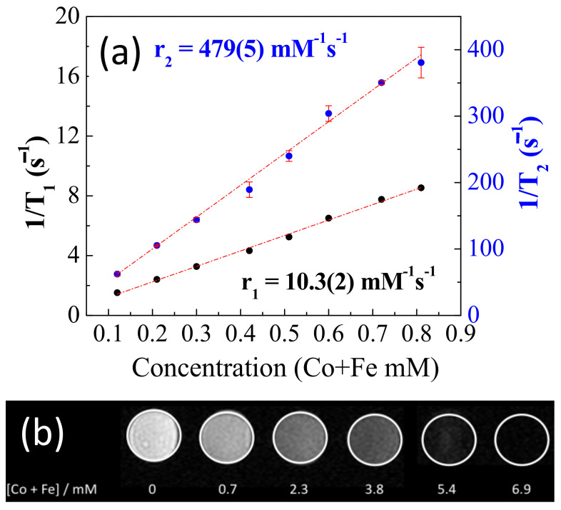
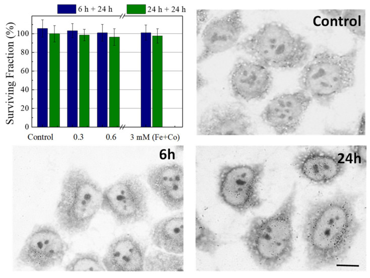
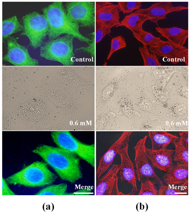
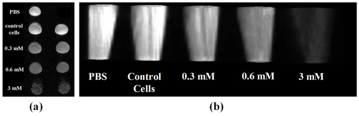
Uniform cobalt ferrite nanoparticles have been synthesized using an electrochemical synthesis method in aqueous media. Their colloidal, magnetic, and relaxometric properties have been analyzed. The novelty of this synthesis relies on the use of iron and cobalt foils as precursors, which assures the reproducibility of the iron and cobalt ratio in the structure. A stable and biocompatible targeting conjugate nanoparticle-folic acid (NP-FA) was developed that was capable of targeting FA receptor positivity in HeLa (human cervical cancer) cancer cells. The biocompatibility of NP-FA was assessed in vitro in HeLa cells using the MTT assay, and morphological analysis of the cytoskeleton was performed. A high level of NP-FA binding to HeLa cells was confirmed through qualitative in vitro targeting studies. A value of 479 Fe+Co mM-1s-1 of transverse relaxivity (r2) was obtained in colloidal suspension. In addition, in vitro analysis in HeLa cells also showed an important effect in negative T2 contrast. Therefore, the results show that NP-FA can be a potential biomaterial for use in bio medical trials, especially as a contrast agent in magnetic resonance imaging (MRI).
Keywords: cobalt ferrite; contrast agent; folic acid; internalization; nanoparticles; targeting.
Conflict of interest statement
The authors declare no conflict of interest.
Figures

References
-
- Liu C., Zou B., Rondinone A.J., Zhang Z.J. Chemical control of superparamagnetic properties of magnesium and cobalt spinel ferrite nanoparticles through atomic level magnetic couplings. J. Am. Chem. Soc. 2000;122:6263–6267. doi: 10.1021/ja000784g. - DOI
-
- Gallo-Cordova Á., Espinosa A., Serrano A., Gutiérrez L., Menéndez N., Del Puerto Morales M., Mazario E. New insights into the structural analysis of maghemite and (MFe2O4, M = Co, Zn) ferrite nanoparticles synthesized by a microwave-assisted polyol process. Mater. Chem. Front. 2020;4:3063–3073. doi: 10.1039/D0QM00460J. - DOI
-
- Stein C.R., Bezerra M.T.S., Holanda G.H.A., André-Filho J., Morais P.C. Structural and magnetic properties of cobalt ferrite nanoparticles synthesized by co-precipitation at increasing temperatures. AIP Adv. 2017;8:056303. doi: 10.1063/1.5006321. - DOI
-
- Lasheras X., Insausti M., Gil de Muro I., Garaio E., Plazaola F., Moros M., De Matteis L., de la Fuente J.M., Lezama L. Chemical Synthesis and Magnetic Properties of Monodisperse Nickel Ferrite Nanoparticles for Biomedical Applications. J. Phys. Chem. C. 2016;120:3492–3500. doi: 10.1021/acs.jpcc.5b10216. - DOI
Grants and funding
LinkOut - more resources
Full Text Sources
Other Literature Sources
Miscellaneous
