. 2021 Feb 5;11(2):250.
doi: 10.3390/diagnostics11020250.

Deep Learning for Diagnosis of Paranasal Sinusitis Using Multi-View Radiographs

Affiliations

Deep Learning for Diagnosis of Paranasal Sinusitis Using Multi-View Radiographs

Yejin Jeon et al. Diagnostics (Basel). .

Abstract

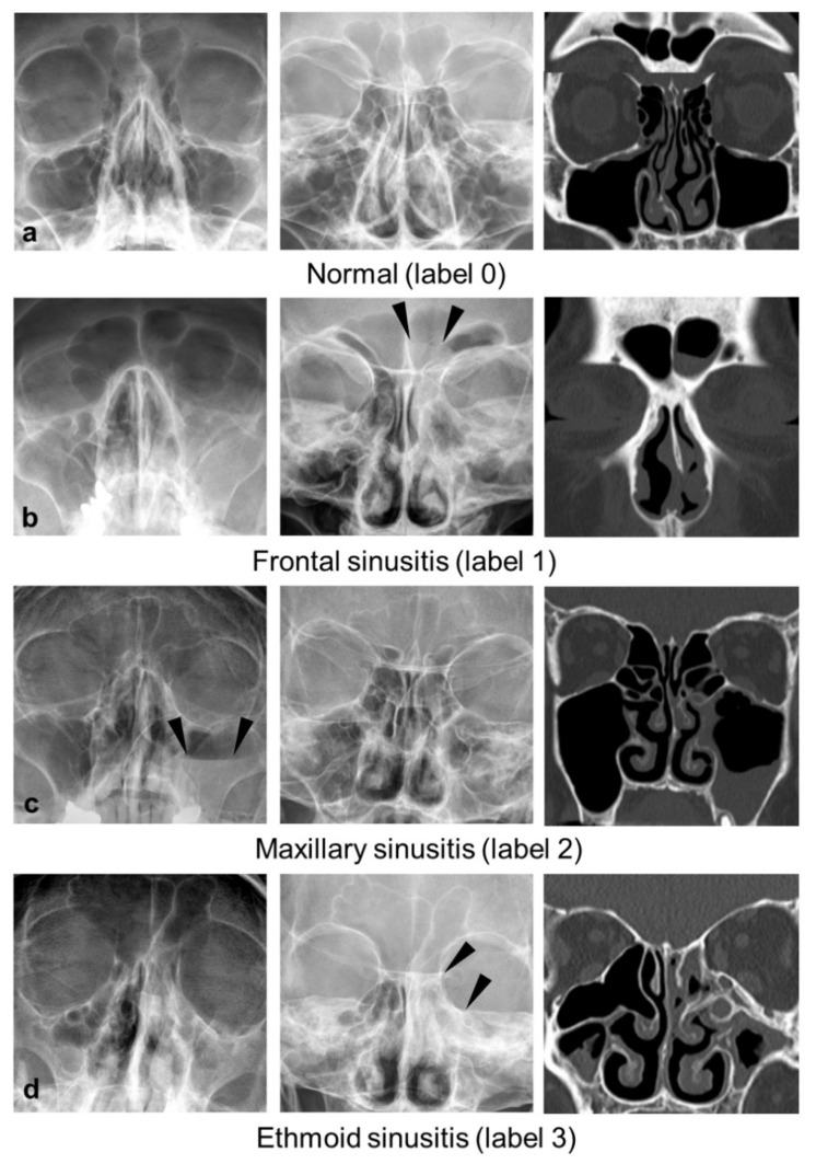
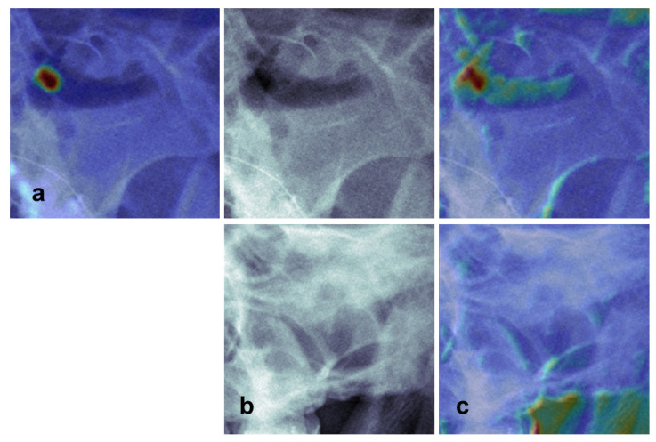
Accurate image interpretation of Waters' and Caldwell view radiographs used for sinusitis screening is challenging. Therefore, we developed a deep learning algorithm for diagnosing frontal, ethmoid, and maxillary sinusitis on both Waters' and Caldwell views. The datasets were selected for the training and validation set (n = 1403, sinusitis% = 34.3%) and the test set (n = 132, sinusitis% = 29.5%) by temporal separation. The algorithm can simultaneously detect and classify each paranasal sinus using both Waters' and Caldwell views without manual cropping. Single- and multi-view models were compared. Our proposed algorithm satisfactorily diagnosed frontal, ethmoid, and maxillary sinusitis on both Waters' and Caldwell views (area under the curve (AUC), 0.71 (95% confidence interval, 0.62-0.80), 0.78 (0.72-0.85), and 0.88 (0.84-0.92), respectively). The one-sided DeLong's test was used to compare the AUCs, and the Obuchowski-Rockette model was used to pool the AUCs of the radiologists. The algorithm yielded a higher AUC than radiologists for ethmoid and maxillary sinusitis (p = 0.012 and 0.013, respectively). The multi-view model also exhibited a higher AUC than the single Waters' view model for maxillary sinusitis (p = 0.038). Therefore, our algorithm showed diagnostic performances comparable to radiologists and enhanced the value of radiography as a first-line imaging modality in assessing multiple sinusitis.

Keywords: artificial intelligence; deep learning; machine learning; multi-view radiographs; paranasal sinusitis.


Conflict of interest statement

The authors declare no conflict of interest.

Figures

Figure 1
Figure 1
Flowchart of clinical datasets for training, validation, and test.
Figure 2
Figure 2
Representative cases with normal (label 0, a), frontal sinusitis (label 1, b), maxillary sinusitis (label 2, c), and ethmoid sinusitis (label 3, d) at each view (Waters’ view: left, Caldwell view: middle, corresponding coronal image of CT, right). For frontal (b) and ethmoid sinusitis (d), mucosal thickening (label 1) and total opacification (label 3) are not well visualized in Waters’ view, whereas Caldwell view provides the best projection for evaluation (arrowheads). In the case of maxillary sinusitis (c), Waters’ view provides a better view of the air-fluid level (label 2, arrowheads) than Caldwell view.
Figure 3
Figure 3
Overview of the proposed network architecture. The network consists of a detector (Mdet), which localizes sinuses with a bounding box, and a classifier (Mcls), which classifies sinusitis with 4-leveled labels. It combines the information of both Caldwell and Waters’ views. For the modified model, which uses only the primary view (denoted as single primary view model), the network path of the second row is removed (vice versa for the single secondary view model, which uses only the secondary view).
Figure 4
Figure 4
Receiver operating characteristic (ROC) curves and the area under the ROC curves (AUCs) of the proposed multi-view model and reviewers in the observer performance study.
Figure 5
Figure 5
Comparison of the class activation maps (CAMs) of the single primary view and multi-view model. (a) CAM of the single primary view model (Waters’ view). (b) Original radiographs showing maxillary sinusitis with air-fluid level (upper row, Waters’ view; lower row, Caldwell view) (c), CAM of the multi-view model (upper row, Waters’ view; lower row, Caldwell view). Single primary view model (a) misclassified as mucosal thickening because the area above the horizontal fluid line is not recognized. In the case of the multi-view model (c), the prediction is correct as the activated area is more expanded on Waters’ view (upper row), while no clear activation is found in the sinus area on the secondary view (lower row).

References

    1. Anand V.K. Epidemiology and economic impact of rhinosinusitis. Ann. Otol. Rhinol. Laryngol. Suppl. 2004;193:3–5. doi: 10.1177/00034894041130S502. - DOI - PubMed
    1. Meltzer E.O., Hamilos D.L. Rhinosinusitis diagnosis and management for the clinician: A synopsis of recent consensus guidelines. Mayo Clin. Proc. 2011;86:427–443. doi: 10.4065/mcp.2010.0392. - DOI - PMC - PubMed
    1. Rosenfeld R.M., Piccirillo J.F., Chandrasekhar S.S., Brook I., Ashok Kumar K., Kramper M., Orlandi R.R., Palmer J.N., Patel Z.M., Peters A., et al. Clinical practice guideline (update): Adult sinusitis. Otolaryngol. Head Neck Surg. 2015;152:S1–S39. doi: 10.1177/0194599815572097. - DOI - PubMed
    1. Aaløkken T.M., Hagtvedt T., Dalen I., Kolbenstvedt A. Conventional sinus radiography compared with CT in the diagnosis of acute sinusitis. Dentomaxillofac. Radiol. 2003;32:60–62. doi: 10.1259/dmfr/65139094. - DOI - PubMed
    1. David M. Imaging in sinus diseases. In: Kennedy D.W., Bolger W.E., Zinreich S.J., editors. Diseases of the Sinuses: Diagnosis and Management. B.C. Decker; Hamilton, ON, Canada: 2001. pp. 132–133.

LinkOut - more resources