Review
. 2021 Feb 9;14(1):11.
doi: 10.1186/s13072-021-00384-w.

Epigenetic regulation of retinal development

Affiliations
Review

Epigenetic regulation of retinal development

Reza Raeisossadati et al. Epigenetics Chromatin. .

Abstract

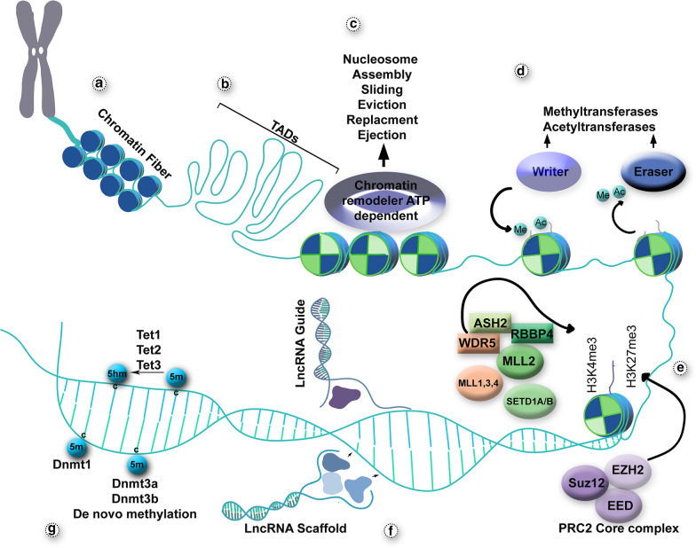
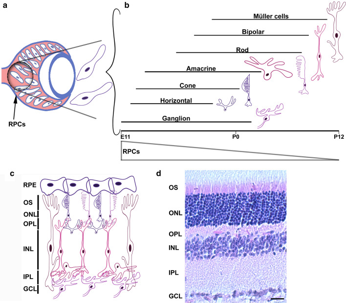
In the developing vertebrate retina, retinal progenitor cells (RPCs) proliferate and give rise to terminally differentiated neurons with exquisite spatio-temporal precision. Lineage commitment, fate determination and terminal differentiation are controlled by intricate crosstalk between the genome and epigenome. Indeed, epigenetic regulation plays pivotal roles in numerous cell fate specification and differentiation events in the retina. Moreover, aberrant chromatin structure can contribute to developmental disorders and retinal pathologies. In this review, we highlight recent advances in our understanding of epigenetic regulation in the retina. We also provide insight into several aspects of epigenetic-related regulation that should be investigated in future studies of retinal development and disease. Importantly, focusing on these mechanisms could contribute to the development of novel treatment strategies targeting a variety of retinal disorders.

Keywords: Chromatin; DNA methylation; Development; Epigenetics; Histone; Retina; lncRNA.


Conflict of interest statement

The authors declare that they have no competing interests.

Figures

Fig. 1
Fig. 1
Schematic depiction of the various layers of epigenetic regulation involved in cell fate determination and differentiation events. a Chromatin folding can be considered as the first layer of transcriptional regulation; nucleosomes are assembled and form 30-nm fibers. b Topologically associating domains (TADs) are genomic regions spanning 0.2 to 1 Mb considered as defined regulatory and structural units. c Binding of ATP-dependent chromatin remodelers to chromatin exposes sites for transcriptional regulation. d Histone tails are subjected to different covalent modifications mainly on their lysine residues. Enzymes known as “writers” (acetyltransferases and methyltransferases) are responsible for adding these modifications, while “erasers” (deacetylases and demethylases) remove them. e Various proteins and protein complexes are involved in establishing bivalent histone marks; the most abundant subunits are components of the PRC2 core complex, which catalyze H3K27 methylation and MLLs, which catalyze H3K4 methylation. f lncRNAs fine-tune gene expression in several ways, such as guiding proteins to a specific location in the genome or acting as a scaffold for other regulatory components. g DNA methyltransferases, such as Dnmt1, Dnmt3a and Dnmt3b add or maintain methyl groups on cytosine residues (5-methylcytosine), while members of the ten-eleven translocation (Tet) family catalyze the oxidation of 5-methylcytosine to 5-hydroxymethylcytosine, which can then be further converted to other intermediates or removed to demethylate the DNA
Fig. 2
Fig. 2
Vertebrate retinal development. a Cartoon of the developing retina. Early in development, the retina is composed of multipotent retinal progenitor cells (RPCs). b Over time, RPCs give rise to the seven cell types of the mature retina and they do so with precise spatio-temporal precision. Retinal ganglion cells are generated first, followed by horizontal cells, cones, amacrine cells, rods, bipolar cells and finally Muller glia. As development proceeds, the competency of RPCs to give rise to each of these cell types becomes further restricted. c Cartoon of the laminar architecture of the mature retina, wherein differentiated neurons are precisely organized into three principal cellular layers: the outer nuclear layer (ONL), inner nuclear layer (INL) and ganglion cell layer (GCL). These layers are separated by the synaptic layers: the outer plexiform layer (OPL) and inner plexiform later (IPL). The retinal pigmented epithelium (RPE) lies at the posterior of the eye, supporting the retina. d Histological section (H + E stained) of a 13-week-old mouse retina highlighting these retinal layers. Scale bar = 10 um

References

    1. Cepko C. Intrinsically different retinal progenitor cells produce specific types of progeny. Nat Rev Neurosci. 2014;15(9):615–627. doi: 10.1038/nrn3767. - DOI - PubMed
    1. Flynn RA, Chang HY. Long noncoding RNAs in cell-fate programming and reprogramming. Cell Stem Cell. 2014;14(6):752–761. doi: 10.1016/j.stem.2014.05.014. - DOI - PMC - PubMed
    1. Perino M, Veenstra GJ. Chromatin control of developmental dynamics and plasticity. Dev Cell. 2016;38(6):610–620. doi: 10.1016/j.devcel.2016.08.004. - DOI - PubMed
    1. Aldiri I, et al. The dynamic epigenetic landscape of the retina during development, reprogramming, and tumorigenesis. Neuron. 2017;94(3):550–568.e10. doi: 10.1016/j.neuron.2017.04.022. - DOI - PMC - PubMed
    1. Buono L, Martinez-Morales JR. Retina development in vertebrates: systems biology approaches to understanding genetic programs: on the contribution of next-generation sequencing methods to the characterization of the regulatory networks controlling vertebrate eye development. BioEssays. 2020;42(4):e1900187. doi: 10.1002/bies.201900187. - DOI - PubMed

Publication types