. 2021 Jun;70(6):1250-1264.
doi: 10.2337/db20-0790. Epub 2021 Feb 9.

Exercise Training Promotes Sex-Specific Adaptations in Mouse Inguinal White Adipose Tissue

Affiliations

Exercise Training Promotes Sex-Specific Adaptations in Mouse Inguinal White Adipose Tissue

Pasquale Nigro et al. Diabetes. 2021 Jun.

Abstract

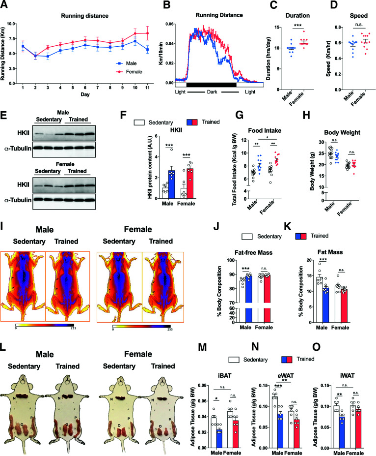
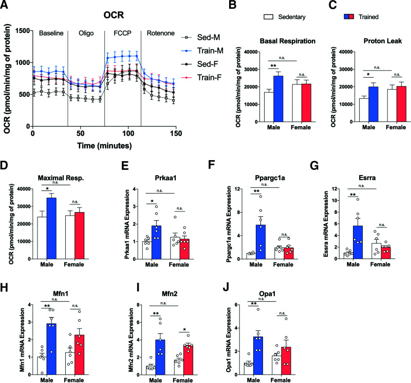
Recent studies demonstrate that adaptations to white adipose tissue (WAT) are important components of the beneficial effects of exercise training on metabolic health. Exercise training favorably alters the phenotype of subcutaneous inguinal WAT (iWAT) in male mice, including decreasing fat mass, improving mitochondrial function, inducing beiging, and stimulating the secretion of adipokines. In this study, we find that despite performing more voluntary wheel running compared with males, these adaptations do not occur in the iWAT of female mice. Consistent with sex-specific adaptations, we report that mRNA expression of androgen receptor coactivators is upregulated in iWAT from trained male mice and that testosterone treatment of primary adipocytes derived from the iWAT of male, but not female mice, phenocopies exercise-induced metabolic adaptations. Sex specificity also occurs in the secretome profile, as we identify cysteine-rich secretory protein 1 (Crisp1) as a novel adipokine that is only secreted from male iWAT in response to exercise. Crisp1 expression is upregulated by testosterone and functions to increase glucose and fatty acid uptake. Our finding that adaptations to iWAT with exercise training are dramatically greater in male mice has potential clinical implications for understanding the different metabolic response to exercise training in males and females and demonstrates the importance of investigating both sexes in studies of adipose tissue biology.


Figures

Figure 1
Figure 1
Exercise training decreases body weight and fat mass in male, but not female mice. Daily voluntary wheel running (VWR) distance (A) and daily frequency actogram (B) measured in male and female mice (n = 12–14/group). VWR indices such as duration (C) and speed (D) for male and female mice (n = 12–14/group). Representative immunoblots for HKII protein content in triceps muscle of sedentary (unfilled) and trained (colored) male and female (E) mice and relative quantification (F) (n = 6/group). Food consumption (G) and body weight (H) after 11 days in sedentary and trained male and female mice (n = 14/group). I: Representative DXA scans of sedentary and trained male (left) and female (right) mice; black arrows indicate subcutaneous fat depots. Relative quantification of fat-free (J) and fat (K) mass in sedentary and trained male and female mice (n = 7–8/group). Representative images (L) of three fat depots iBAT (M), eWAT (N), and iWAT (O) with relative adipose tissue mass weight in sedentary (unfilled) and trained (colored) male and female mice (n = 6–8/group). Data are expressed as mean ± SEM. n.s. indicates no significant difference. *P < 0.05; **P < 0.01; ***P < 0.001. A.U., arbitrary unit; BW, body weight.
Figure 2
Figure 2
Exercise training is more effective in reducing fat mass in male than in female mice. Representative images of hematoxylin and eosin staining (A), cell size distribution (B), and average diameter (C) of iWAT in sedentary and trained male and female mice (n = 4 mice/group; calculated from 10 fields/mouse). Scale bars, 100 μm. mRNA expression of Leptin (Lep) (D), Lipe (E), and Pnpla2 (F) in iWAT of sedentary and trained male and female mice (n = 6/group). Cell proliferation rate of ASCs evaluated by MTS incorporation assay (G) (n = 6/group), representative images of sedentary and trained male and female mice differentiated ASCs (H) and quantitative Oil Red O staining of differentiated cells (I) (n = 6 mice/group). Scale bars, 100 μm. Data are expressed as mean ± SEM. n.s. indicates no significant difference. *P < 0.05; **P < 0.01; ***P < 0.001 as indicated. Sed-F, sedentary female; Sed-M, sedentary male; Train-F, trained female; Train-M, trained male.
Figure 3
Figure 3
Exercise training induces beiging and thermogenesis in iWAT of male, but not female, mice. Ucp1 mRNA expression in iWAT (A) of sedentary (unfilled) and trained (colored) male and female mice (n = 6/group), representative images of UCP1 protein by Western blot (B), and epifluorescence microscopy (C) of sedentary and trained male (left) and female (right) mice with relative immunofluorescence intensity quantification (D) (n = 4/group; calculated from three fields/mouse). Scale bars, 50 μm. mRNA expression of beiging-related genes Cidea (E), Dio2 (F), Prdm16 (G), and Tle3 (H) in iWAT of sedentary and trained male and female mice (n = 6/group). Representative thermal images (I) for iWAT of sedentary and trained male (top) and female (bottom) mice with relative quantification of top 10% mean surface area temperature (J) (n = 9/group). Data are expressed as mean ± SEM. n.s. indicates no significant difference. *P < 0.05; **P < 0.01; ***P < 0.001 A.U., arbitrary unit; ROI, region of interest.
Figure 4
Figure 4
Exercise training increases OCR only in iWAT of male mice, altering mitochondrial biogenesis and dynamics. Change in OCR in iWAT (A) of sedentary and trained male and female mice, basal respiration (B), proton leak (C), and maximal respiration (Resp.) (D) (n = 6/group). mRNA expression of genes involved in mitochondrial biogenesis: Prkaa1 (E), Ppargc1a (F), and Esrra (G) (n = 6/group). mRNA expression of genes involved in mitochondrial fusion: Mfn1 (H), Mfn2 (I), and Opa1 (J) n = 6/group). Data are expressed as mean ± SEM, n.s. indicates no significant difference. *P < 0.05; **P < 0.01. FCCP, carbonyl cyanide-4-phenylhydrazone; Oligo, oligomycin; Sed-F, sedentary female; Sed-M, sedentary male; Train-F, trained female; Train-M, trained male.
Figure 5
Figure 5
Exercise training increases the androgen receptor activity in iWAT of male mice. A–E: Network motif analysis by Network Analyst (A) on the most upregulated genes (FDR <0.01) with 11 days of voluntary wheel running in male iWAT (Gene Expression Omnibus data set GSE68161). List of genes regulated by AR generated by RegNetwork database (B) and relative pathway enrichment analysis using the Kyoto Encyclopedia of Genes and Genomes (KEGG) (C), GO-BP (D), and GO-CC (E) databases. F: Jaspar analysis for Ucp1 promoter region using Eukaryotic Promoter Database-Swiss Institute of Bioinformatics (SIB) website; androgen response element consensus sequences are represented as red bars. G: Quantitative chromatin immunoprecipitation PCR experiment performed on DNA samples precipitated with antibody against AR after treatment with 10 μmol/L testosterone and 10 μmol/L DHT (n = 3/group). Change in mRNA expression level for Ucp1 (H), Prkaa1 (I), Ppargc1a (J), and Esrra (K) on iWAT after 24 h of 10 mmol/L testosterone treatment (n = 6/group). Data are expressed as mean ± SEM. n.s. indicates no significant difference. *P < 0.05; ***P < 0.001. PPAR, peroxisome proliferator–activated receptor.
Figure 6
Figure 6
CRISP1 is a novel adipokine induced by exercise training and testosterone treatment in iWAT of male but not female mice. Crisp1 mRNA expression in sedentary (unfilled) and trained (colored) iWAT of male and female mice (A) (n = 6/group), correlation analysis (B) with daily running distance for male mice (n = 6), and immunoblotting for CRISP1 (C) in iWAT of male (top) and female (bottom) sedentary and trained mice. D and E: CRISP1 concentrations (conc.) in conditioned media of iWAT from sedentary and trained male and female mice (n = 3/group). F: Serum CRISP1 concentrations (n = 14/group). G: Crisp1 mRNA expression in SVF and mature adipocytes relative to male SVF (n = 3/group). H: Crisp1 mRNA expression in mature adipocytes isolated from sedentary (unfilled) and trained (colored) iWAT of male and female mice, expressed relative to male sedentary mice (n = 5/group). Change in Crisp1 (I) and Ar (J) mRNA expression along differentiation of primary ASCs of male and female mice (n = 3/group). Change in Crisp1 mRNA expression in iWAT of male and female treated with 10 μmol/L testosterone (K) and CRISP1 protein content (L) in the conditioned media from iWAT of male and female mice treated with 10 μmol/L testosterone (Testo) and 200 nmol/L flutamide (Fluta) for 24 h (n = 6/group). Data are expressed as mean ± SEM, n.s. indicates no significant difference. *P < 0.05; **P < 0.01; ***P < 0.001. A.U., arbitrary unit; N.D., not determined.
Figure 7
Figure 7
CRISP1 induces UCP1 expression and regulates fatty acid (FA) and glucose metabolism in adipocytes. A: Ucp1 mRNA expression in primary ASCs isolated from iWAT of male and female mice with 10–100 ng/mL CRISP1 during differentiation, collected at day 2 and day 6 (n = 3/group). Western blot image of UCP1 protein (B) and relative quantification (C) in primary ASCs differentiated with CRISP1 (10–100 ng/mL) at day 6. mRNA expression of Ppargc1a (D) and lipolytic genes Lipe (E) and Pnpla2 (F) in primary ASCs treated with 10–100 ng/mL CRISP1 during differentiation collected at day 2 and day 6 (n = 3/group). G: Representative images Oil Red O of primary ASCs from male (top) and female (bottom) differentiated with CRISP1 (10–100 ng/mL) at day 6. Scale bars, 100 μm. FA uptake (H) and oxidation (I) on 3T3-L1 treated with and without CRISP1 (10 and 100 ng/mL) for 24 h (n = 3/group). Glucose uptake analysis by [3H]2-deoxyglucose (2DG) (J) and by glycolysis stress test (K) on 3T3-L1 treated with vehicle (PBS one time) and CRISP1 (100 ng/mL) for 5 h (n = 3/group). Insulin (100 nmol/L, 1 h) was used as positive control. L: mRNA expression of Glut4 in primary ASCs at day 2 and day 6 of differentiation with and without CRISP1 (n = 3/group). Data are expressed as mean ± SEM. n.s. indicates no significant difference. *P < 0.05; **P < 0.01; ***P < 0.001. A.U., arbitrary unit; ECAR, extracellular acidification rate.
Figure 8
Figure 8
Proposal model for beiging process and CRISP1 upregulation promoted by exercise training in male iWAT. Voluntary wheel running (VWR) for 11 days increases beiging program (Ucp1) and fatty acid (FA) metabolism (Acox1, Acsl1, etc.) expression through direct participation of AR transcriptional activity that also supports the expression of CRISP1, a novel adipokine with autocrine function that stimulates glucose and FA metabolism and advocates beiging process. Figure created with BioRender (biorender.com).

Comment in

References

    1. Kusminski CM, Bickel PE, Scherer PE. Targeting adipose tissue in the treatment of obesity-associated diabetes. Nat Rev Drug Discov 2016;15:639–660 - PubMed
    1. Wu J, Boström P, Sparks LM, et al. Beige adipocytes are a distinct type of thermogenic fat cell in mouse and human. Cell 2012;150:366–376 - PMC - PubMed
    1. Gollisch KSC, Brandauer J, Jessen N, et al. Effects of exercise training on subcutaneous and visceral adipose tissue in normal- and high-fat diet-fed rats. Am J Physiol Endocrinol Metab 2009;297:E495–E504 - PMC - PubMed
    1. Craig BW, Hammons GT, Garthwaite SM, Jarett L, Holloszy JO. Adaptation of fat cells to exercise: response of glucose uptake and oxidation to insulin. J Appl Physiol 1981;51:1500–1506 - PubMed
    1. Boström P, Wu J, Jedrychowski MP, et al. A PGC1-α-dependent myokine that drives brown-fat-like development of white fat and thermogenesis. Nature 2012;481:463–468 - PMC - PubMed

Publication types