Multicenter Study
. 2021 Jun;11(6):1454-1467.
doi: 10.1158/2159-8290.CD-20-1050. Epub 2021 Feb 9.

Mutations in the RAS/MAPK Pathway Drive Replication Repair-Deficient Hypermutated Tumors and Confer Sensitivity to MEK Inhibition

Affiliations
Multicenter Study

Mutations in the RAS/MAPK Pathway Drive Replication Repair-Deficient Hypermutated Tumors and Confer Sensitivity to MEK Inhibition

Brittany B Campbell et al. Cancer Discov. 2021 Jun.

Abstract

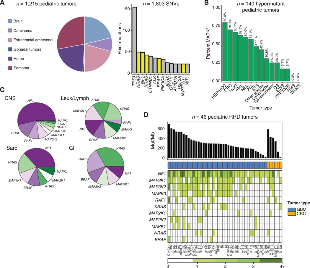
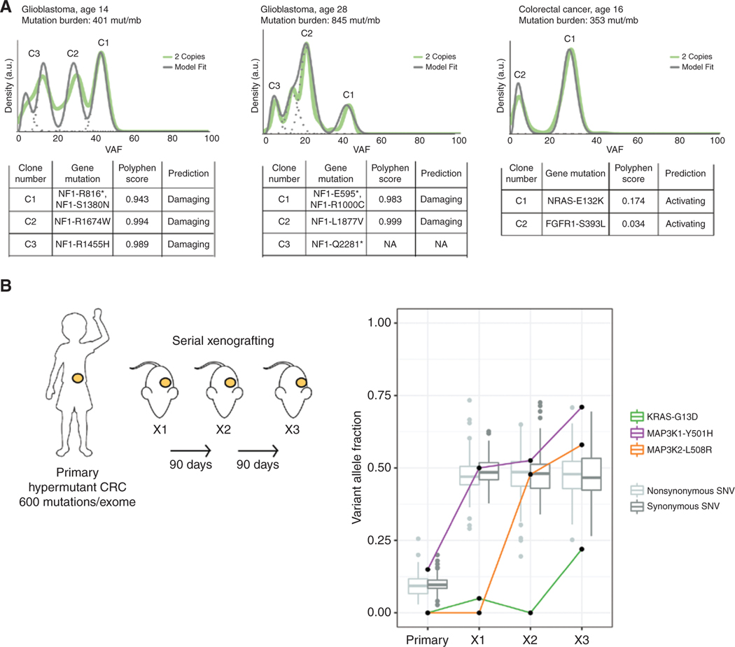
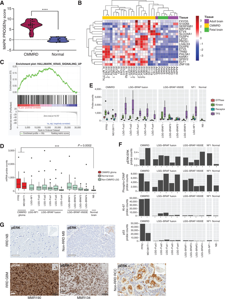
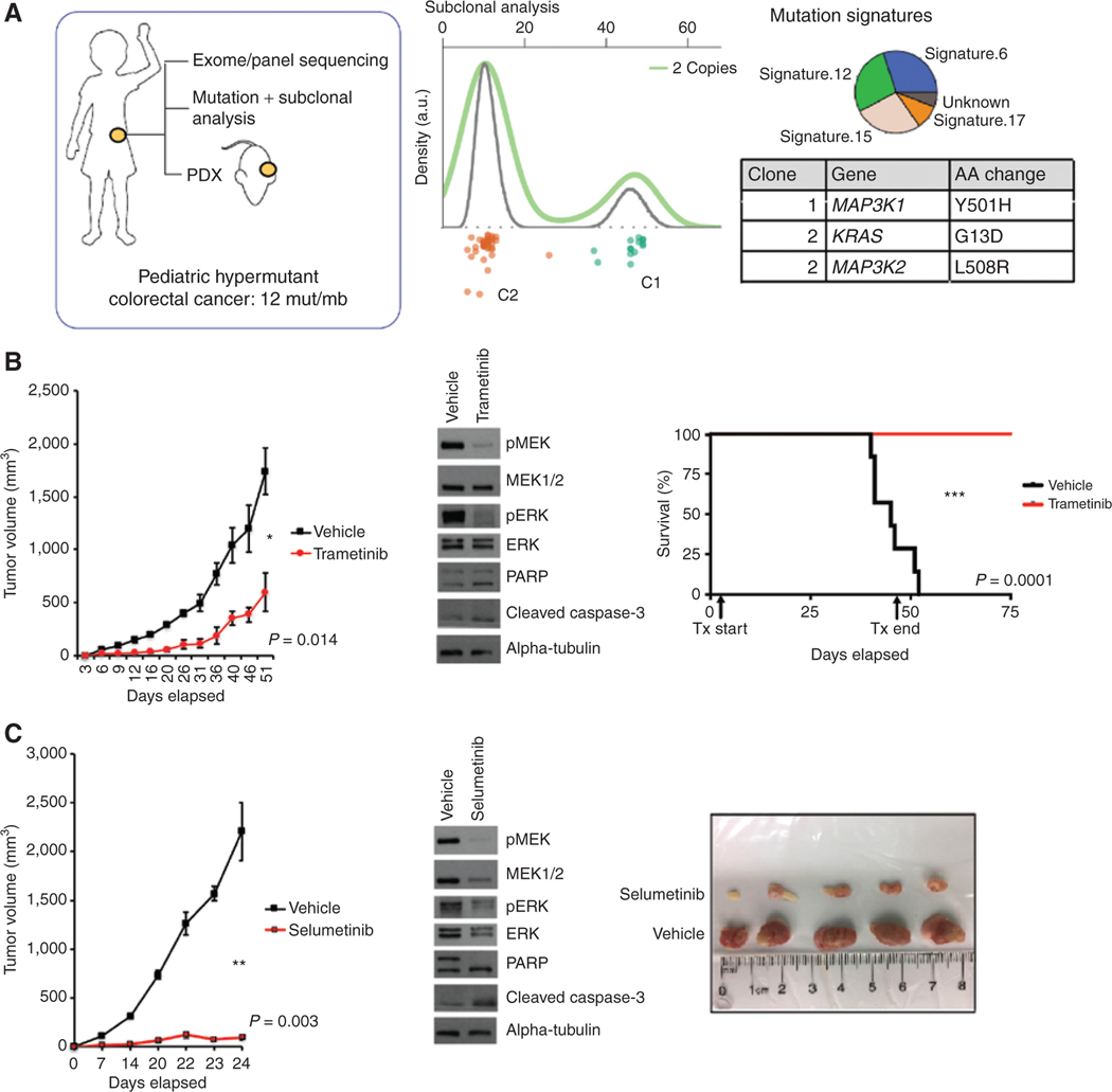
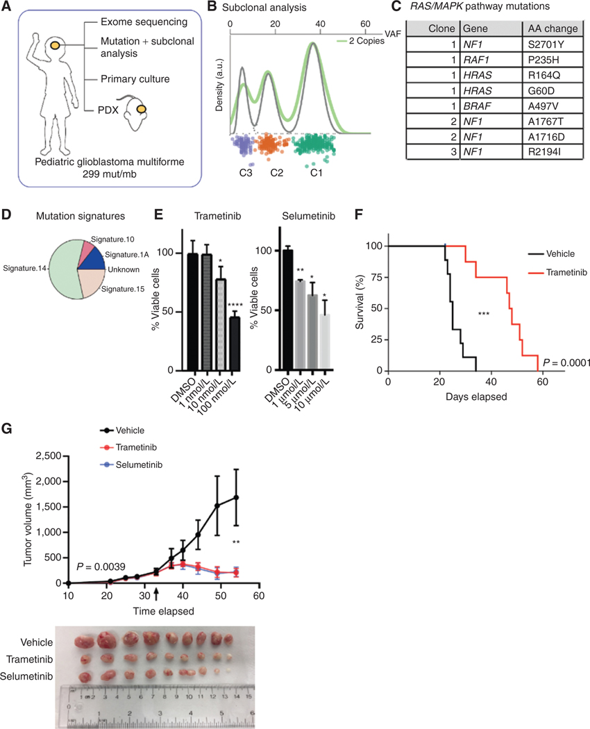
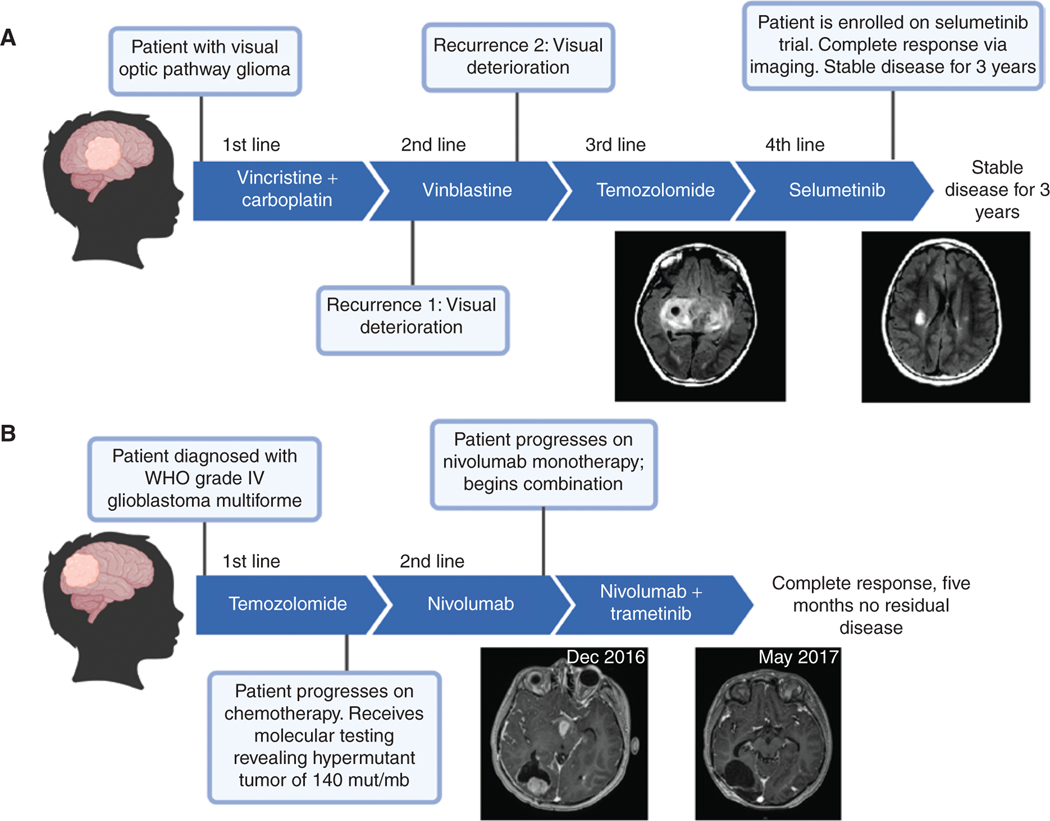
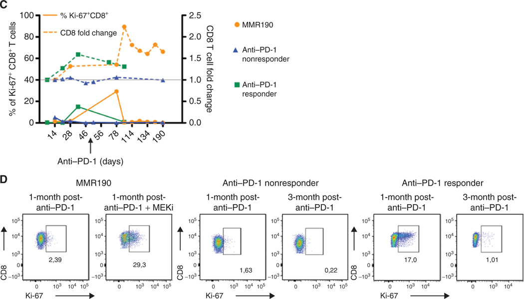
The RAS/MAPK pathway is an emerging targeted pathway across a spectrum of both adult and pediatric cancers. Typically, this is associated with a single, well-characterized point mutation in an oncogene. Hypermutant tumors that harbor many somatic mutations may obscure the interpretation of such targetable genomic events. We find that replication repair-deficient (RRD) cancers, which are universally hypermutant and affect children born with RRD cancer predisposition, are enriched for RAS/MAPK mutations (P = 10-8). These mutations are not random, exist in subclones, and increase in allelic frequency over time. The RAS/MAPK pathway is activated both transcriptionally and at the protein level in patient-derived RRD tumors, and these tumors responded to MEK inhibition in vitro and in vivo. Treatment of patients with RAS/MAPK hypermutant gliomas reveals durable responses to MEK inhibition. Our observations suggest that hypermutant tumors may be addicted to oncogenic pathways, resulting in favorable response to targeted therapies. SIGNIFICANCE: Tumors harboring a single RAS/MAPK driver mutation are targeted individually for therapeutic purposes. We find that in RRD hypermutant cancers, mutations in the RAS/MAPK pathway are enriched, highly expressed, and result in sensitivity to MEK inhibitors. Targeting an oncogenic pathway may provide therapeutic options for these hypermutant polyclonal cancers.This article is highlighted in the In This Issue feature, p. 1307.


Conflict of interest statement

Authors’ Disclosures

No disclosures were reported by the other authors.

Figures

Figure 1.
Figure 1.
Prevalence of RAS/MAPK genetic events across 1,215 pediatric cancers. A, Pie graph indicates the tissue of origin of 1,215 pediatric cancers. 1,803 SNVs were detected across the entire cohort; RAS/MAPK-activating events are indicated in yellow. B, Prevalence of RAS/MAPK mutations in hypermutant pediatric tumors, stratified by tumor type. HEP/HCC, hepatocellular carcinoma; CRC, colorectal cancer; HGG, high grade glioma; AML, acute myeloid leukemia; NBL, neuroblastoma; ALL, acute lymphocytic leukemia; STS, soft tissue sarcoma; OS, osteosarcoma; EWS, Ewing sarcoma; PNET/MB, primitive neuro-ectodermal tumors/medulloblastoma; RMS, rhabdomyosarcoma; WLMS, Wilms tumor. C, Pie graphs demonstrating the distribution of the 11 most commonly mutated RAS/MAPK genes across different tissues of origin. D, Exome sequencing of 46 glioblastomas (GBM) and colorectal cancers (CRC) from patients with germline mutations in MMR and POLE. Top, tumor mutation burden in mutations/mb; middle, tumor type and number of nonsynonymous, protein-coding events in 11 commonly mutated RAS/MAPK genes.
Figure 2.
Figure 2.
Prevalence of RAS/MAPK mutations in polyclonal and temporal sampling of RRD tumors. A, Clonal analysis of three heavily mutated tumors and the respective RAS/MAPK mutations in each clone. PolyPhen scores for each mutation distinguish damaging mutations in negative regulators, in contrast to benign or activating mutations in gain-of-function genes B, Schematic of serial xenografting experiment performed on one hypermutant patient-derived colorectal cancer (CRC) xenograft (left). Changes in the VAF of RAS/MAPK genes mutated in the primary and subsequent xenografts. All mutations increase in tumor fraction, suggesting positive selection, whereas other variants in the primary remain stable (right).
Figure 3.
Figure 3.
Assessment of RAS/MAPK pathway activation in hypermutant RRD gliomas. A, Violin plot of RNA-seq–derived MAPK pathway PROGENy signature scores of CMMRD GBMs (n = 21) compared with normal fetal (n = 4) and adult (n = 5) brains (P < 0.0001; Welch t test). Lines indicate median and quartile values. B, Unsupervised clustering of all samples in A based on expression of an 18-gene RAS transcriptional output signature. C, GSEA enrichment plot for genes upregulated by KRAS activation in CMMRD GBM vs. normal brain samples in A and B. NES is reported (FDR = 0). D, NanoString counts of single mRNA molecules in 20 RAS/MAPK pathway–related probes, including EGFR, BRAF, KRAS, MAP3K1, FOS, and JUN, in CMMRD GBM compared with tissue-matched BRAFV600E mutant low-grade gliomas. E, NanoString counts of single mRNA molecules in 20 RAS/MAPK pathway–related probes, stratified by protein-type. Transcription factor targets of the RAS/MAPK pathway were the most upregulated. F, NanoString counts of single protein molecules involved in RAS/MAPK pathway activation, including phospho-ERK and phospho-CRAF. Ki-67 protein levels are shown to highlight the distinguishing growth characteristics of LGG versus GBM. For box plots D–E, median and quartile values are depicted. G, Positive IHC staining for phospho-ERK (pERK) on two representative CMMRD brain tumors harboring several RAS/MAPK pathway alterations (bottom left; MMR190 and MMR134), compared with a CMMRD normal postmortem brain sample and non-RRD pediatric medulloblastoma demonstrating negative staining (top), and a human renal cell carcinoma (RCC) sample demonstrating nuclear and cytoplasmic staining of phospho-ERK in tubular epithelial structures surrounded by negatively staining lymphocytes (bottom right). Scale bars, 50 μm. ****, P < 0.0001.
Figure 4.
Figure 4.
Genomic characterization and subsequent xenografting and treatment of an ultrahypermutant childhood colorectal cancer. A, Schematic of tumor sequencing analysis performed on a patient-derived hypermutant colorectal cancer. Targeted panel sequencing revealed two major clones. A secondary KRASG13D known RAS/MAPK driver pathway mutation was acquired in the secondary clone. Mutation signatures within the tumor reveal a characteristic pattern of RRD. B, Tumor growth experiments in response to MEK inhibition therapy (trametinib) for flank-implanted colorectal cancer xenograft. Tumor volume was measured biweekly in trametinib treated vs. vehicle group. Western blot analysis of tissues and the conclusion of the experiment reveal a decrease in phospho-MEK and phospho-ERK. A second repeated experiment to demonstrate survival differences is shown. C, Tumor growth experiments in response to MEK inhibition therapy using selumetinib. Mice were sacrificed after 24 days to display differences in tumor sizes.
Figure 5.
Figure 5.
Genomic characterization and subsequent xenografting and treatment of an ultrahypermutant childhood glioblastoma. A, Schematic of experiments performed on a CMMRD pediatric glioblastoma. B, Subclonal analysis reveals multiple mutations per clone. C, Identity of RAS/MAPK pathway–related alterations in this tumor. D, Mutational signature analysis reveals the source of mutations as RRD. E, Primary culturing of cells derived from this tumor, without prior culturing, and treatment with either trametinib or selumetinib. F, Survival experiment following flank implantation of this ultrahypermutant GBM and subsequent treatment in vivo with trametinib. G, A second set of flank engraftment experiments were conducted to assess tumor growth following flank implantation and treatment with trametinib or selumetinib. Arrow indicates time at which treatment commenced.
Figure 6.
Figure 6.
Clinical response of patients with replication repair–deficient high-grade gliomas to MEK inhibitors. Previous failed treatment courses and sustained responses to selumetinib (A) and trametinib (B), respectively. C, Correlation between immune activation and clinical response to MEK inhibition in patient 2 (MMR190). Percentage of CD3+CD8+ PBMCs expressing Ki-67 (left axis) and fold change in total CD3+CD8+ PBMCs (right axis) over time in patients with GBM tumors that showed response to anti–PD-1 monotherapy, no response to anti–PD-1 monotherapy, and response to combination anti–PD-1 and the MEK inhibitor trametinib (MMR190—patient 2 from 6B). Arrow indicates start of trametinib administration. D, Ki-67 expression on CD3+CD8+ PBMCs one month following anti–PD-1 treatment initiation, and one month following combination anti–PD-1/MEK inhibition (MMR190) or three months following anti–PD-1 initiation (anti–PD-1 responder and nonresponder). MEKi = trametinib.
Figure 6.
Figure 6.
Clinical response of patients with replication repair–deficient high-grade gliomas to MEK inhibitors. Previous failed treatment courses and sustained responses to selumetinib (A) and trametinib (B), respectively. C, Correlation between immune activation and clinical response to MEK inhibition in patient 2 (MMR190). Percentage of CD3+CD8+ PBMCs expressing Ki-67 (left axis) and fold change in total CD3+CD8+ PBMCs (right axis) over time in patients with GBM tumors that showed response to anti–PD-1 monotherapy, no response to anti–PD-1 monotherapy, and response to combination anti–PD-1 and the MEK inhibitor trametinib (MMR190—patient 2 from 6B). Arrow indicates start of trametinib administration. D, Ki-67 expression on CD3+CD8+ PBMCs one month following anti–PD-1 treatment initiation, and one month following combination anti–PD-1/MEK inhibition (MMR190) or three months following anti–PD-1 initiation (anti–PD-1 responder and nonresponder). MEKi = trametinib.

References

    1. Campbell BB, Light N, Fabrizio D, Zatzman M, Fuligni F, de Borja R, et al.Comprehensive analysis of hypermutation in human cancer. Cell 2017;171:1042–56. - PMC - PubMed
    1. Gargiulo P, Della Pepa C, Berardi S, Califano D, Scala S, Buonaguro L, et al.Tumor genotype and immune microenvironment in POLE-ultramutated and MSI-hypermutated endometrial cancers: new candidates for checkpoint blockade immunotherapy? Cancer Treat Rev 2016;48:61–8. - PubMed
    1. Pritchard CC, Morrissey C, Kumar A, Zhang X, Smith C, Coleman I, et al.Complex MSH2 and MSH6 mutations in hypermutated microsatellite unstable advanced prostate cancer. Nat Commun 2014;5:4988. - PMC - PubMed
    1. Hunter C, Smith R, Cahill DP, Stephens P, Stevens C, Teague J, et al.A hypermutation phenotype and somatic MSH6 mutations in recurrent human malignant gliomas after alkylator chemotherapy. Cancer Res 2006;66:3987–91. - PMC - PubMed
    1. Touat M, Li YY, Boynton AN, Spurr LF, Iorgulescu JB, Bohrson CL, et al.Mechanisms and therapeutic implications of hypermutation in gliomas. Nature 2020;580:517–23. - PMC - PubMed

Publication types

MeSH terms

Substances