Case Reports
. 2021 Feb 9;14(2):e240070.
doi: 10.1136/bcr-2020-240070.

Endoscopic management of a giant dentigerous cyst

Affiliations
Case Reports

Endoscopic management of a giant dentigerous cyst

Vinay Raj Thattarakkal et al. BMJ Case Rep. .

Abstract

Dentigerous cyst is one of the most common developmental cyst of the jaw which accounts for approximately 20%-30% of bone cyst in the head and neck region. Most common site is the third molar of the mandible. However, maxillary involvement is not uncommon. The clinical presentation of this depends mainly on the size and anatomical compromise that occur due to compression. This case highlights the role of endoscopic approach in the management of large expansible cyst of maxilla involving the palate, thus preserving the anatomy and reducing the morbidity associated with an open procedure.

Keywords: dentistry and oral medicine; ear; nose and throat; oral and maxillofacial surgery.


Conflict of interest statement

Competing interests: None declared.

Figures

Figure 1
Figure 1
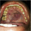
Intraoral clinical photograph of the oral cavity shows a solitary swelling seen in the centre of hard palate.
Figure 2
Figure 2
Diagnostic nasal endoscopy shows smooth pink mass in the right nasal cavity pushing the septum to opposite side.
Figure 3
Figure 3
CT of nose and paranasal sinus axial and coronal section shows hypodense lesion completely filling right maxillary sinus and extending to the nasal cavities and causing considerable bony erosions.
Figure 4
Figure 4
Intraoperative photograph shows the cyst was incised.
Figure 5
Figure 5
Intraoperative photograph shows the complete removal of cyst.
Figure 6
Figure 6
HPE shows a cyst lined by non-keratinised stratified squamous enamel epithelium.
Figure 7
Figure 7
Postoperative photograph shows the completeness of removal.

References

    1. Kirtaniya BC, Sachdev V, Singla A, et al. . Marsupialization: a conservative approach for treating dentigerous cyst in children in the mixed dentition. J Indian Soc Pedod Prev Dent 2010;28:203–8. 10.4103/0970-4388.73795 - DOI - PubMed
    1. Aher V, Chander PM, Chikkalingaiah RG, et al. . Dentigerous cysts in four quadrants: a rare and first reported case. J Surg Tech Case Rep 2013;5:21–6. 10.4103/2006-8808.118607 - DOI - PMC - PubMed
    1. Benn A, Altini M. Dentigerous cysts of inflammatory origin. A clinicopathologic study. Oral Surg Oral MedOral Pathol Oral Radiol Endod 1996;81:203–9. 10.1016/S1079-2104(96)80416-1 - DOI - PubMed
    1. Tilakraj TN, Kiran NK, Mukunda KS, et al. . Non syndromic unilateral dentigerous cyst in a 4-year-old child: a rare case report. Contemp Clin Dent 2011;2:398–401. 10.4103/0976-237X.91813 - DOI - PMC - PubMed
    1. Ide F, Kikuchi K, Miyazaki Y, et al. . Archegonous cystic Odontoma is not necessarily primordial. Head Neck Pathol 2016;10:418–21. 10.1007/s12105-016-0684-y - DOI - PMC - PubMed

Publication types