. 2021;97(2):89-102.
doi: 10.2183/pjab.97.005.

JF1/B6F1 Ngly1-/- mouse as an isogenic animal model of NGLY1 deficiency

Affiliations

JF1/B6F1 Ngly1-/- mouse as an isogenic animal model of NGLY1 deficiency

Makoto Asahina et al. Proc Jpn Acad Ser B Phys Biol Sci. 2021.

Abstract

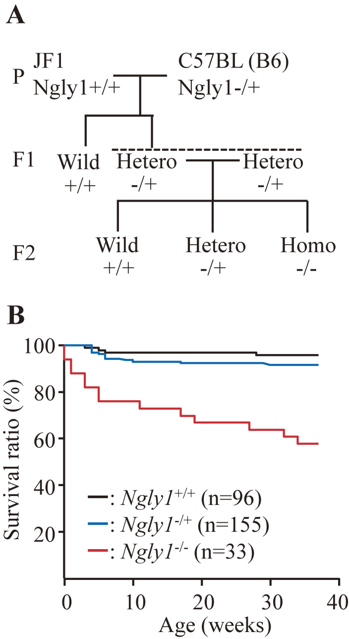
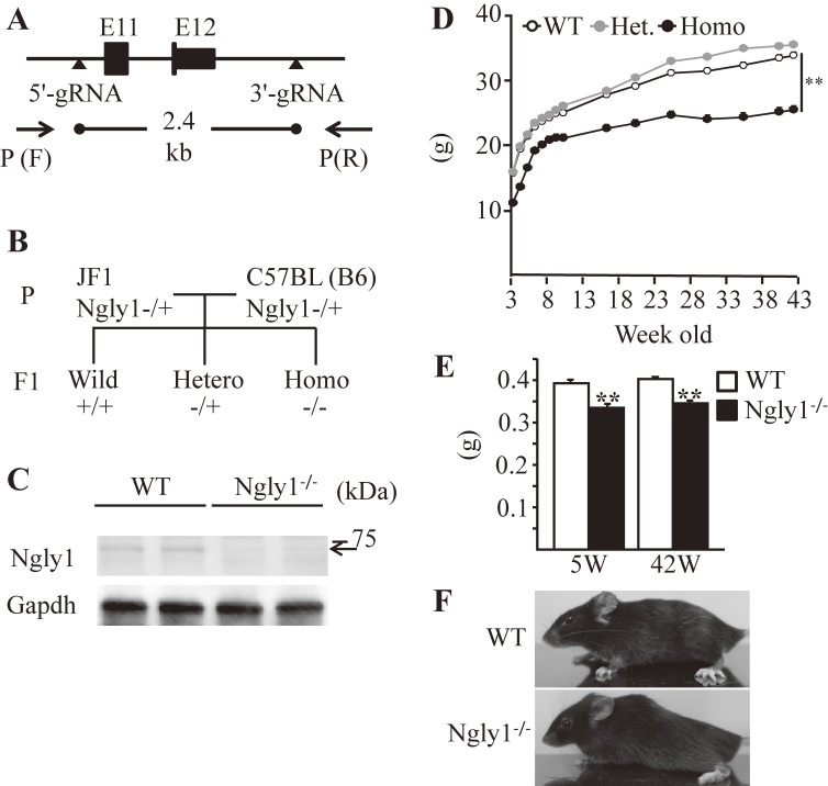
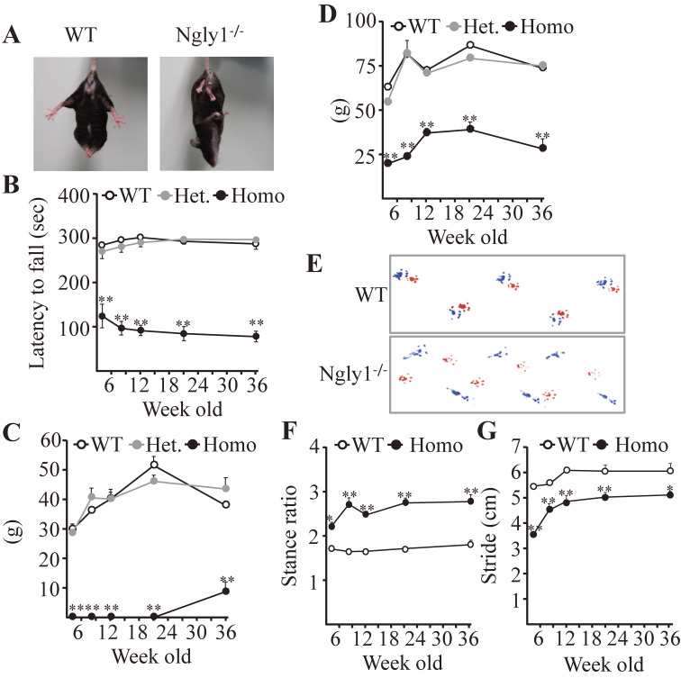
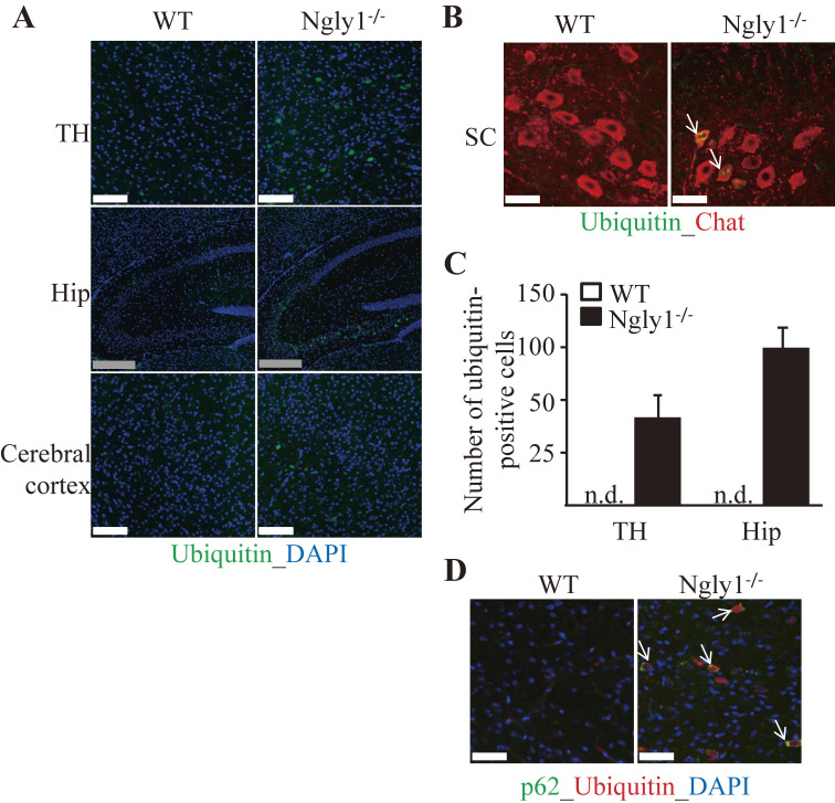
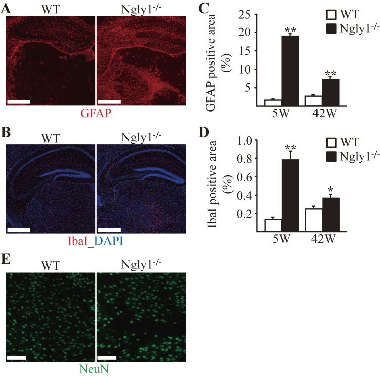
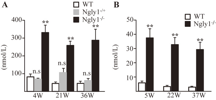
N-Glycanase 1 (NGLY1) deficiency is a congenital disorder caused by mutations in the NGLY1 gene. Because systemic Ngly1-/- mice with a C57BL/6 (B6) background are embryonically lethal, studies on the mechanism of NGLY1 deficiency using mice have been problematic. In this study, B6-Ngly1-/+ mice were crossed with Japanese wild mice-originated Japanese fancy mouse 1 (JF1) mice to produce viable F2 Ngly1-/- mice from (JF1×B6)F1 Ngly1-/+ mice. Systemic Ngly1-/- mice with a JF1 mouse background were also embryonically lethal. Hybrid F1 Ngly1-/- (JF1/B6F1) mice, however, showed developmental delay and motor dysfunction, similar to that in human patients. JF1/B6F1 Ngly1-/- mice showed increased levels of plasma and urinary aspartylglycosamine, a potential biomarker for NGLY1 deficiency. JF1/B6F1 Ngly1-/- mice are a useful isogenic animal model for the preclinical testing of therapeutic options and understanding the precise pathogenic mechanisms responsible for NGLY1 deficiency.

Keywords: NGLY1 deficiency; animal model; aspartylglycosamine; congenital neurological disorder; motor dysfunction; neuroinflammation.


Figures

Figure 1.
Figure 1.
Production and survival ratio of (JF1/B6F1 Ngly1−/+)F2 mice. (A) Schematic figure showing the generation of (JF1/B6F1 Ngly1−/+)F2 mice. B6-Ngly1−/+ mice were crossed with JF1 mice to produce JF1/B6F1 Ngly1−/+ mice. JF1/B6F1 Ngly1−/+ mice were further crossed to produce F2 mice. (B) Survival curve for F2 mice.
Figure 2.
Figure 2.
Generation, appearance, and body weights of JF1/B6F1 Ngly1−/− mice. (A) Schematic figure showing the generation of JF1-Ngly1−/− mouse using CRISPR/Cas9 genome editing technology. (B) Production of JF1/B6F1 Ngly1-deficient mouse. (C) Western blotting analysis of NGLY1 expression in the brains of Ngly1−/− and their littermate WT mice. (D) Body weight of male JF1/B6F1 Ngly1−/−, Ngly1−/+, and WT mice (n = 16–18). Mice were weighed after weaning (at 4 weeks of age). (E) Brain weights at 5 and 42 weeks of age in male mice. (F) Representative scoliosis in male 42-week-old Ngly1−/− and age-matched WT mice. Mean ± SEM (n = 6–8, each genotype). **P < 0.01 (Student’s t-test).
Figure 3.
Figure 3.
Motor function in male JF1/B6F1 Ngly1−/− mice compared to their littermate WT mice. (A) Abnormal hindlimb clasping of mice when suspended by the tail. (B) Rotarod test for motor coordination of mice. The time until dropping occurred from the accelerating rod is shown. (C and D) Grip-strength test for the assessment of forelimb (C) or forelimb and hindlimb (D) muscle force. (E–G) Gait analysis: Representative paw placement records of 36-week-old mice (red for forepaws and blue for hind paws; E), stance ratios (F), and stride lengths (G). Mean ± SEM. The number of mice examined was 10 to 12 each. **P < 0.01; *P < 0.05.
Figure 4.
Figure 4.
Accumulation of ubiquitinated proteins in the CNS of JF1/B6F1 Ngly1−/− mice. (A and B) Ubiquitin-positive proteins in JF1/B6F1 Ngly1−/− mice. Immunohistochemistry of hippocampus (Hip), thalamus (TH), cerebral cortex, and spinal cord (SC) sections from JF1/B6F1 Ngly1−/− and WT mice at 5 weeks of age, stained with an anti-ubiquitin antibody. Nuclei were stained with 4′,6-diamidino-2-phenylindole (DAPI). (A) White scale bar, 100 µm; gray scale bar, 250 µm. (B) Arrows indicate ubiquitin-positive cells positive for ChAT, a marker of motor neurons. Scale bar, 50 µm. (C) Number of ubiquitin-positive cells in the hippocampus and thalamus in JF1/B6F1 Ngly1−/− and WT mice at 5 weeks of age. Mean ± SEM (n = 5 or 6). (D) Colocalization of ubiquitin and p62 in JF1/B6F1 Ngly1−/− mice at 5 weeks of age. Arrows indicate ubiquitin- and p62-positive cells. Scale bar, 50 µm.
Figure 5.
Figure 5.
Glial activation in the thalamus and hippocampus of JF1/B6F1 Ngly1−/− mice. Immunohistochemistry for GFAP (A) and IbaI (B) in the thalamus and hippocampus of JF1/B6F1 Ngly1−/− mice and their littermate WT mice at 5 weeks of age. Scale bar, 0.5 mm. Nuclei were stained with DAPI. (C and D) Quantitative analyses showing the area occupied by GFAP-positive (C) or IbaI-positive (D) areas in the thalamus of WT and Ngly1−/− mice. Mean ± SEM (n = 4–6). *P < 0.05; **P < 0.01. (E) Immunohistochemistry of the thalamus from JF1/B6F1 Ngly1−/− and their littermate WT mice at 5 weeks of age, stained with anti-NeuN, a mature neuron marker (green). Nuclei were stained with DAPI. Scale bar, 100 µm.
Figure 6.
Figure 6.
Increased AsnGlcNAc levels in plasma (A) and urine (B) of JF1/B6F1 Ngly1−/− mice. Plasma and urine of five mice in each genotype were analyzed. **P < 0.01, WT mice compared to hetero- or homo-KO mice.

References

    1. Suzuki T., Park H., Hollingsworth N.M., Sternglanz R., Lennarz W.J. (2000) PNG1, a yeast gene encoding a highly conserved peptide:N-glycanase. J. Cell Biol. 149, 1039–1052. - PMC - PubMed
    1. Suzuki T., Huang C., Fujihira H. (2016) The cytoplasmic peptide:N-glycanase (NGLY1) — Structure, expression and cellular functions. Gene 577, 1–7. - PMC - PubMed
    1. Hirsch C., Blom D., Ploegh H.L. (2003) A role for N-glycanase in the cytosolic turnover of glycoproteins. EMBO J. 22, 1036–1046. - PMC - PubMed
    1. Huang C., Harada Y., Hosomi A., Masahara-Negishi Y., Seino J., Fujihira H., et al. (2015) Endo-β-N-acetylglucosaminidase forms N-GlcNAc protein aggregates during ER-associated degradation in Ngly1-defective cells. Proc. Natl. Acad. Sci. U.S.A. 112, 1398–1403. - PMC - PubMed
    1. Suzuki T. (2015) The cytoplasmic peptide:N-glycanase (Ngly1) — Basic science encounters a human genetic disorder. J. Biochem. 157, 23–24. - PubMed

MeSH terms

Substances

Supplementary concepts