. 2021 Jan 30:2021:1250858.
doi: 10.1155/2021/1250858. eCollection 2021.

BGP-15 Protects against Heart Failure by Enhanced Mitochondrial Biogenesis and Decreased Fibrotic Remodelling in Spontaneously Hypertensive Rats

Affiliations

BGP-15 Protects against Heart Failure by Enhanced Mitochondrial Biogenesis and Decreased Fibrotic Remodelling in Spontaneously Hypertensive Rats

Orsolya Horvath et al. Oxid Med Cell Longev. .

Abstract

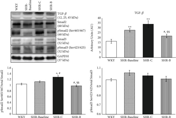
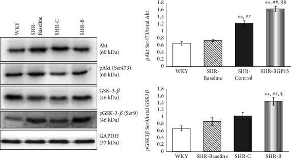
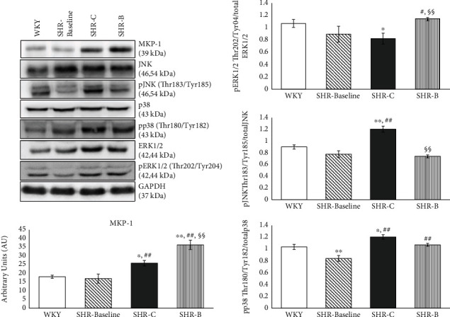
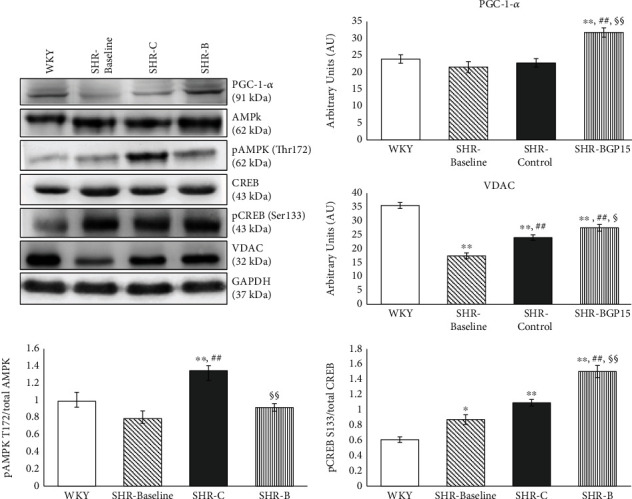
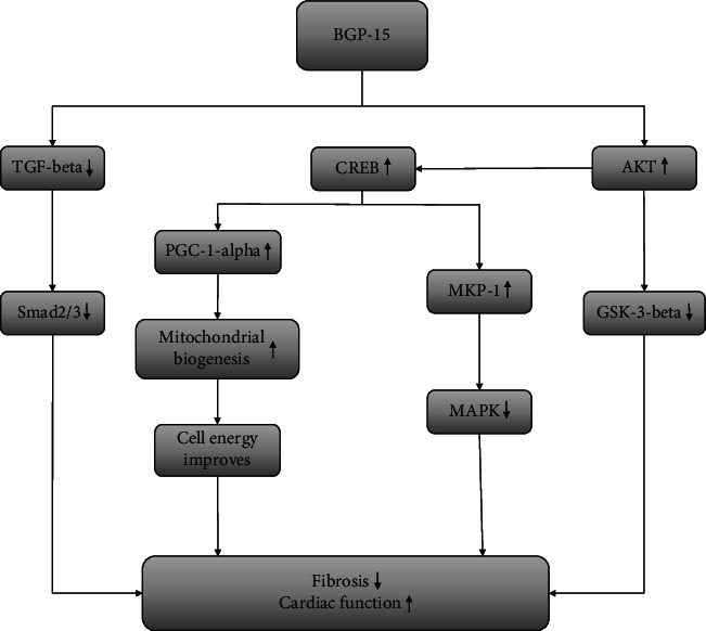
Heart failure (HF) is a complex clinical syndrome with poor clinical outcomes despite the growing number of therapeutic approaches. It is characterized by interstitial fibrosis, cardiomyocyte hypertrophy, activation of various intracellular signalling pathways, and damage of the mitochondrial network. Mitochondria are responsible for supplying the energy demand of cardiomyocytes; therefore, the damage of the mitochondrial network causes cellular dysfunction and finally leads to cell death. BGP-15, a hydroxylamine derivative, is an insulin-sensitizer molecule and has a wide range of cytoprotective effects in animal as well as in human studies. Our recent work was aimed at examining the effects of BGP-15 in a chronic hypertension-induced heart failure model. 15-month-old male SHRs were used in our experiment. The SHR-Baseline group represented the starting point (n = 7). Animals received BGP-15 (SHR-B, n = 7) or placebo (SHR-C, n = 7) for 18 weeks. WKY rats were used as age-matched normotensive controls (n = 7). The heart function was monitored by echocardiography. Histological preparations were made from cardiac tissue. The levels of signalling proteins were determined by Western blot. At the end of the study, systolic and diastolic cardiac function was preserved in the BGP-treated animals. BGP-15 decreased the interstitial collagen deposition via decreasing the activity of TGFβ/Smad signalling factors and prevented the cardiomyocyte hypertrophy in hypertensive animals. BGP-15 enhanced the prosurvival signalling pathways (Akt/Gsk3β). The treatment increased the activity of MKP1 and decreased the activity of p38 and JNK signalling routes. The mitochondrial mass of cardiomyocytes was also increased in BGP-15-treated SHR animals due to the activation of mitochondrial biogenesis. The mitigation of remodelling processes and the preserved systolic cardiac function in hypertension-induced heart failure can be a result-at least partly-of the enhanced mitochondrial biogenesis caused by BGP-15.


Conflict of interest statement

On behalf of all authors, the corresponding author states that there is no conflict of interest.

Figures

Figure 1
Figure 1
Effect of BGP-15 treatment on the extent of interstitial fibrosis, collagen type I deposition, and on the diameter of cardiomyocytes. Representative histological sections stained with Picrosirius red (a) (n = 7). Scale bar: 150 μm, magnification: 10-fold. Densitometric evaluation of the sections is shown (b). p < 0.05 vs. WKY, ∗∗p < 0.01 vs. WKY, ##p < 0.01 vs. SHR-Baseline, §§p < 0.01 vs. SHR-C. Representative histological sections detected with collagen type I immunohistochemistry (c) (n = 7). Scale bar: 100 μm, magnification: 10-fold. Densitometric evaluation of the sections is shown (d). ∗∗p < 0.01 vs. WKY, ##p < 0.01 vs. SHR-C, §§p < 0.01 vs. SHR-C. Representative histological sections stained with Picrosirius red (e) (n = 7). Scale bar: 50 μm, magnification: 10-fold. The average cellular diameter in the different groups is shown (f). ∗∗p < 0.01 vs. WKY, #p < 0.05 vs. SHR-Baseline, §§p < 0.01 vs. SHR-C. WKY: age-matched normotensive Wistar-Kyoto rats; SHR-Baseline: 15-month-old spontaneously hypertensive rats; SHR-C: 19-month-old spontaneously hypertensive rats received placebo for 18 weeks; SHR-B: 19-month-old spontaneously hypertensive rats received BGP-15 for 18 weeks.
Figure 2
Figure 2
Effect of BGP-15 treatment on the TGFβ/Smad signalling pathway. Representative Western blot analysis of TGFβ, Smad2, Smad3, and phosphorylation and densitometric evaluation are shown. GAPDH was used as a loading control. WKY: age-matched normotensive Wistar-Kyoto rats, n = 7; SHR-Baseline: 15-month-old spontaneously hypertensive rats, n = 7; SHR-C: nontreated spontaneously hypertensive rats, n = 7; SHR-B: spontaneously hypertensive rats receiving BGP-15 for 18 weeks, n = 7. Values are mean ± SEM. p < 0.05 vs. WKY, ∗∗p < 0.01 vs. WKY, #p < 0.05 vs. SHR-Baseline, §§p < 0.01 vs. SHR-C.
Figure 3
Figure 3
Effect of BGP-15 treatment on the phosphorylation of Akt-1Ser473 and GSK-3βSer9. Representative Western blot analysis of Akt-1 and GSK-3β phosphorylation and densitometric evaluation are shown. GAPDH was used as a loading control. WKY: age-matched normotensive Wistar-Kyoto rats, n = 7; SHR-Baseline: 15-month-old spontaneously hypertensive rats, n = 7; SHR-C: nontreated spontaneously hypertensive rats, n = 7; SHR-B: spontaneously hypertensive rats receiving BGP-15 for 18 weeks, n = 7. Values are mean ± SEM. ∗∗p < 0.01 vs. WKY, ##p < 0.01 vs. SHR-Baseline, §p < 0.05 vs. SHR-C, §§p < 0.01 vs. SHR-C.
Figure 4
Figure 4
Effect of the BGP-15 treatment on the phosphorylation state of MAP kinases and on MKP-1. Representative Western blot analysis of MKP-1 as well as ERK1/2, p38, and JNK phosphorylation. Densitometric evaluation is also shown. GAPDH was used as a loading control. WKY: age-matched normotensive Wistar-Kyoto rats, n = 7; SHR-Baseline: 15-month-old spontaneously hypertensive rats, n = 7; SHR-C: nontreated spontaneously hypertensive rats, n = 7; SHR-B: spontaneously hypertensive rats receiving BGP-15 for 18 weeks, n = 7. Values are mean ± SEM. p < 0.05 vs. WKY, ∗∗p < 0.01 vs. WKY, ##p < 0.01 vs. SHR-Baseline, §§p < 0.01 vs. SHR-C.
Figure 5
Figure 5
Effect of the BGP-15 treatment on the regulation of mitochondrial biogenesis. Representative Western blot analysis of PGC-1α, VDAC, CREB, AMPK, and phosphorylation of CREB and AMPK. Densitometric evaluation is also shown. GAPDH was used as a loading control. WKY: age-matched normotensive Wistar-Kyoto rats, n = 7; SHR-Baseline: 15-month-old spontaneously hypertensive rats, n = 7; SHR-C: nontreated spontaneously hypertensive rats, n = 7; SHR-B: spontaneously hypertensive rats receiving BGP-15 for 18 weeks, n = 7. Values are mean ± SEM. p < 0.05 vs. WKY, ∗∗p < 0.01 vs. WKY, ##p < 0.01 vs. SHR-Baseline, §p < 0.05 vs. SHR-C, §§p < 0.01 vs. SHR-C.
Figure 6
Figure 6
The suspected mechanism of BGP-15 treatment in a hypertension-induced heart failure model. BGP-15 has a beneficial effect against hypertension-induced cardiac remodelling and cardiac fibrosis. The BGP-15 treatment decreases the activity of TGFβ/Smad and MAPK signalling factors and in this way prevents against hypertension-induced interstitial collagen deposition. BGP-15 favourably influences the prosurvival signalling pathways. Moreover, the mitochondrial biogenesis is activated due to BGP-15 administration, thereby resulting in an increase in mitochondrial mass.

References

    1. Benjamin E. J., Muntner P., Alonso A., et al. Heart Disease and Stroke Statistics—2019 update: a report from the American Heart Association. Circulation. 2019;139:e56–e528. doi: 10.1161/CIR.0000000000000659. - DOI - PubMed
    1. Kemp C. D., Conte J. V. The pathophysiology of heart failure. Cardiovascular Pathology. 2012;21(5):365–371. doi: 10.1016/j.carpath.2011.11.007. - DOI - PubMed
    1. Jarraya F. Treatment of hypertension: which goal for which patient? Advances in Experimental Medicine and Biology. 2016;956:117–127. doi: 10.1007/5584_2016_97. - DOI - PubMed
    1. Kong P., Christia P., Frangogiannis N. G. The pathogenesis of cardiac fibrosis. Cellular and Molecular Life Sciences. 2014;71(4):549–574. doi: 10.1007/s00018-013-1349-6. - DOI - PMC - PubMed
    1. Travers J. G., Kamal F. A., Robbins J., Yutzey K. E., Blaxall B. C. Cardiac fibrosis: the fibroblast awakens. Circulation Research. 2016;118(6):1021–1040. doi: 10.1161/CIRCRESAHA.115.306565. - DOI - PMC - PubMed

MeSH terms