. 2021 Apr 1;320(4):L568-L582.
doi: 10.1152/ajplung.00472.2020. Epub 2021 Feb 10.

YAP expression in endothelial cells prevents ventilator-induced lung injury

Affiliations

YAP expression in endothelial cells prevents ventilator-induced lung injury

Kai Su et al. Am J Physiol Lung Cell Mol Physiol. .

Abstract

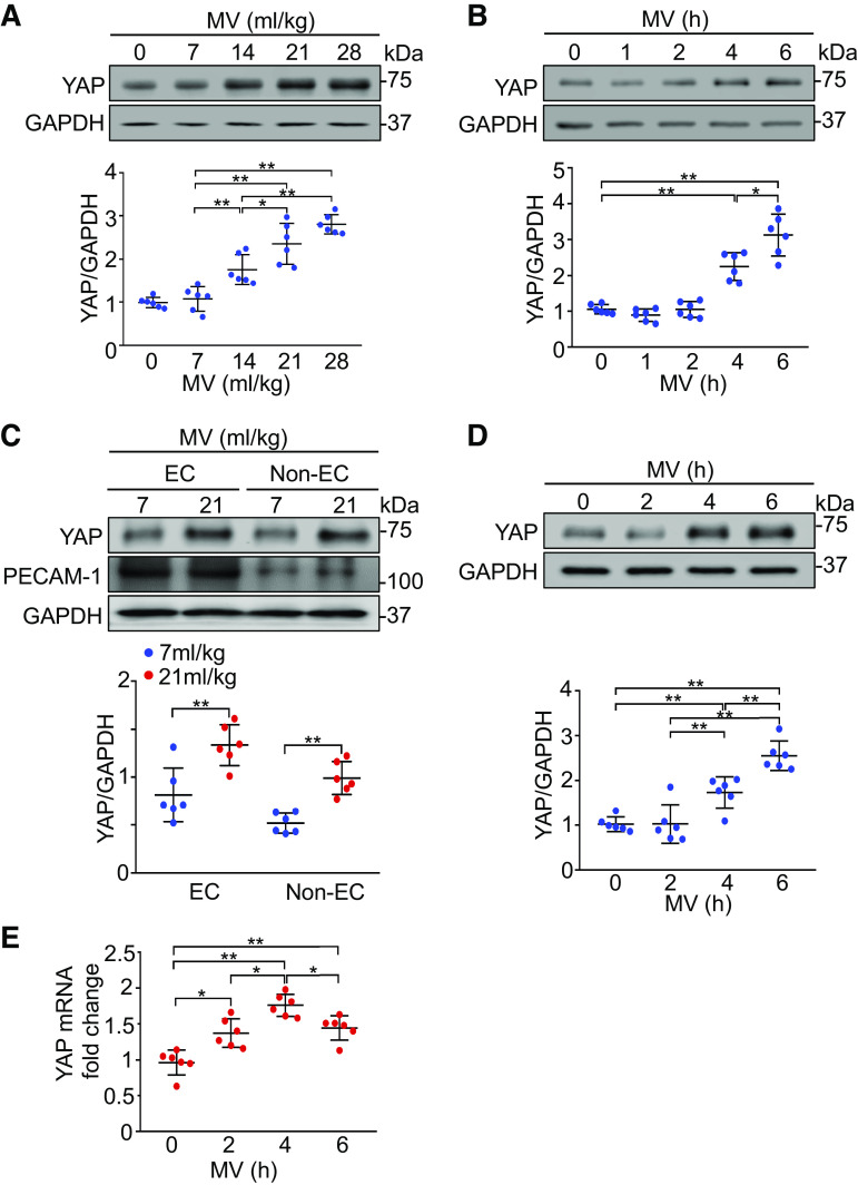
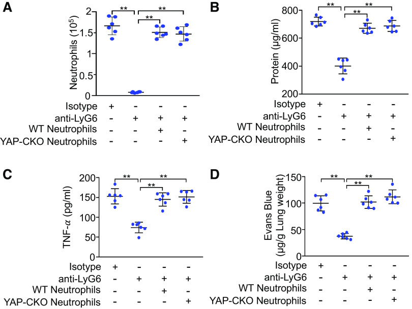
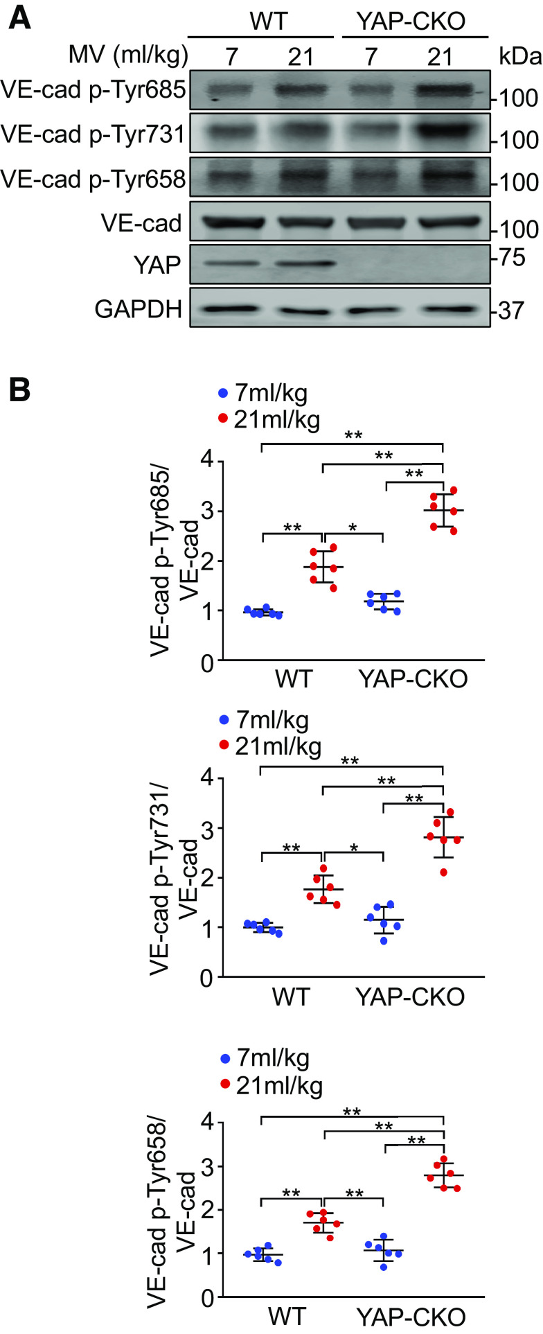
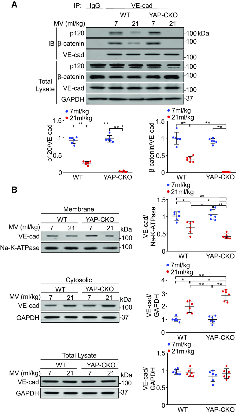
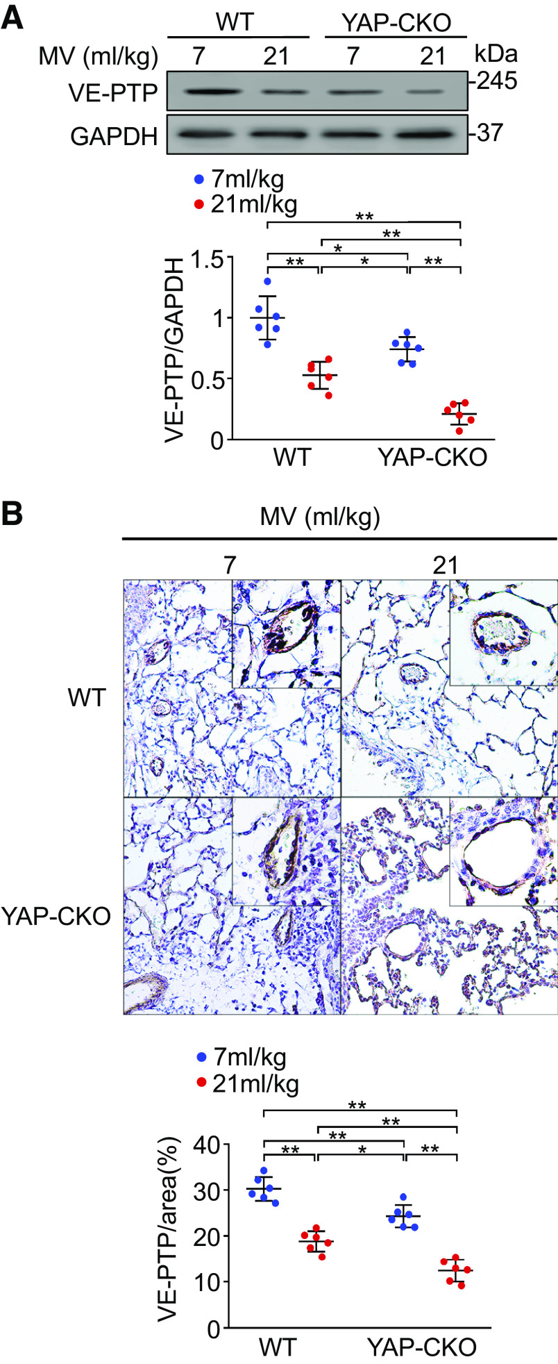
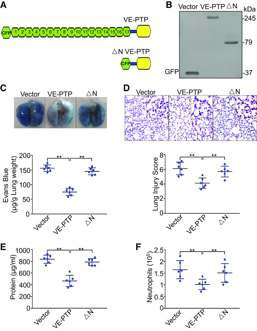
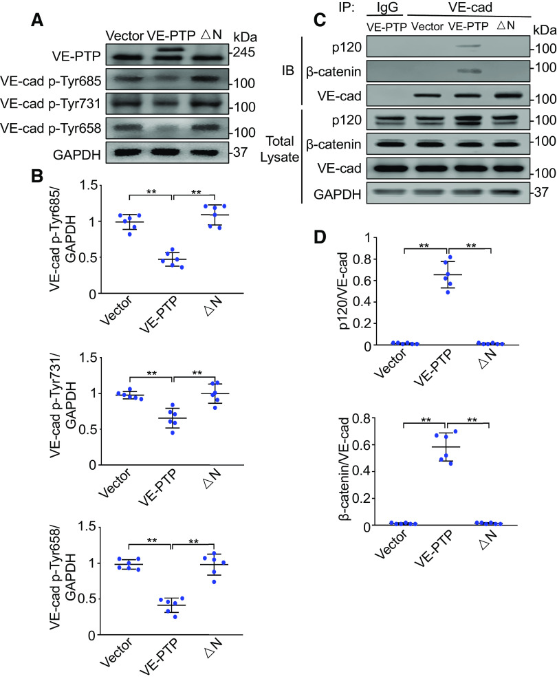
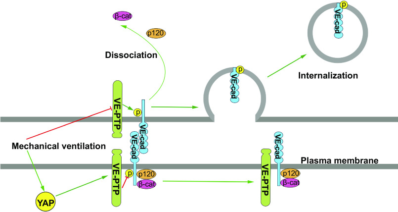
Ventilator-induced lung injury is associated with an increase in mortality in patients with respiratory dysfunction, although mechanical ventilation is an essential intervention implemented in the intensive care unit. Intrinsic molecular mechanisms for minimizing lung inflammatory injury during mechanical ventilation remain poorly defined. We hypothesize that Yes-associated protein (YAP) expression in endothelial cells protects the lung against ventilator-induced injury. Wild-type and endothelial-specific YAP-deficient mice were subjected to a low (7 mL/kg) or high (21 mL/kg) tidal volume (VT) ventilation for 4 h. Infiltration of inflammatory cells into the lung, vascular permeability, lung histopathology, and the levels of inflammatory cytokines were measured. Here, we showed that mechanical ventilation with high VT upregulated YAP protein expression in pulmonary endothelial cells. Endothelial-specific YAP knockout mice following high VT ventilation exhibited increased neutrophil counts and protein content in bronchoalveolar lavage fluid, Evans blue leakage, and histological lung injury compared with wild-type littermate controls. Deletion of YAP in endothelial cells exaggerated vascular endothelial (VE)-cadherin phosphorylation, downregulation of vascular endothelial protein tyrosine phosphatase (VE-PTP), and dissociation of VE-cadherin and catenins following mechanical ventilation. Importantly, exogenous expression of wild-type VE-PTP in the pulmonary vasculature rescued YAP ablation-induced increases in neutrophil counts and protein content in bronchoalveolar lavage fluid, vascular leakage, and histological lung injury as well as VE-cadherin phosphorylation and dissociation from catenins following ventilation. These data demonstrate that YAP expression in endothelial cells suppresses lung inflammatory response and edema formation by modulating VE-PTP-mediated VE-cadherin phosphorylation and thus plays a protective role in ventilator-induced lung injury.

Keywords: Yes-associated protein; inflammation; vascular endothelial protein tyrosine phosphatase; vascular endothelial-cadherin; ventilator-induced lung injury.


Conflict of interest statement

No conflicts of interest, financial or otherwise, are declared by the authors.

Figures

Figure 1.
Figure 1.
Mechanical ventilation (MV) induces YAP expression in mouse lungs and endothelial cells (ECs). A: effects of different tidal volumes on expression of YAP in the lung. Mice were ventilated at indicated tidal volumes for 4 h. Top: representative Western blots for YAP expression; bottom: protein quantification (normalized to GAPDH) by densitometry. n =6 lungs (from three male and three female mice). *P < 0.05, **P < 0.01, one-way ANOVA with Bonferroni post hoc test. B: time course of YAP expression in the lung following ventilation. Mice were ventilated at a tidal volume of 21 mL/kg for the indicated times. Top: representative Western blots for YAP expression; bottom: protein quantification (normalized to GAPDH) by densitometry. n = 6 lungs (from three male and three female mice). *P < 0.05, **P < 0.01 versus 14 mL/kg group, one-way ANOVA with Bonferroni post hoc test. C: YAP expression in mouse lung EC following ventilation. Lung endothelial cells (ECs) and nonendothelial cells (non-ECs) were isolated from mice ventilated at a tidal volume of 21 mL/kg for 4 h. Top: representative Western blots for YAP expression; middle and bottom: protein quantification (normalized to GAPDH) by densitometry. n =6 lungs (from 3 male and 3 female mice). **P < 0.01, independent two-sample t test. D: time course of YAP expression in lung endothelial cells following ventilation. Lung endothelial cells were isolated from mice ventilated at a tidal volume of 21 mL/kg for the indicated times. Top: representative Western blots for total YAP expression; bottom: protein quantification (normalized to GAPDH) by densitometry. n =6 lungs (from 3 male and 3 female mice). *P < 0.05, **P < 0.01, one-way ANOVA with Bonferroni post hoc test. E: quantitative analysis of YAP mRNA levels in lung endothelial cells by real-time PCR and normalized to control. n =6 lungs (from 3 male and 3 female mice). *P < 0.05, **P < 0.01, one-way ANOVA with Bonferroni post hoc test. Data represent means ± SD. YAP, Yes-associated protein.
Figure 2.
Figure 2.
YAP deletion in endothelial cells increases lung inflammatory injury following mechanical ventilation (MV). Wild-type (WT) and endothelial cell-specific YAP knockout mice (YAP-CKO) were ventilated at indicated tidal volumes for 4 h. A: pulmonary vascular permeability measured by Evans blue dye extravasation. Top: representative lung appearance after Evans blue dye administration. Bottom: quantitative analysis of Evans blue-labeled albumin extravasation. n =6/group. **P < 0.001, two-way ANOVA with Bonferroni post hoc test. B: protein concentrations in bronchoalveolar lavage fluid. C: hematoxylin and eosin staining of sections of lungs. Top: representative lung histology. Magnification, ×20; inset, ×40. Bottom: quantification of histopathological lung injury scores. n =6/group (3 male and 3 female mice). **P < 0.001, two-way ANOVA with Bonferroni post hoc test. All images for control and treatment groups were collected at the same time under the same conditions. D: neutrophil count in bronchoalveolar lavage fluid by cytospin analysis. n =6/group (3 male and 3 female mice). *P < 0.05, **P < 0.001, two-way ANOVA with Bonferroni post hoc test. E: MPO activity in the lung tissues. **P < 0.001, two-way ANOVA with Bonferroni post hoc test. F and G: levels of TNF-α (F) and IL-6 (G) in the bronchoalveolar lavage fluid of ventilated WT and endothelial cell-specific YAP knockout mice (YAP-CKO). n = 6/group (three male and three female mice). **P < 0.001, two-way ANOVA with Bonferroni post hoc test. Data represent means ± SD. YAP, Yes-associated protein.
Figure 3.
Figure 3.
Effects of neutrophil depletion and repletion on lung inflammatory injury. Endothelial cell-specific YAP knockout mice (YAP-CKO) were intraperitonially injected with isotype control immunoglobulin (Ig) G2a antibody or monoclonal anti-Ly6G antibody for 3 consecutive days. Neutrophil repletion was performed in neutropenic mice with neutrophils (2 × 106 cells/mouse, iv) isolated from wild-type (WT) or endothelial cell-specific YAP-CKO mice. All mice were ventilated with a tidal volume of 21 mL/kg for 4 h. A: neutrophil count in bronchoalveolar lavage fluid. B: protein concentrations in bronchoalveolar lavage fluid. C: levels of TNF-α in the bronchoalveolar lavage fluid. D: pulmonary vascular permeability measured by Evans blue dye extravasation. n = 6/group (three male and three female mice). **P < 0.001, two-way ANOVA with Bonferroni post hoc test. Data represent means ± SD. YAP, Yes-associated protein.
Figure 4.
Figure 4.
YAP deletion in endothelial cells increases tyrosine phosphorylation of VE-cadherin (VE-cad) following mechanical ventilation (MV). Wild-type (WT) and endothelial cell-specific YAP knockout mice (YAP-CKO) were ventilated at indicated tidal volumes for 4 h and endothelial cells were isolated from the lung. A: representative Western blots for expression of total VE-cadherin (VE-cad), phospho-Tyr685-VE-cad (VE-cad-Tyr685), VE-cad-Tyr731, and VE-cad-Tyr658 in endothelial cells. B: protein quantification (normalized to VE-cad) by densitometry. n =6 lungs (from 3 male and 3 female mice). *P < 0.05, **P < 0.001, two-way ANOVA with Bonferroni post hoc test. Data represent means ± SD. YAP, Yes-associated protein.
Figure 5.
Figure 5.
YAP ablation enhances the disassociation of VE-cadherin (VE-cad) from catenins and VE-cadherin internalization during mechanical ventilation (MV). Wild-type (WT) and endothelial cell-specific YAP knockout mice (YAP-CKO) littermates were ventilated at indicated tidal volumes for 4 h and endothelial cells were isolated from the lung. A: effect of YAP deletion on the association of VE-cadherin (VE-cad) with catenins. Top: the association between VE-cadherin and p120-catenin (p120) or between VE-cadherin and β-catenin was detected by immunoprecipitation with anti-VE-cadherin antibody followed by immunoblotting for anti-p120- and anti-β-catenin antibodies. Bottom: protein quantification (normalized to VE-cad) by densitometry. n =6 lungs (from 3 male and 3 female mice). **P < 0.001, two-way ANOVA with Bonferroni post hoc test. B: effects of YAP depletion on subcellular localization of VE-cadherin. Left: immunoblot showing the levels of VE-cadherin expression in membrane and cytosolic fractions; right: protein quantification by densitometry. Bar graph shows the relative abundance of VE-cadherin protein (normalized to that of loading controls) from six independent experiments (lungs) (from 3 male and 3 female mice). *P < 0.05, **P < 0.001, two-way ANOVA with Bonferroni post hoc test. Data represent means ± SD. YAP, Yes-associated protein.
Figure 6.
Figure 6.
Effects of YAP deletion on VE-PTP expression in lung endothelial cells. A: VE-PTP expression in lung endothelial cells analyzed by Western blot. Endothelial cells were isolated from the lungs of wild-type (WT) and endothelial cell-specific YAP knockout mice (YAP-CKO) littermates mechanically ventilated with at indicated tidal volumes (MV) for 4 h. Top: levels of VE-PTP in endothelial cells were determined by Western blot analysis. Bottom: protein quantification (normalized to GAPDH) by densitometry. B: immunohistochemical staining of VE-PTP in lung sections. Top: low-power (×20) and inset high-power (×40) magnified images of immunohistochemical staining for VE-PTP in the lungs of WT and YAP-CKO mice. Bottom: quantification of VE-PTP. n =6 lungs (5 slices/lungs) (from 3 male and 3 female mice). All images for control and treatment groups were collected at the same time under the same conditions. *P < 0.05, **P < 0.01, two-way ANOVA with Bonferroni post hoc test. Data represent means ± SD. VE-PTP, vascular endothelial protein tyrosine phosphatase; YAP, Yes-associated protein.
Figure 7.
Figure 7.
Exogenous expression of VE-PTP in pulmonary vasculature rescues YAP deficiency-mediated lung injury and inflammation. Endothelial cell-specific YAP knockout mice (YAP-CKO) overexpressing full-length VE-PTP and VE-PTP mutant (△N) were ventilated with a tidal volume of 21 mL/kg for 4 h. A: schematic representation of full length VE-PTP and △N VE-PTP (lacking extracellular domain) mutant. B: verification of overexpression of VE-PTP and VE-PTP mutant (△N) in lung endothelial cells of endothelial cell-specific YAP knockout mice (YAP-CKO) by Western blot. Green fluorescent protein (GFP) for detection of cyan fluorescent protein-tagged VE-PTP and VE-PTP mutant (△N). C: pulmonary vascular permeability measured by Evans blue dye extravasation. Top: representative lung appearance after Evans blue dye administration. Bottom: quantitative analysis of Evans blue-labeled albumin extravasation. n =6/group (3 male and 3 female mice). **P < 0.001, one-way ANOVA with Bonferroni post hoc test. D: hematoxylin and eosin staining of lung sections. Top: representative lung histology. Magnification, ×20; inset, ×40. Bottom: quantification of histopathological lung injury scores. n =6/group (3 male and 3 female mice). **P < 0.001, one-way ANOVA with Bonferroni post hoc test. All images for control and treatment groups were collected at the same time under the same conditions. E: protein concentrations in bronchoalveolar lavage fluid. F: neutrophil count in bronchoalveolar lavage fluid by cytospin analysis. n =6/group (3 male and 3 female mice). **P < 0.001, one-way ANOVA with Bonferroni post hoc test. Data represent means ± SD. VE-PTP, vascular endothelial protein tyrosine phosphatase; YAP, Yes-associated protein.
Figure 8.
Figure 8.
Exogenous expression of VE-PTP in pulmonary vasculature prevents VE-cadherin (VE-cad) phosphorylation and dissociation of VE-cadherin with catenins. Full-length VE-PTP and VE-PTP mutant (△N) cDNAs were delivered to YAP-CKO mice via cationic liposomes. Lung microvascular endothelial cells were isolated from these transfected mice. A: effects of overexpressed VE-PTP and VE-PTP mutant on tyrosine phosphorylation of VE-cadherin (VE-cad) in the endothelial cells of YAP-CKO mice. B: protein quantification (normalized to GAPDH) by densitometry. n =6 lungs (from 3 male and 3 female mice). *P < 0.05, **P < 0.001, one-way ANOVA with Bonferroni post hoc test. C: association of VE-cadherin with p120- or β-catenin detected by immunoprecipitation after overexpression of VE-PTP and VE-PTP mutant (△N) in the YAP-CKO mice. D: protein quantification (normalized to VE-cadherin, VE-cad) by densitometry. n =6 lung (from 3 male and 3 female mice). **P < 0.001, one-way ANOVA with Bonferroni post hoc test. Data represent means ± SD. VE-PTP, vascular endothelial protein tyrosine phosphatase; YAP, Yes-associated protein.
Figure 9.
Figure 9.
Model of Yes-associated protein (YAP)-mediated protection against ventilator-induced lung injury. Yes-associated protein (YAP) expression is increased in lung endothelial cells after mechanical ventilation. YAP controls vascular endothelial protein tyrosine phosphatase (VE-PTP) expression, which inhibits tyrosine phosphorylation (p) of VE-cadherin (VE-cad), preventing dissociation of VE-cadherin from p120-catenin (p120) and β-catenin (β-cat), thus stabilizing vascular endothelial barrier function. YAP deletion in endothelial cells causes a decrease in VE-PTP expression, which increases phosphorylation of VE-cadherin, dissociation of p120-catenin and β-catenin from VE-cadherin, and subsequent VE-cadherin internalization from adherens junctions, which leads to enhanced vascular hyperpermeability and neutrophil infiltration.

Similar articles

Cited by

References

    1. Acute Respiratory Distress Syndrome Network, Brower RG, Matthay MA, Morris A, Schoenfeld D, Thompson BT, Wheeler A. Ventilation with lower tidal volumes as compared with traditional tidal volumes for acute lung injury and the acute respiratory distress syndrome. N Engl J Med 342: 1301–1308, 2000. doi:10.1056/nejm200005043421801. - DOI - PubMed
    1. Futier E, Constantin J-M, Paugam-Burtz C, Pascal J, Eurin M, Neuschwander A, Marret E, Beaussier M, Gutton C, Lefrant J-Y, Allaouchiche B, Verzilli D, Leone M, De Jong A, Bazin JE, Pereira B, Jaber S, IMPROVE Study Group. A trial of intraoperative low-tidal-volume ventilation in abdominal surgery. N Engl J Med 369: 428–437, 2013. doi:10.1056/NEJMoa1301082. - DOI - PubMed
    1. Guay J, Ochroch EA. Intraoperative use of low volume ventilation to decrease postoperative mortality, mechanical ventilation, lengths of stay and lung injury in patients without acute lung injury. Cochrane Database Syst Rev, 7: CD011151, 2015. doi:10.1002/14651858.cd011151.pub2. - DOI - PubMed
    1. Neto AS, Cardoso SO, Manetta JA, Pereira VG, Espósito DC, Pasqualucci MO, Damasceno MC, Schultz MJ. Association between use of lung-protective ventilation with lower tidal volumes and clinical outcomes among patients without acute respiratory distress syndrome: a meta-analysis. JAMA 308: 1651–1659, 2012. doi:10.1001/jama.2012.13730. - DOI - PubMed
    1. Slutsky AS, Ranieri VM. Ventilator-induced lung injury. N Engl J Med 369: 2126–2136, 2013. [Erratum in N Engl J Med 370: 1668–1669, 2014]. doi:10.1056/NEJMra1208707. - DOI - PubMed

Publication types

MeSH terms

LinkOut - more resources