. 2021 Mar;70(3):359-373.
doi: 10.1007/s00011-021-01443-4. Epub 2021 Feb 10.

Therapeutic treatment of dietary docosahexaenoic acid for particle-induced pulmonary inflammation in Balb/c mice

Affiliations

Therapeutic treatment of dietary docosahexaenoic acid for particle-induced pulmonary inflammation in Balb/c mice

Paige Fletcher et al. Inflamm Res. 2021 Mar.

Abstract

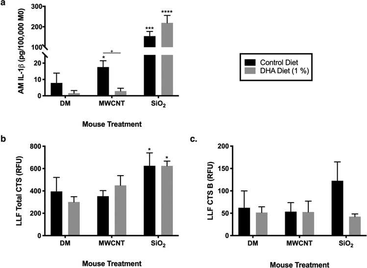
Objective and design: The omega-3 polyunsaturated fatty acid docosahexaenoic acid (DHA) has been reported to suppress inflammation. Pulmonary inflammation can be directly linked to exposure of various occupational and man-made particles leading to pulmonary diseases. Therapeutic treatments are lacking for particle-induced pulmonary inflammation. These studies evaluated DHA as a therapeutic treatment for semi-acute and chronic particle-induced pulmonary inflammation.

Methods: Balb/c mice were oropharyngeal instilled with hydrophobic multi-walled carbon nanotube (MWCNT) or hydrophilic crystalline silica (SiO2) either as one instillation (semi-acute) or once a week for 4 weeks (chronic). One week later, the mice were placed on either a control or 1% DHA-containing diet for 3 weeks (semi-acute) or 12 weeks (chronic). Mice were assessed for inflammatory signaling within the lung lavage fluid, impact on phagolysosomal membrane permeability, shifts of macrophage phenotype gene expression (M1, M2a, M2b, and M2c), and pulmonary histopathology.

Results: DHA increased pulmonary inflammatory markers and lung pathology when mice were exposed to SiO2. There were trending decreases of inflammatory markers for MWCNT-exposed mice with DHA treatment, however, mostly not statistically significant.

Conclusion: The anti-inflammatory benefits of DHA treatment depend upon the type of inflammatory particle, magnitude of inflammation, and duration of treatment.

Keywords: Crystalline silica; Docosahexaenoic acid; Macrophage phenotype; Multi-walled carbon nanotube; Phagolysosomal membrane damage; Pulmonary inflammation.


Conflict of interest statement

5.2 Conflicts of interest/Competing interests

The authors have no conflicts of interest or competing interests to declare.

Figures

Fig. 1
Fig. 1. Pulmonary cells and lung injury analysis in semi-acute model.
Balb/c mice were instilled once with either DM-only, MWCNT, or SiO2 at week 0, kept on the normal mouse chow for 1 week, and then put on either a control or 1% DHA diet for 3 weeks. (a) Total cell counts and (b) percentage of cell types via cytospins of cell differentials were assessed. (c) LDH and (d) protein concentration were assessed in the LLF for lung damage. Data presented as mean ± SEM, n=4–6. *P<0.05, **P<0.01, ***P<0.001, ****P<0.0001 compared to DM, + compared to different diet within the same exposure group. Analyses were done with log-transformed data
Fig. 2
Fig. 2. Cytokine analysis in semi-acute model.
Balb/c mice were instilled once with either DM-only, MWCNT, or SiO2 at week 0, kept on the normal mouse chow for 1 week, and then put on either a control or 1% DHA diet for 3 weeks. Inflammatory cytokines; IFN-γ, IL-1β, TNFα, IL-33, pro-resolving cytokines; IL-10, and IL-13, and IL-6 were analyzed in the LLF. Data presented as mean ± SEM, n=4–6. *P<0.05, **P<0.01, ****P<0.0001 compared to DM, + compared to different diet within the same exposure group. Analyses were done with log-transformed data
Fig. 3
Fig. 3. Pulmonary cells and lung injury analysis in chronic model.
Balb/c mice were instilled with either DM-only, MWCNT, or SiO2 once a week at weeks 0, 1, 2, 3 while on the normal mouse chow and at week 4 were put on a control or 1% DHA diet for 12 weeks. (a) Total cell counts and (b) percentage of cell types via cytospins of cell differentials were assessed. (c) LDH and (d) protein concentration were assessed in the LLF for lung damage. Data presented as mean ± SEM, n=4–7. *P<0.05, **P<0.01, ***P<0.001, ****P<0.0001 compared to DM, + compared to different diet within the same exposure group. Analyses were done with log-transformed data
Fig. 4
Fig. 4. Cytokine analysis in chronic model.
Balb/c mice were instilled with either DM-only, MWCNT, or SiO2 once a week at weeks 0, 1, 2, 3 while on the normal mouse chow and at week 4 were put on a control or 1% DHA diet for 12 weeks. Inflammatory cytokines; IFN-γ, IL-1β, TNFα, IL-33, pro-resolving cytokines; IL-10, and IL-13, and IL-6 were analyzed in the LLF. Data presented as mean ± SEM, n=4–7. *P<0.05, **P<0.01, ***P<0.001, ****P<0.0001 compared to DM, + compared to different diet within the same exposure group. Analyses were done with log-transformed data
Fig. 5
Fig. 5. Assessment of LMP in semi-acute model.
Balb/c mice were instilled once with either DM-only, MWCNT, or SiO2 at week 0, kept on the normal mouse chow for 1 week, and then put on either a control or 1% DHA diet for 3 weeks. LMP was assessed through detection of (a) IL-1β release directly from AMs ex vivo, (b) cathepsin release within the LLF, and (c) cathepsin B release within the LLF. Data presented as mean ± SEM, n=3–6. *P<0.05, ***P<0.001 ****P<0.0001 compared to DM, + compared to different diet within the same exposure group. Analyses were done with log-transformed data. CTS; cathepsin
Fig. 6
Fig. 6. Assessment of LMP in chronic model.
Balb/c mice were instilled with either DM-only, MWCNT, or SiO2 once a week at weeks 0, 1, 2, 3 while on the normal mouse chow and at week 4 were put on a control or 1% DHA diet for 12 weeks. LMP was assessed through detection of (a) IL-1β release directly from AMs ex vivo, (b) cathepsin release within the LLF, and (c) cathepsin B release within the LLF. Data presented as mean ± SEM, n=4–7. *P<0.05, **P<0.01, ***P<0.001, ****P<0.0001 compared to DM, + compared to different diet within the same exposure group. Analyses were done with log-transformed data. CTS; cathepsin
Fig. 7
Fig. 7. Impact of particle exposure and DHA treatment on macrophage phenotype in semi-acute model.
Balb/c mice were instilled once with either DM-only, MWCNT, or SiO2 at week 0, kept on the normal mouse chow for 1 week, and then put on either a control or 1% DHA diet for 3 weeks. Relative gene expression of control diet MWCNT or SiO2 mice were normalized to control diet DM-only mice for assessment of particle effect in (a) lung and (c) spleen tissue. Relative gene expression of 1% DHA diet mice were normalized to corresponding control diet mice for assessment of diet effect in (b) lung and (d) spleen tissue. Each bar within the heat maps consist of 5–7 mice for lung tissue and 4–5 mice for spleen tissue
Fig. 8
Fig. 8. Impact of particle exposure and DHA treatment on macrophage phenotype in chronic model.
Balb/c mice were instilled with either DM-only, MWCNT, or SiO2 once a week at weeks 0, 1, 2, 3 while on the normal mouse chow and at week 4 were put on a control or 1% DHA diet for 12 weeks. Relative gene expression of control diet MWCNT or SiO2 mice were normalized to control diet DM-only mice for assessment of particle effect in (a) lung and (c) spleen tissue. Relative gene expression of 1% DHA diet mice were normalized to corresponding control diet mice for assessment of diet effect in (b) lung and (d) spleen tissue. Each bar within the heat maps consist of 5–7 mice for lung tissue and 4–6 mice for spleen tissue
Fig. 9
Fig. 9. Histopathology analysis of lung tissue in semi-acute model.
Balb/c mice were instilled once with either DM-only, MWCNT, or SiO2 at week 0, kept on the normal mouse chow for 1 week, and then put on either a control or 1% DHA diet for 3 weeks. (a) Pathology was scored blinded by two observers; data presented as median ± SEM. (b) Airway thickness was measured blinded; data presented as mean ± SEM. (c) Representative images (10x) of Masson’s trichrome-stained lung airway sections. n=3; **P<0.01 compared to DM. Arrows indicate particle encapsulation, blue staining indicates collagen deposition
Fig. 10
Fig. 10. Histopathology analysis of lung tissue in chronic model.
Balb/c mice were instilled with either DM-only, MWCNT, or SiO2 once a week at weeks 0, 1, 2, 3 while on the normal mouse chow and at week 4 were put on a control or 1% DHA diet for 12 weeks. (a) Pathology was scored blinded by two observers; data presented as median ± SEM. (b) Airway thickness was measured blinded; data presented as mean ± SEM. (c) Representative images (10x) of Masson’s trichrome-stained lung airway sections. n=3; *P<0.05, **P<0.01, ***P<0.001 compared to DM, + compared to different diet within the same exposure group. Arrows indicate particle encapsulation, arrow head indicates increased lung airway thickness, and blue staining indicates collagen deposition

References

    1. Calder PC Omega-3 fatty acids and inflammatory processes: From molecules to man. Biochemical Society Transactions. 2017;45:1105–1115. 10.1042/BST20160474 - DOI - PubMed
    1. Molendi-Coste O, Legry V, & Leclercq IA Why and How Meet n-3 PUFA Dietary Recommendations? Gastroenterology Research and Practice. 2011;364040. 10.1155/2011/364040 - DOI - PMC - PubMed
    1. Swanson D, Block R, & Mousa SA Omega-3 Fatty Acids EPA and DHA: Health Benefits Throughout Life. Advances in Nutrition. 2012;3:1–7. 10.3945/an.111.000893 - DOI - PMC - PubMed
    1. Bates MA, Brandenberger C, Langohr II, Kumagai K, Lock AL, Harkema JR, Holian A, & Pestka JJ Silica-Triggered Autoimmunity in Lupus-Prone Mice Blocked by Docosahexaenoic Acid Consumption. PLOS ONE. 2016;11(8):e0160622. 10.1371/journal.pone.0160622 - DOI - PMC - PubMed
    1. Li X-Y, Hao L, Liu Y-H, Chen C-Y, Pai VJ, & Kang JX Protection against fine particle-induced pulmonary and systemic inflammation by omega-3 polyunsaturated fatty acids. Biochimica Et Biophysica Acta. General Subjects. 2017;1861:577–584. 10.1016/j.bbagen.2016.12.018 - DOI - PubMed

MeSH terms

LinkOut - more resources