

Characterization of a Murine Model System to Study MicroRNA-147 During Inflammatory Organ Injury
- PMID: 33566257
- PMCID: PMC7873671
- DOI: 10.1007/s10753-021-01427-w
Characterization of a Murine Model System to Study MicroRNA-147 During Inflammatory Organ Injury
Abstract
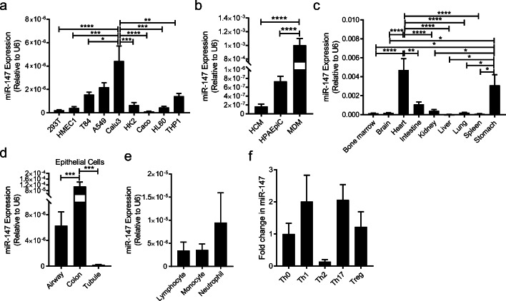
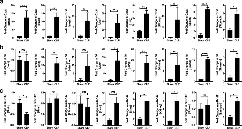
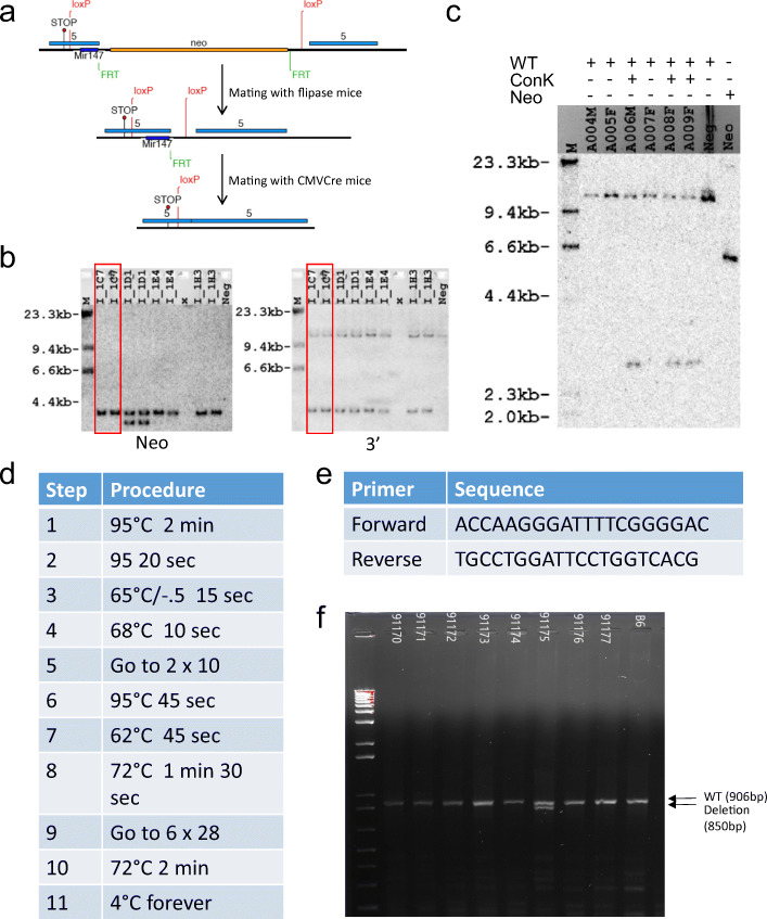
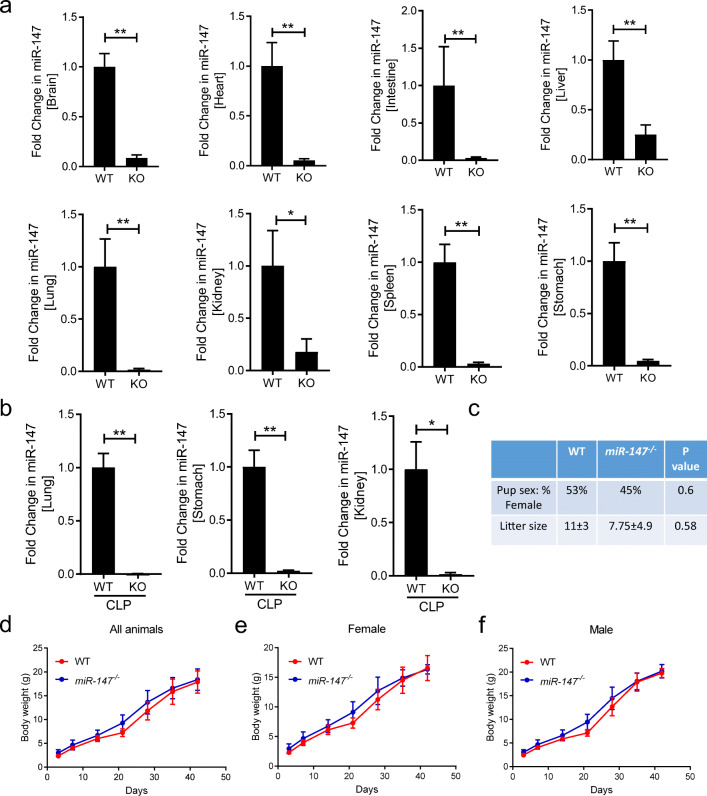
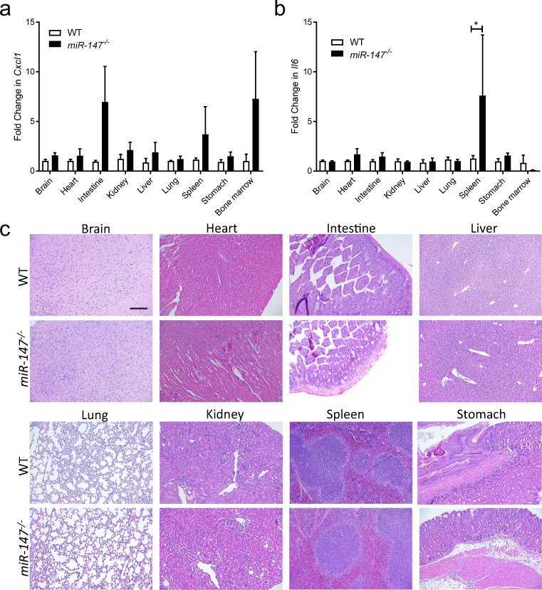
Inflammatory organ injury and sepsis have profound impacts on the morbidity and mortality of surgical and critical care patients. MicroRNAs are small RNAs composed of 20-25 nucleotides that have a significant contribution to gene regulation. MicroRNA-147 (miR-147), in particular, has been shown to have an emerging role in different physiological functions such as cell cycle regulation and inflammatory responses. However, animal model systems to study tissue-specific functions of miR-147 during inflammatory conditions in vivo are lacking. In the present study, we characterize miR-147 expression in different organs and cell types. Next, we generated a transgenic mouse line with a floxed miR-147 gene. Subsequently, we used this mouse line to generate mice with whole-body deletion of miR-147 (miR-147 -/-) by crossing "floxed" miR-147 mice with transgenic mice expressing Cre recombinase in all tissues (CMVcre mice). Systematic analysis of miR-147 -/- mice demonstrates normal growth, development, and off-spring. In addition, deletion of the target gene in different organs was successful at baseline or during inflammation, including the heart, intestine, stomach, liver, spleen, bone marrow, lungs, kidneys, or stomach. Moreover, miR-147 -/- mice have identical baseline inflammatory gene expression compared to C57BL/6 mice, except elevated IL-6 expression in the spleen (7.5 fold, p < 0.05). Taken together, our data show the successful development of a transgenic animal model for tissue and cell-specific deletion of miR-147 that can be used to study the functional roles of miR-147 during inflammatory organ injury.
Keywords: Inflammation; MicroRNA; MicroRNA-147; Organ injury; Transgenic mouse model.
© 2021. The Author(s), under exclusive licence to Springer Science+Business Media, LLC part of Springer Nature.
Conflict of interest statement
The authors declare no competing interests.
Figures
Similar articles
-
miR-27a is up regulated and promotes inflammatory response in sepsis.Cell Immunol. 2014 Aug;290(2):190-5. doi: 10.1016/j.cellimm.2014.06.006. Epub 2014 Jul 10. Cell Immunol. 2014. PMID: 25043848
-
MicroRNA-34a Inhibition Alleviates Lung Injury in Cecal Ligation and Puncture Induced Septic Mice.Front Immunol. 2020 Aug 13;11:1829. doi: 10.3389/fimmu.2020.01829. eCollection 2020. Front Immunol. 2020. PMID: 32903604 Free PMC article.
-
microRNA-98 protects sepsis mice from cardiac dysfunction, liver and lung injury by negatively regulating HMGA2 through inhibiting NF-κB signaling pathway.Cell Cycle. 2019 Aug;18(16):1948-1964. doi: 10.1080/15384101.2019.1635869. Epub 2019 Jul 7. Cell Cycle. 2019. Retraction in: Cell Cycle. 2022 Dec;21(23):2552. doi: 10.1080/15384101.2022.2097800. PMID: 31234706 Free PMC article. Retracted.
-
MicroRNA-181b Inhibits Inflammatory Response and Reduces Myocardial Injury in Sepsis by Downregulating HMGB1.Inflammation. 2021 Aug;44(4):1263-1273. doi: 10.1007/s10753-020-01411-w. Epub 2021 Jun 2. Inflammation. 2021. PMID: 34076811
-
MicroRNA-155: Regulation of Immune Cells in Sepsis.Mediators Inflamm. 2021 Jan 8;2021:8874854. doi: 10.1155/2021/8874854. eCollection 2021. Mediators Inflamm. 2021. PMID: 33505221 Free PMC article. Review.
Cited by
-
Deficiency of peroxisome proliferator-activated receptor α attenuates apoptosis and promotes migration of vascular smooth muscle cells.Biochem Biophys Rep. 2021 Jul 28;27:101091. doi: 10.1016/j.bbrep.2021.101091. eCollection 2021 Sep. Biochem Biophys Rep. 2021. PMID: 34381883 Free PMC article.
-
Co-regulation of circadian clock genes and microRNAs in bone metabolism.J Zhejiang Univ Sci B. 2022 Jul 15;23(7):529-546. doi: 10.1631/jzus.B2100958. J Zhejiang Univ Sci B. 2022. PMID: 35794684 Free PMC article. Review.
-
Alternative adenosine Receptor activation: The netrin-Adora2b link.Front Pharmacol. 2022 Jul 15;13:944994. doi: 10.3389/fphar.2022.944994. eCollection 2022. Front Pharmacol. 2022. PMID: 35910389 Free PMC article. Review.
-
Expression of MicroRNAs in Sepsis-Related Organ Dysfunction: A Systematic Review.Int J Mol Sci. 2022 Aug 19;23(16):9354. doi: 10.3390/ijms23169354. Int J Mol Sci. 2022. PMID: 36012630 Free PMC article.
References
-
- Blanch L, Fernandez R, Mancebo J. Acute respiratory distress syndrome. The New England Journal of Medicine. 1995;332(24):1649. - PubMed
MeSH terms
Substances
Grants and funding
LinkOut - more resources
Full Text Sources
Other Literature Sources
Medical
Molecular Biology Databases
