. 2021 Mar;35(3):e21380.
doi: 10.1096/fj.202002087R.

Interleukin-11 (IL-11) receptor cleavage by the rhomboid protease RHBDL2 induces IL-11 trans-signaling

Affiliations

Interleukin-11 (IL-11) receptor cleavage by the rhomboid protease RHBDL2 induces IL-11 trans-signaling

Lydia Koch et al. FASEB J. 2021 Mar.

Abstract

Interleukin-11 (IL-11) is a pleiotropic cytokine with both pro- and anti-inflammatory properties. It activates its target cells via binding to the membrane-bound IL-11 receptor (IL-11R), which then recruits a homodimer of the ubiquitously expressed, signal-transducing receptor gp130. Besides this classic signaling pathway, IL-11 can also bind to soluble forms of the IL-11R (sIL-11R), and IL-11/sIL-11R complexes activate cells via the induction of gp130 homodimerization (trans-signaling). We have previously reported that the metalloprotease ADAM10 cleaves the membrane-bound IL-11R and thereby generates sIL-11R. In this study, we identify the rhomboid intramembrane protease RHBDL2 as a so far unrecognized alternative sheddase that can efficiently trigger IL-11R secretion. We determine the cleavage site used by RHBDL2, which is located in the extracellular part of the receptor in close proximity to the plasma membrane, between Ala-370 and Ser-371. Furthermore, we identify critical amino acid residues within the transmembrane helix that are required for IL-11R proteolysis. We also show that ectopically expressed RHBDL2 is able to cleave the IL-11R within the early secretory pathway and not only at the plasma membrane, indicating that its subcellular localization plays a central role in controlling its activity. Moreover, RHBDL2-derived sIL-11R is biologically active and able to perform IL-11 trans-signaling. Finally, we show that the human mutation IL-11R-A370V does not impede IL-11 classic signaling, but prevents RHBDL2-mediated IL-11R cleavage.

Keywords: RHBDL2; cytokine; interleukin-11; protease; rhomboid.


Conflict of interest statement

CG has received funding support from Corvidia Therapeutics (Waltham, MA, USA).

Figures
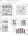
FIGURE 1
FIGURE 1
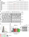
The IL‐11R is a substrate for RHDBL2. A, Juxtamembrane and transmembrane region of human EGF (from Trp‐1026 to Arg‐1064) and human IL‐11R (from Ser‐362 to Gly‐400). The RHBDL2 cleavage site within EGF with an alanine residue at the P1 positions (highlighted red) is indicated with a black arrowhead, the corresponding potential RHDBL2 cleavage sites within the IL‐11R are indicated with black arrowheads and a question marks. The alanine residue at the P1 positions are highlighted in bold and red font. The amino acid residues belonging to the TMHs are underlined. B, HEK293 cells were co‐transfected with expression plasmids encoding either RHDBL1, RHBDL2, RHBDL3, or RHBDL4 and an expression plasmid encoding either human IL‐11R or control as indicated. Proteolysis of the IL‐11R and expression of the four rhomboid proteases was analyzed by western blotting of cell lysates. Actinin was visualized as loading control. sIL‐11R was precipitated from cell culture supernatant by TCA precipitation and also analyzed by western blot. C, HEK293 cells were transfected with IL‐11R, RHDBL2 wt, the inactive variant RHBDL2‐SA, or IL‐11R in combination with both RHDBL2 variants as indicated. The experiment was performed as described for panel b. D, HEK293 cells were co‐transfected with expression plasmids encoding either RHDBL1, RHBDL2, RHBDL3, or RHBDL4 and an expression plasmid encoding human IL‐6R as indicated. Proteolysis of the IL‐6R was analyzed by western blotting of cell lysates. Actinin was visualized as loading control. sIL‐6R was precipitated from cell culture supernatant by TCA precipitation and also analyzed by western blot. E, HEK293 cells were co‐transfected with expression plasmids encoding IL‐11R and RHDBL2. Cells were treated with the rhomboid inhibitor DCI at a concentration of 20 µM overnight when indicated. Afterward, cells and supernatant were analyzed as described for panel b. F, HEK293 cells were co‐transfected with an expression plasmid encoding IL‐11R or co‐transfected with expression plasmids encoding IL‐11R and RHDBL2. The experiment was in principle performed as described for panels b and e, but cells were treated with the broad‐spectrum metalloprotease inhibitor marimastat (MM, 10 µM) or the ADAM10 inhibitor GI254023X (GI, 3 µM) for 4 hours were indicated. G, 1.8 × 10⁶ HEK293 cells were seeded in a 10 cm dish and transiently transfected with expression plasmids encoding IL‐11R and RHBDL2. One day later, cells were detached and transferred into a 96‐well plate in 100 µL medium. Ten hours later, different amounts of marimastat (0.01‐10 µM) or DCI (0.02‐20 µM) in triplicate were added overnight and cell viability determined the following day. SN: supernatant
FIGURE 2
FIGURE 2
RHDBL2 cleaves the IL‐11R after Ala‐370, but not after Ala‐367. A, Juxtamembrane and transmembrane region of the human IL‐11R (from Gln‐351 to Gly‐400) is depicted as IL‐11R wt. The alanine residues representing two potential RHBDL2 cleavage sites are shown in bold font. Below the wild‐type sequence, all other deletion variants and point mutations used in the experiments shown in this figure are listed. Modifications of the amino acid sequence in comparison to the IL‐11R wild type are marked in red and bold font. B, All IL‐11R variants listed in panel a were transiently transfected in HEK293 cells, either together with RHBDL2 or with mock control. Expression and proteolysis of the different IL‐11R variants and expression of RHBDL2 were analyzed by western blotting of cell lysates. Actinin was visualized as loading control. sIL‐11R was precipitated from cell culture supernatant by TCA precipitation and also analyzed by western blotting. C, D, Cell surface expression of the different IL‐11R variants used in the experiment described in panel B was analyzed by flow cytometry. Shown are one representative experiment in panel C and the mean ± SD from three independent experiments in panel D. Expression of IL‐11R wild type was set to 1 and the expression of all other variants calculated accordingly. E, HEK293 cells were transiently transfected with expression plasmids encoding IL‐11R wild type, IL‐11R‐A370F, or left untransfected. Two days after transfection, medium was removed, cells were washed and stimulated in serum‐free medium with 1 µM of the ionophore ionomycin for 60 minutes to activate ADAM10 or with DMSO as control. Proteolysis of the IL‐11R variants was analyzed by western blot of cell lysates. Actinin was visualized as loading control. sIL‐11R was precipitated from cell culture supernatant by TCA precipitation and also analyzed by western blot. SN: supernatant
FIGURE 3
FIGURE 3
Helix‐destabilizing amino acid residues within the TMH of the IL‐11R are required for RHBDL2‐mediated cleavage. A, Juxtamembrane and transmembrane region of the human IL‐11R (from Gln‐351 to Gly‐400) is depicted as IL‐11R wt. The RHBDL2 cleavage motif is shown in bold blue font. Potential destabilizing amino acid residues within the TMH are indicated in bold red font. A structural representation of the TMH with the N‐terminal cleavage motif is shown above. Below the wild‐type sequence, all point mutations used in the experiments shown in this figure are listed. Modifications of the amino acid sequence in comparison to the IL‐11R wild type are marked in red and bold font. B, All IL‐11R variants listed in panel a were transiently transfected in HEK293 cells, either together with RHBDL2 or with mock control. Expression and proteolysis of the different IL‐11R variants and expression of RHBDL2 were analyzed by western blotting of cell lysates. Actinin was visualized as loading control. sIL‐11R was precipitated from cell culture supernatant by TCA precipitation and also analyzed by western blot. C, D, Cell surface expression of the different IL‐11R variants used in the experiment described in panel B was analyzed by flow cytometry. Shown are one representative experiment in panel C and the mean ± SD from three independent experiments in panel D. Expression of IL‐11R wild type was set to 1 and the expression of all other variants calculated accordingly. SN: supernatant
FIGURE 4
FIGURE 4
RHDBL2 can cleave the IL‐11R in the early secretory pathway. A, HeLa cells were co‐transfected with expression plasmids encoding IL‐11R wild type and RHBDL2. Localization of both proteins was determined via fluorescence microscopy. B, IL‐11R wild type, IL‐11R with a C‐terminal ER retention signal (IL‐11R‐KKSS) and the patient mutation IL‐11R‐R296W were transiently transfected in HEK293 cells, either together with RHBDL2 or with mock control. Expression and proteolysis of the different IL‐11R variants and expression of RHBDL2 were analyzed by western blotting of cell lysates. Actinin was visualized as loading control. sIL‐11R was precipitated from cell culture supernatant by TCA precipitation and also analyzed by western blot. C, IL‐11R wild type was transiently transfected in HEK293 cells either with mock control or different RHBDL2 variants (RHBDL2 wt, RHBDL2‐SA, RHBDL2‐KDEL, and RHBDL2‐KDEL‐SA). The experiment was then conducted as described in panel b. SN: supernatant
FIGURE 5
FIGURE 5
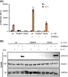
RHDBL2‐derived sIL‐11R is biologically active. A, Equal amounts of Ba/F3‐gp130 cells were incubated in triplicate with supernatants of HEK293 cells that were transfected with either IL‐11R wt, IL‐11R‐R296W, or IL‐11R‐KKSS, either together with control or with RHBDL2. Supernatant was either supplemented with recombinant IL‐11 (500 ng/mL) or left untreated. Cells were incubated for 2 days and cell viability determined as described in Materials and Methods. Shown are the mean ± SD from one experiment out of three independent experiments with similar outcome. B, Equal amounts of Ba/F3‐gp130 cells were stimulated for 15 minutes with the supernatants described in the legend of panel A and recombinant IL‐11 was added were indicated. Phosphorylation of STAT3 was determined in cell lysates via western blot. Total STAT3 levels and GAPDH were visualized to ensure equal protein loading
FIGURE 6
FIGURE 6
The IL‐11R variant IL‐11R‐A370V is biologically active, but not cleaved by RHBDL2. A, Juxtamembrane and transmembrane region of the human IL‐11R (from Gln‐351 to Gly‐400) is depicted as IL‐11R wt. The mutation A370V is indicated in bold red font. B, IL‐11R wild type and the variant IL‐11R‐A370V were transiently transfected in HEK293 cells, either together with RHBDL2 or with mock control. Expression and proteolysis of the different IL‐11R variants and expression of RHBDL2 were analyzed by western blotting of cell lysates. Actinin was visualized as loading control. sIL‐11R was precipitated from cell culture supernatant by TCA precipitation and also analyzed by western blot. C, Equal amounts of Ba/F3‐gp130 cells were stimulated for 15 minutes with the supernatants described in the legend of panel b and recombinant IL‐11 was added were indicated. Phosphorylation of STAT3 was determined in cell lysates via western blot. Total STAT3 levels and GAPDH were visualized to ensure equal protein loading. D, Cell surface amounts of the IL‐11R on Ba/F3‐gp130, Ba/F3‐gp130‐IL‐11R, and Ba/F3‐gp130‐IL‐11R‐A370V cells were determined via flow cytometry. FACS plots depict one experiment out of three independent experiments. Control staining (ctl) was performed without the primary antibody. E, Equal amounts of Ba/F3‐gp130‐IL‐11R‐A370V cells were incubated in triplicate with increasing amounts (0.001‐100 ng/mL) of either recombinant IL‐11 or Hyper‐IL‐6 as indicated. Cells were incubated for 2 days and cell viability determined as described in Materials and Methods. Shown are the mean ± SD from one experiment out of three independent experiments with similar outcome. F, Equal amounts of Ba/F3‐gp130‐IL‐11R and Ba/F3‐gp130‐IL‐11R‐A370V cells were stimulated for 15 minutes with 10 ng/mL Hyper‐IL‐6, 10 ng/mL IL‐11, or left unstimulated. Phosphorylation of STAT3 was determined in cell lysates via western blot. Total STAT3 levels and GAPDH were visualized to ensure equal protein loading. SN: supernatant

References

    1. Garbers C, Scheller J. Interleukin‐6 and interleukin‐11: same same but different. Biol Chem. 2013;394:1145‐1161. - PubMed
    1. Putoczki T, Ernst M. More than a sidekick: the IL‐6 family cytokine IL‐11 links inflammation to cancer. J Leukoc Biol. 2010;88:1109‐1117. - PubMed
    1. Garbers C, Hermanns H, Schaper F, et al. Plasticity and cross‐talk of Interleukin 6‐type cytokines. Cytokine Growth Factor Rev. 2012;23:85‐97. - PubMed
    1. Putoczki TL, Ernst M. IL‐11 signaling as a therapeutic target for cancer. Immunotherapy. 2015;7:441‐453. - PubMed
    1. Suen Y, Chang M, Lee SM, Buzby JS, Cairo MS. Regulation of interleukin‐11 protein and mRNA expression in neonatal and adult fibroblasts and endothelial cells. Blood. 1994;84:4125‐4134. - PubMed

Publication types

LinkOut - more resources