. 2021 Apr 1;32(7):521-537.
doi: 10.1091/mbc.E20-11-0720. Epub 2021 Feb 10.

Inner-nuclear-membrane-associated degradation employs Dfm1-independent retrotranslocation and alleviates misfolded transmembrane-protein toxicity

Affiliations

Inner-nuclear-membrane-associated degradation employs Dfm1-independent retrotranslocation and alleviates misfolded transmembrane-protein toxicity

Matthew P Flagg et al. Mol Biol Cell. .

Abstract

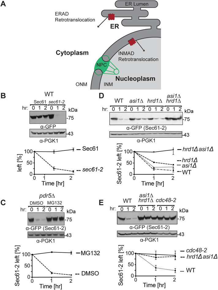
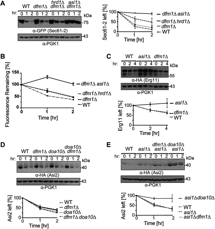
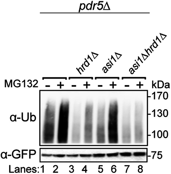
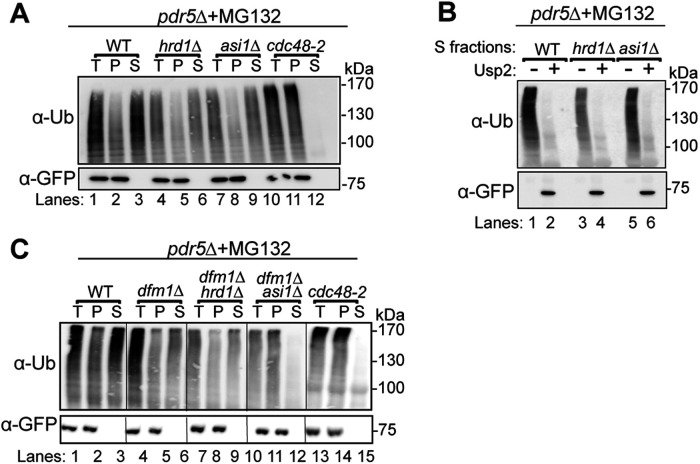
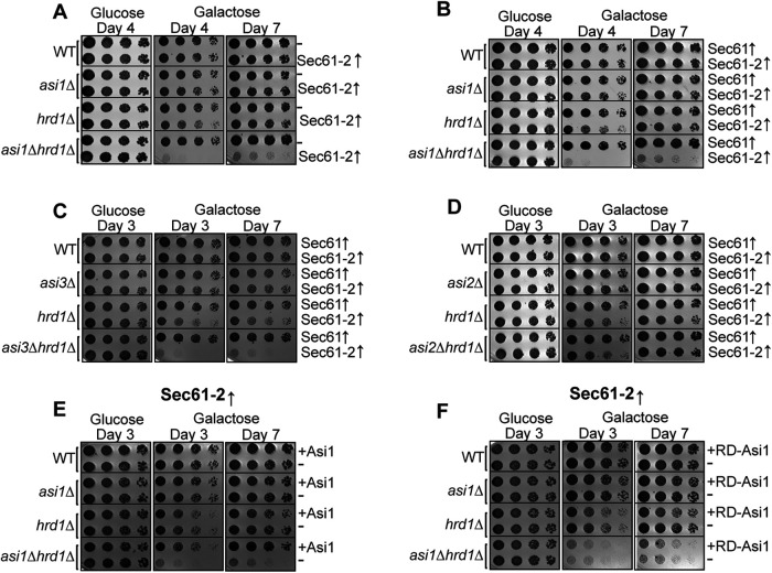
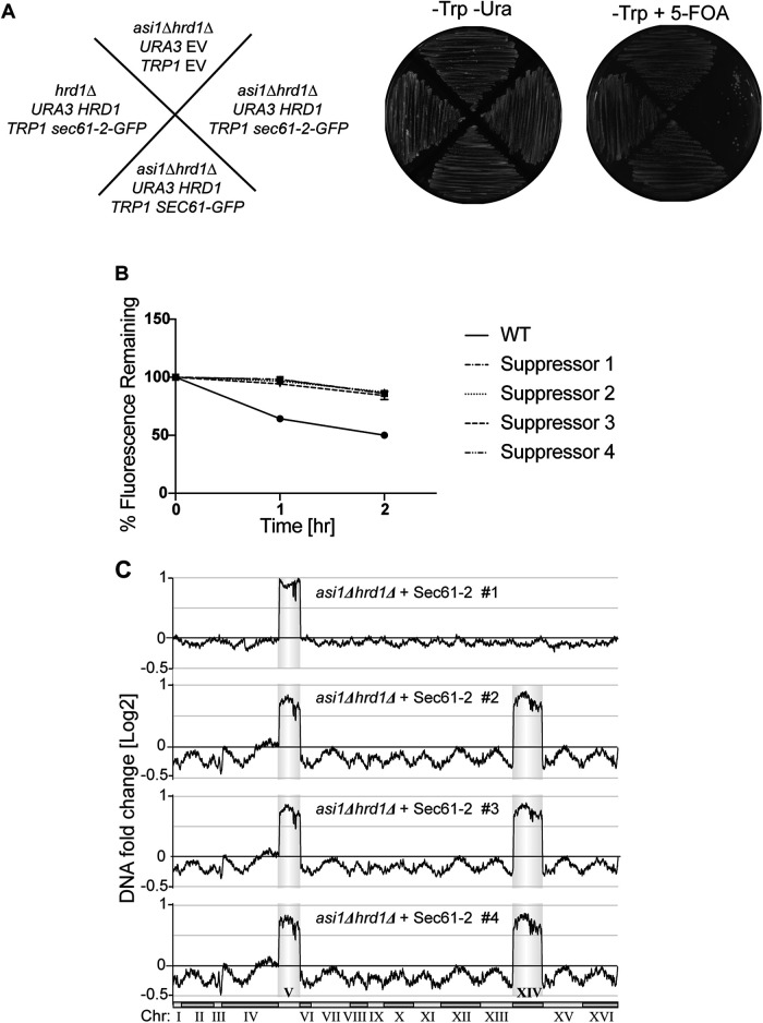
Before their delivery to and degradation by the 26S proteasome, misfolded transmembrane proteins of the endoplasmic reticulum (ER) and inner-nuclear membrane (INM) must be extracted from lipid bilayers. This extraction process, known as retrotranslocation, requires both quality-control E3 ubiquitin ligases and dislocation factors that diminish the energetic cost of dislodging the transmembrane segments of a protein. Recently, we showed that retrotranslocation of all ER transmembrane proteins requires the Dfm1 rhomboid pseudoprotease. However, we did not investigate whether Dfm1 also mediated retrotranslocation of transmembrane substrates in the INM, which is contiguous with the ER but functionally separated from it by nucleoporins. Here, we show that canonical retrotranslocation occurs during INM-associated degradation (INMAD) but proceeds independently of Dfm1. Despite this independence, ER-associated degradation (ERAD)-M and INMAD cooperate to mitigate proteotoxicity. We show a novel misfolded-transmembrane-protein toxicity that elicits genetic suppression, demonstrating the cell's ability to tolerate a toxic burden of misfolded transmembrane proteins without functional INMAD or ERAD-M. This strikingly contrasted the suppression of the dfm1Δ null, which leads to the resumption of ERAD-M through HRD-complex remodeling. Thus, we conclude that INM retrotranslocation proceeds through a novel, private channel that can be studied by virtue of its role in alleviating membrane-associated proteotoxicity.


Figures

FIGURE 1:
FIGURE 1:
Sec61-2-GFP is quality-control substrate of Hrd1 and Asi1. (A) Depiction of the contiguous ER and INM. A subset of ER proteins can diffuse through the nuclear pore complex (NPC) into the INM. Both the 26S proteosome and Cdc48 can access the nucleoplasm through nucleoporins, and cell physiology thus supports ERAD retrotranslocation into the cytoplasm and INMAD retrotranslocation into the nucleoplasm. (B) Sec61-GFP is stable, whereas sec61-2 GFP is a degraded. Isogenic strains expressing Sec61-GFP or Sec61-2-GFP were grown into log phase, and the degradation of each protein was measured using cycloheximide chase (CHX). After the addition of CHX, cells were collected and lysed at the indicated times. Lysates were analyzed by SDS–PAGE and immunoblotting with α-GFP and α-Pgk1. Densitometry was performed using ImageJ, and the α-GFP signal was normalized to α-Pgk1 signal. t = 0 was taken as 100% ,and data plotted are mean ± SD from three experiments. (C) Sec61-2-GFP is stabilized by the proteasome inhibitor MG132. A pdr5Δ strain expressing Sec61-2-GFP was grown into log phase and then treated with either MG132 (25 µg/ml) or DMSO. Degradation was then measured by CHX. After the addition of CHX, cells were collected and lysed at the indicated times. Lysates were analyzed by SDS–PAGE and immunoblotting with α-GFP and α-Pgk1. Data plotted are mean ± SD from three experiments. (D) Sec61-2-GFP degradation depends on both Hrd1 and Asi1. WT, hrd1Δ, asi1Δ, and hrd1Δasi1Δ strains expressing Sec61-2-GFP were subjected to CHX. After the addition of CHX, cells were collected and lysed at the indicated times. Lysates were analyzed by SDS–PAGE and immunoblotting with α-GFP and α-Pgk1. Data plotted are mean ± SD from three experiments. (E) Sec61-2-GFP degradation requires the Cdc48 ATPase. WT, hrd1Δasi1Δ, and retrotranslocation-deficient cdc48-2 strains expressing Sec61-2-GFP were subjected to CHX. After the addition of CHX, cells were collected and lysed at the indicated times. Lysates were analyzed by SDS–PAGE and immunoblotting with α-GFP and α-Pgk1. Data plotted are mean ± SD from three experiments.
FIGURE 2:
FIGURE 2:
INMAD proceeds independently of Dfm1. (A) Dfm1 acts downstream of Hrd1 and in parallel with the Asi complex. WT, dfm1Δ, hrd1Δdfm1Δ, and asi1Δdfm1Δ strains expressing Sec61-2-GFP were subjected to CHX. After the addition of CHX, cells were collected and lysed at the indicated times. Lysates were analyzed by SDS–PAGE and immunoblotting with α-GFP and α-Pgk1. Data plotted are mean ± SD from three experiments. (B) Sec61-2-GFP degradation is recapitulated by flow cytometry. WT, dfm1Δ, hrd1Δdfm1Δ, and asi1Δdfm1Δ strains expressing Sec61-2-GFP were subjected to CHX. After the addition of CHX, cells were assayed for fluorescence by flow cytometry, and at each time point, the mean fluorescence of 10,000 cells was measured. t = 0 was taken as 100%, and data plotted are the mean ± SD from three experiments. (C) Erg11-3HA degradation is Dfm1 independent. WT, dfm1Δ, and asi1Δ strains expressing Erg11-3HA were subjected to CHX. After the addition of CHX, cells were collected and lysed at the indicated times. Lysates were analyzed by SDS–PAGE and immunoblotting with α-HA and α-Pgk1. Data plotted are mean ± SD from three experiments. (D) HA-Asi2 is stabilized in neither dfm1Δ nor doa10Δ strains. WT, dfm1Δ, and doa10Δ strains were subjected to CHX. After the addition of CHX, cells were collected and lysed at the indicated times. Lysates were analyzed by SDS–PAGE and immunoblotting with α-HA and α-Pgk1. Data plotted are mean ± SD from three experiments. (E) HA-Asi2 degradation by Doa10 and the Asi complex is Dfm1 independent. WT, asi1Δ, asi1Δdfm1Δ, and asi1Δdoa10Δ strains were subjected to CHX. After the addition of CHX, cells were collected and lysed at the indicated times. Lysates were analyzed by SDS–PAGE and immunoblotting with α-HA and α-Pgk1. Data plotted are mean ± SD from three experiments.
FIGURE 3:
FIGURE 3:
Both Asi1 and Hrd1 ubiquitinate Sec61-2-GFP in vivo. The indicated strains expressing Sec61-2-GFP were grown into log phase and treated with MG132 or a vehicle control (DMSO). Cells were lysed, and microsomes were collected and immunoprecipitated with α-GFP. Samples were then subjected to SDS–PAGE and immunoblot by α-ubiquitin and α-GFP. One of three biological replicates is shown.
FIGURE 4:
FIGURE 4:
Retrotranslocation of full-length Sec61-2-GFP. (A) In vivo retrotranslocation of Sec61-2-GFP through both Hrd1 and Asi channels. WT, hrd1Δ, asi1Δ, and cdc48-2 strains expressing Sec61-2-GFP were grown into log phase and treated with MG132 (25 µg/ml). Crude lysates were ultracentrifuged to separate Sec61-2-GFP that has been retrotranslocated into the soluble fraction (S) and Sec61-2-GFP that has not been retrotranslocated from membrane (P). Sec61-2-GFP was immunoprecipitated from both fractions and then analyzed by SDS–PAGE and immunoblotting with α-GFP and α-ubiquitin. One representative of three biological replicates is shown. (B) In vivo retrotranslocated Sec61-2-GFP is full length. WT, hrd1Δ, asi1Δ, and cdc48-2 strains expressing Sec61-2-GFP were grown into log phase and treated with MG132 (25 µg/ml). Crude lysates were ultracentrifuged to separate Sec61-2-GFP to collect retrotranslocated Sec61-2-GFP from soluble fractions. Solubilized Sec61-2-GFP was immunoprecipitated and then either treated with either buffer (–) or the catalytic core of the deubiquitinase Usp2 (+). Samples were analyzed by SDS–PAGE and immunoblotted with α-GFP and α-ubiquitin. One representative of three biological replicates is shown. (C) In vivo retrotranslocation of Sec61-2-GFP through Asi1 is Dfm1 independent. WT, dfm1Δ, dfm1Δhrd1Δ, dfm1Δasi1Δ, and cdc48-2 strains expressing Sec61-2-GFP were grown into log phase and treated with MG132 (25 µg/ml). Crude lysates were ultracentrifuged to separate Sec61-2-GFP that has been retrotranslocated into the soluble fraction (S) and Sec61-2-GFP that has not been retrotranslocated from membrane (P). Sec61-2-GFP was immunoprecipitated from both fractions and then analyzed by SDS–PAGE and immunoblotting with α-GFP and α-ubiquitin. One representative of three biological replicates is shown.
FIGURE 5:
FIGURE 5:
Sec61-2-GFP is lethal to cells lacking INMAD and ERAD. (A, B) Galactose-induced Sec61-2-GFP expression is lethal to asi1Δhrd1Δ cells. WT, asi1Δ, hrd1Δ, and asi1Δhrd1Δ cells bearing empty vector (–), GAL-driven Sec61-GFP, or GAL-driven Sec61-2-GFP were monitored for growth by dilution assay. Fivefold dilutions of each strain were spotted onto glucose- or galactose-containing plates to induce Sec61-GFP and Sec61-2-GFP overexpression. Plates were incubated at 30°C and imaged at the indicated times. One representative of three biological replicates is shown. (C) Galactose-induced Sec61-2-GFP expression is also lethal to asi3Δhrd1Δ cells. WT, asi3Δ, hrd1Δ, and asi1Δhrd1Δ cells bearing GAL-driven Sec61-GFP or GAL-driven Sec61-2-GFP were monitored for growth by dilution assay. Fivefold dilutions of each strain were spotted onto glucose- or galactose-containing plates to induce Sec61-GFP and Sec61-2-GFP overexpression. Plates were incubated at 30°C and imaged at the indicated times. One representative of three biological replicates is shown. (D) Galactose-induced Sec61-2-GFP expression is not lethal to asi2Δhrd1Δ cells. WT, asi3Δ, hrd1Δ, and asi2Δhrd1Δ cells bearing GAL-driven Sec61-GFP or GAL-driven Sec61-2-GFP were monitored for growth by dilution assay. Fivefold dilutions of each strain were spotted onto glucose- or galactose-containing plates to induce Sec61-GFP and Sec61-2-GFP overexpression. Plates were incubated at 30°C and imaged at the indicated times. One representative of three biological replicates is shown. (E, F) Asi1 catalytic activity is required to prevent Sec61-2-GFP lethality. WT, asi1Δ, hrd1Δ, and asi1Δhrd1Δ cells bearing GAL-driven Sec61-2-GFP were cotransformed with empty vector (–), WT ASI1, or RING-dead ASI1 (RD-Asi1). These strains were then monitored for growth by dilution assay. Fivefold dilutions of each strain were spotted onto glucose- or galactose-containing plates to induce Sec61-GFP and Sec61-2-GFP overexpression. Plates were incubated at 30°C and imaged at the indicated times. One representative of three biological replicates is shown.
FIGURE 6:
FIGURE 6:
Suppressees of Sec61-2-GFP lethality are ChrV and XIV aneuploids. (A) Constitutive overexpression of Sec61-2-GFP is lethal to asi1Δhrd1Δ cells. Left, schematic denoting the genotypes of each strain tested before exposure to 5-FOA. Center and right, the indicated strains were streaked onto plates that either selected (-Trp -Ura) or counterselected the URA3 plasmids. Plates were incubated at 30°C for 2 d before imaging. One representative of three biological replicates is shown. (B) Lethality suppressees cannot degrade the Sec61-2-GFP. Four suppressees and a WT strain expressing Sec61-2-GFP were subjected to CHX chase. After the addition of CHX, cells were assayed for fluorescence by flow cytometry, and at each time point, the mean fluorescence of 10,000 cells was measured. t = 0 was taken as 100%, and data plotted are the mean ± SD from three experiments. (C) Genome profiling reveals duplications of ChrV and XIV in suppressees. Chromosome profiles of whole-genome sequencing are mapped across the yeast genome. Copy number is indicated on the y-axis, and the chromosome number is indicated on the x-axis. Reads from each of four suppressees are shown.

Similar articles

Cited by

References

    1. Antebi A, Fink GR (1992). The yeast Ca(2+)-ATPase homologue, PMR1, is required for normal Golgi function and localizes in a novel Golgi-like distribution. Mol Biol Cell 3, 633–654. - PMC - PubMed
    1. Balakrishnan R, Park J, Karra K, Hitz BC, Binkley G, Hong EL, Sullivan J, Micklem G, Cherry JM (2012). YeastMine—an integrated data warehouse for Saccharomyces cerevisiae data as a multipurpose tool-kit. Database (Oxford) 2012, bar062. - PMC - PubMed
    1. Baldridge RD, Rapoport TA (2016). Autoubiquitination of the Hrd1 ligase triggers protein retrotranslocation in ERAD. Cell 166, 394–407. - PMC - PubMed
    1. Biederer T, Volkwein C, Sommer T (1996). Degradation of subunits of the Sec61p complex, an integral component of the ER membrane, by the ubiquitin-proteasome pathway. EMBO J 15, 2069–2076. - PMC - PubMed
    1. Boban M, Pantazopoulou M, Schick A, Ljungdahl PO, Foisner R (2014). A nuclear ubiquitin-proteasome pathway targets the inner nuclear membrane protein Asi2 for degradation. J Cell Sci 127, 3603–3613. - PMC - PubMed

Publication types

MeSH terms