. 2021 Feb 8:116:e200443.
doi: 10.1590/0074-02760200443. eCollection 2021.

Morphology and morphogenesis of SARS-CoV-2 in Vero-E6 cells

Affiliations

Morphology and morphogenesis of SARS-CoV-2 in Vero-E6 cells

Debora Ferreira Barreto-Vieira et al. Mem Inst Oswaldo Cruz. .

Abstract

Background: The coronaviruses (CoVs) called the attention of the world for causing outbreaks of severe acute respiratory syndrome (SARS-CoV), in Asia in 2002-03, and respiratory disease in the Middle East (MERS-CoV), in 2012. In December 2019, yet again a new coronavirus (SARS-CoV-2) first identified in Wuhan, China, was associated with a severe respiratory infection, known today as COVID-19. This new virus quickly spread throughout China and 30 additional countries. As result, the World Health Organization (WHO) elevated the status of the COVID-19 outbreak from emergency of international concern to pandemic on March 11, 2020. The impact of COVID-19 on public health and economy fueled a worldwide race to approve therapeutic and prophylactic agents, but so far, there are no specific antiviral drugs or vaccines available. In current scenario, the development of in vitro systems for viral mass production and for testing antiviral and vaccine candidates proves to be an urgent matter.

Objective: The objective of this paper is study the biology of SARS-CoV-2 in Vero-E6 cells at the ultrastructural level.

Methods: In this study, we documented, by transmission electron microscopy and real-time reverse transcription polymerase chain reaction (RT-PCR), the infection of Vero-E6 cells with SARS-CoV-2 samples isolated from Brazilian patients.

Findings: The infected cells presented cytopathic effects and SARS-CoV-2 particles were observed attached to the cell surface and inside cytoplasmic vesicles. The entry of the virus into cells occurred through the endocytic pathway or by fusion of the viral envelope with the cell membrane. Assembled nucleocapsids were verified inside rough endoplasmic reticulum cisterns (RER). Viral maturation seemed to occur by budding of viral particles from the RER into smooth membrane vesicles.

Main conclusions: Therefore, the susceptibility of Vero-E6 cells to SARS-CoV-2 infection and the viral pathway inside the cells were demonstrated by ultrastructural analysis.


Figures

Fig. 1:
Fig. 1:. ultrastructural analyses of Vero-E6 cells by electron microscopy. (A) Uninfected cell presenting no morphological alterations. (B-H) Vero-E6 cell, 72 h post infection (hpi) with severe acute respiratory syndrome (SARS-CoV-2), presenting numerous filopodia (head arrow) and vesicles (V). SARS-CoV-2 particles (arrow), nucleus (N), cell cytoplasm (CC).
Fig. 2:
Fig. 2:. ultrastructural alterations in Vero-E6 72 h post infection (hpi) with severe acute respiratory syndrome (SARS-CoV-2). (A) Uninfected cell presenting no morphological alterations in mitochondria and in rough endoplasmic reticulum cistern (RER). (B-E) Alterations and degeneration of mitochondria (*). (F) Rough endoplasmic reticulum cistern with more electron-dense ribosomes. (B, G) Thickening of the rough endoplasmic reticulum cistern (star). (G) Presence of clathrin-coated vesicles (arrow heads, inset). Nucleus (N), cell cytoplasm (CC).
Fig. 3:
Fig. 3:. intense smooth vesicle proliferation (V) in Vero-E6 72 h post infection (hpi) with severe acute respiratory syndrome (SARS-CoV-2). (A-D) Vesicle (V) proliferation. Virus particle (B, arrow), nucleus (N). (E) Cell cytoplasm presenting numerous myelin figures (concentric membrane arrays) (*).
Fig. 4:
Fig. 4:. (A-C) alterations of the nucleus (N) chromatin profile in Vero-E6 72 h post infection (hpi) with severe acute respiratory syndrome (SARS-CoV-2). (D) Viral nucleocapsid associated with the nucleus membrane (arrow). Cell cytoplasm (CC).
Fig. 5:
Fig. 5:. attachment and entry of severe acute respiratory syndrome (SARS-CoV-2) in Vero-E6 cells 72 h post infection (hpi). (A, B, E) Several virus particles (arrows) attached to cell membranes were observed. (C, D) Entry of virus particles into cells by the endocytic pathway and by fusion of virus envelopes with cell membranes (head arrows) (B). (E) Nucleocapsids inside swollen rough endoplasmic reticulum cistern (circles). Cell cytoplasm (CC), endocytic vesicles (EV), nucleus (N).
Fig. 6:
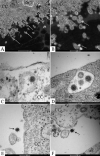
Fig. 6:. release of severe acute respiratory syndrome (SARS-CoV-2) in Vero-E6 cells 72 h post infection (hpi). (A-E) Virions (arrow) attached to cell filopodia (arrow) and inside smooth vesicles (asterisk) at the periphery of the cell and with your membrane fused with membrane cell were observed. (E-F) Virus particles presenting spherical morphology, displaying spikes, and a diameter between 80 and 100 nm. Cell cytoplasm (CC), nucleus (N).

References

    1. Lin L, Lianfeng Lu , Wei Cao , Taisheng Li Hypothesis for potential pathogenesis of SARS-CoV-2 infection - a review of immune changes in patients with viral pneumonia. Emerg Microbes Infect. 2020;9(1):727–732. - PMC - PubMed
    1. Sun P, Qie S, Liu Z, Ren J, Li K, Xi J. Clinical characteristics of 50 466 hospitalized patients with 2019-nCoV infection. J Med Virol. 2020;92(7):716–718. - PMC - PubMed
    1. Lai CC, Tzu-Ping S, Wen-ChienKo HJ, Po-RenHsueh Severe acute respiratory syndrome coronavirus 2 (SARS-CoV-2) and coronavirus disease-2019 (COVID-19) The epidemic and the challenges. Int J Antimicrob Agents. 2020;55(3):105924–105924. - PMC - PubMed
    1. Lu H, Stratton CW, Tang YW. Outbreak of pneumonia of unknown etiology in Wuhan China the mystery and the miracle. J Med Virol. 2020;92(4):401–402. - PMC - PubMed
    1. Li Q, Xuhua G, Peng W, Xiaoye W, Lei Z, Yeqing T. Early transmission dynamics in Wuhan, China, of novel coronavirus-infected pneumonia. N Engl J Med. 2020;382(13):1199–1207. - PMC - PubMed