

Case Reports
doi: 10.36660/abc.20190485.
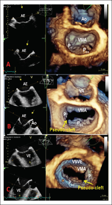
Three-dimensional Echocardiography Reveals the True Enemy in a Young Male with ST-Elevation Myocardial Infarction and Severe Mitral Regurgitation: Posterior Mitral Valve "Pseudo-Cleft" and Prolapse
[Article in
English,
Portuguese]
Affiliations
- PMID: 33567002
- PMCID: PMC8118632
- DOI: 10.36660/abc.20190485
Item in Clipboard
Case Reports
Three-dimensional Echocardiography Reveals the True Enemy in a Young Male with ST-Elevation Myocardial Infarction and Severe Mitral Regurgitation: Posterior Mitral Valve "Pseudo-Cleft" and Prolapse
[Article in
English,
Portuguese]
Arq Bras Cardiol.
2021 Feb.
No abstract available
Conflict of interest statement
Declaro não haver conflito de interesses pertinentes.
Figures


References
-
- Lang RM, Mor-Avi V, Sugeng L, Nieman PS, Sahn DJ. Three-dimensional echocardiography: the benefits of the additional dimension. J Am Coll Cardiol. 2006;48(10):2053–2069. - PubMed
- 1. Lang RM, Mor-Avi V, Sugeng L, Nieman PS, Sahn DJ. Three-dimensional echocardiography: the benefits of the additional dimension. J Am Coll Cardiol. 2006;48(10):2053-69. - PubMed
-
- Amin A, Davis M, Auseon A. Isolated cleft posterior mitral valve leaflet: an uncommon cause of mitral regurgitation. European journal of echocardiography : J Am Soc Echocardiogr. 2009;10(1):173–174. - PubMed
- 2. Amin A, Davis M, Auseon A. Isolated cleft posterior mitral valve leaflet: an uncommon cause of mitral regurgitation. European journal of echocardiography : J Am Soc Echocardiogr. 2009;10(1):173-4. - PubMed
-
- Miglioranza MH, Muraru D, Mihaila S, Haertel JC, Iliceto S, Badano LP. Isolated Anterior Mitral Valve Leaflet Cleft: 3D Transthoracic Echocardiography-Guided Surgical Strategy. Arq Bras Cardiol. 2015;104(5):e49–e52. - PMC - PubMed
- 3. Miglioranza MH, Muraru D, Mihaila S, Haertel JC, Iliceto S, Badano LP. Isolated Anterior Mitral Valve Leaflet Cleft: 3D Transthoracic Echocardiography-Guided Surgical Strategy. Arq Bras Cardiol. 2015;104(5):e49-52. - PMC - PubMed
-
- Narang A, Addetia K, Weinert L, Yamat M, Shah AP, Blair JE, et al. Diagnosis of Isolated Cleft Mitral Valve Using Three-Dimensional Echocardiography. J Am Soc Echocardiogr. 2018;31(11):1161–1167. - PMC - PubMed
- 4. Narang A, Addetia K, Weinert L, Yamat M, Shah AP, Blair JE, et al. Diagnosis of Isolated Cleft Mitral Valve Using Three-Dimensional Echocardiography. J Am Soc Echocardiogr. 2018;31(11):1161-7. - PMC - PubMed
-
- McEnany MT, English TA, Ross DN. The congenitally cleft posterior mitral valve leaflet. An anticedent to mitral regurgitation. Ann Thorac Surg. 1973;16(3):281–292. - PubMed
- 5. McEnany MT, English TA, Ross DN. The congenitally cleft posterior mitral valve leaflet. An anticedent to mitral regurgitation. Ann Thorac Surg. 1973;16(3):281-92. - PubMed
Publication types
MeSH terms
LinkOut - more resources
Full Text Sources
Other Literature Sources
Medical
